
THE IMMUNE SYSTEMLec. 1
HOST DEFENCE
. Whether the organisms invade and cause disease
is determined by the balance of the pathogenicity
of the organism (i.e. the virulence factors that it
has ) and the integrity of the host defence
mechanisms..
The immune system
protect the host from pathogens
while minimising damage to self tissue.
the immune system not only protects against
infection, but can lead to autoimmune diseases.
so Dysfunction or deficiency of the immune response
leads to a wide variety of diseases, involving every
organ system in the body.
Immunity is often divided into two types
1- innate and
2-adaptive (acquired or specific), although in
practice these overlap and interlink
...

1-INNATE IMMUNITY
Innate immunity provides the immediately active,
first line, non-specific host defence mechanisms. It
includes
A-
physical (e.g. epithelial cells,skin and
mucous
membrane,coughreflex,mucociliaryCiliary
paralysis (smoking, primary ciliarydyskinesis
syndromes)
,Increased mucus production
(asthma) ,
washing,tear,saliva,urine)
B-
chemical (e.g. 'natural' antimicrobial
substances like defenses at surface barrierseg.
Gastric acid secreation)
C- Biological Colonization resistance provided
by nonpathogenic commensal Organisims of
skin and gut
d-those
directly activated by infectious agents,
tissue damage or tumours , )
consist of
1-Cellular such as phagocytic cells (neutrophils,
and monocytes in the blood; macrophages

including dendritic cells in tissues, natural killer
cell, eosinophils, mast cells and basophils)
2-humoral components (e.g. complement,
acute-phase reactants, cytokines).
Initiation of the inflammatory response:
Phagocytosiss
Phagocytes ('eating cells') are specialised cells which
ingest and kill microorganisms,.
They include
neutrophils, monocytes and macrophages,
and are
particularly important for defence against bacterial
and fungal infections
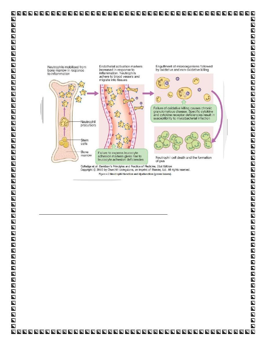
neutrophils travel within the blood They are short-
lived cells with a half-life of 6 hours
. A series of
events leads to the recruitment and activation of
these cells at the site of tissue damage.
Cell recruitment:
Recruitment of cells of the immune system
(phagocytes and lymphocytes) to tissue sites
involves

1-cellular adhesion molecules (CAM) ). The
main ones are the
a-intercellular adhesion
molecules (ICAM),
b-integrins,and
c-selectins
Adhesion molecules associate with cytoskeletal
components to cause cytoskeletal reorganization,
resulting in migration and spreading, allowing the
cells to move
2-ChemoattractantsCells move towards the
site of inflammation in response to
chemoattractants (chemicals which attract
cells) at sites of infection or tissue damage.
The cells pass between endothelial cells into the
tissues by the formation of foot-like processes
(pseudopodia) that push through the intercellular
spaces; this process is called diapedesis. The cells
continue to move along the chemoattractant
gradient to the site of infection ).
Once the neutrophils have been recruited,
phagocytosis (ingestion) and intracellular
killing of microbes begins. Phagocytosis

occurs by the formation of pseudopodia
(projections of cytoplasmic membrane)
around the organism or particle to be ingested
). Owing to the fluidity of the cell membrane,
the tips eventually fuse to form a membrane-
bound vesicle called a phagosome. This fuses
with the neutrophil cytoplasmic granules,
lysosom ) to form a phagolysosome. Within
this localized environment, killing occurs
Ingestion and killing of organisms is much
more effective if the particle is first coated or
opsonized ('made ready to eat') with a-
specific antibody , b-acute phase protein
and c-complement.
This is because
neutrophils have receptors for the Fc portion
of antibody molecules (FcR), and complement
(CR). Binding of cell-surface receptors to
complement and antibody on the particle both
increases the strength of adhesion and causes
transduction of intracellular signals, which
activate the cell to promote phagocytic and
killing activity.
Opsonins
include acute phase proteins such as
C-reactive protein (CRP), antibodies and
complement. They bind both to the pathogen
and to phagocyte receptors, acting as a bridge
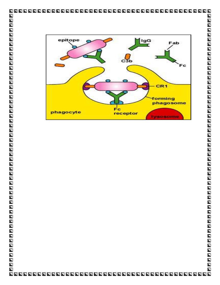
between the two to facilitate phagocytosis (.
Monocytes and macrophages
Monocytes are the precursors of tissue
macrophages
. They are produced in the bone
marrow and exported to the circulation, where they
constitute about 5% of leucocytes. From the blood
stream, they migrate to peripheral tissues where they
differentiate into tissue macrophages and reside for
long periods.
Specialised populations of tissue macrophages
include Kupffer cells in the liver, alveolar
macrophages in the lung, mesangial cells in the
kidney, and microglial cells in the brain.
Macrophages, like neutrophils, are capable of
phagocytosis and killing of microorganisms . Unlike
neutrophils, macrophages do not die after killing
pathogens
Eosinophils in host defence :
eosinophils are most commonly associated with
allergic disease, . Eosinophils have receptors for
IgE which is the major antiparasite antibody,
particularly against nematodes. Eosinophils bind
IgE via the FcεR, and
toxic metabolites are
released from the eosinophil granules
directly
onto the parasite surface

Mast cells and basophils :
Mast cell function appears to be in the
initiation of
inflammatory responses (increased vascular
permeability, bronchoconstriction)
by the release
(following degranulation) of pro-inflammatory
mediators such as
histamine, leukotrienes, platelet-activating factor
(PAF), prostaglandins and some cytokines (e.g. IL-
4).
Basophils are morphologically similar to mast
cells but are found in very small numbers in the
blood.
Dendritic cells/Langerhans' cells
These are derived from the lymphoid and myeloid
cell lines; dendritic cells in the skin are called
Langerhans' cells.
Their major function is to
present antigen to T cells when stimulated
.
Dendritic cells link innate immunity to the

adaptive immune system by being the only cell
that can activate native T cells to initiate an
adaptive immune response
-Natural killer (NK) cells
These non-phagocytic cells have the morphology of
lymphocytes but do not bear the markers for T or B
cells. They are distinguished by the presence of
numerous cytoplasmic granules. They have non-
specific antiviral and antitumour activity, causing
lysis of cells with which they react
-Cytokines
Cytokines are small soluble intercellular
messengers that exert their effect by
binding to specific receptors on target cells..
Cytokines are produced by any cell. Their
biological effect varies according to the
cytokine and the cell involved ,
Function: signal certain cell populations to
activate,
divide or home in on a particular site in the
body.

Cytokines include:
1-Interleukins produced by and signal
between white cells.
2-Chemokines have a chemoattractant
function.
3-Colony-stimulating factors cause
differentiation and proliferation of stem
cells.
4- Tumour necrosis factors. TFN-
α increases
phagocyte function.
5- Interferons
interferons are antiviral agents produced mainly by
fibroblasts
Complement
The complement system comprises a series of at
least 20 glycoproteins that are activated in a
cascade sequence, with proenzymes that undergo
sequential proteolytic cleavage to their active

forms. It is a major part of the innate immune
system
Complement pathway.
Three main pathways of complement
activation exist, termed the classical,
alternative and mannan-binding lectin
(MBL) pathway.
The complement pathways are triggered by different
factors:
Classical pathway
by antigen-antibody immune
complexes, apoptotic cells, C-reactive protein
bound to ligand and certain viruses and bacteria.
Alternative pathway
by bacterial endotoxin,
fungal cell walls, viruses and tumour cells.
Mannan-binding lectin (MBL) pathway
is
activated by microbes with terminal mannose
groups
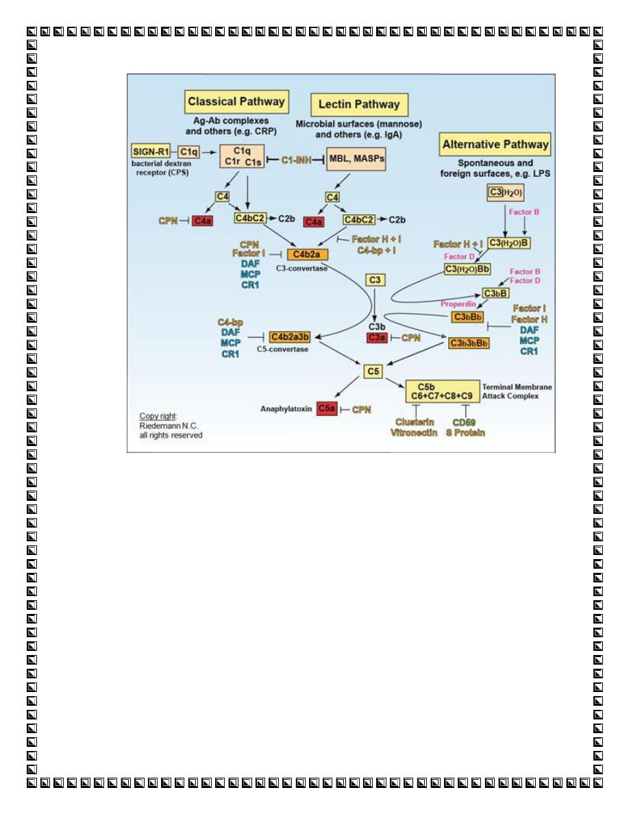
Complement activation is focused at cell membranes.
Host cells are protected from complement-mediated
lysis by
inhibitory surface molecules
, for example
decay accelerating factor (DAF). Most organisms
lack any protective molecules and are therefore
susceptible to complement.

Functions of complement
A-Anti-infective function
:
o
1-opsonization by C3b and C4b
o
2-chemotaxis - attraction of phagocytes
by chemoattractant activation products
o
3-activation of leucocytes by
anaphylatoxins (C5a, C3a and C4a); via
receptors on leucocytes
o
4-lysis of bacteria and cells (C5b-C9).
B-Interplay between innate and adaptive
immune system
. Immunomodulation of B-cell
responses to specific antigen through binding
of complement receptors on B-cell surface,
thus augmenting antibody responses and
immunological memory.
C-Clearance of:
o
immune complexes (C1q, C3 and C4)
.apoptotic cells (C1q, C3 and C4).
ADAPTIVE (SPECIFIC OR
ACQUIRED) IMMUNITY

Consist of
-
cellular(T
–lymphocyteand B-
lymphocyte
)
1-
-
humeral (AntibodY
)
2-
antigen-specific receptor
.
Innate immunity is a rapid non-specific
response
whereas in adaptive immunity The
characters of this response are
:
-
1-the use of antigen-specific receptors on T
lymphocytes (T-cell receptor, TCR) and B
lymphocytes to direct the
response.(response is very focused
)
-
2-The response takes time to develop so
that it cannot provide immediate protection
on first meeting an antigen
.
-
3-the development of memory CELL so
that subsequent exposure leads to a more
rapid response

-
4-Phagocytes only recognize extracellular
organisms, mostly bacteria. In contrast, T
cells are able to combat intracellular
infections, such as viruses, bacteria
(mycobacteria, legionella, listeria, brucella,
salmonella), many fungi and protozoa
.
B-CEL immunoglobulin producing
cell,identified by present of I.G on their
surface
.
These cells comprise approximately 25% of
lymphocytes. B cells divide and are
activated to become plasma cells which
secrete large amounts of antibody
