esophagus
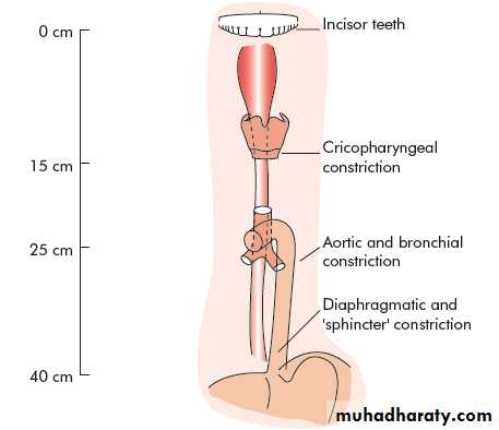
Surgical anatomyThe esophagus is muscular tube 25 cm long.
Extend from cricopharengeus muscle to the cardia of stomach.
The upper part has striated muscle.
Followed by transitional zone.
While the lower half has smooth muscle.
Figure1: endoscopic landmarks from the incisor teeth
Esophagus is innervated by the myenteric (auerbach) plexus, while the meissner plexus is sparse.
physiology
The main function of esophagus is to transfer food from mouth to the stomach.The 1st step in swallowing is the pharyngeal phase(voluntary)
This voluntary swallow initiate the primary peristalsis which is a coordinated esophageal wave under vagal control.
When the primary peristalsis reach the lower sphincter, it relax and opens.
The secondary peristalsis is initiated by the passage of food bolus.
While tertiary peristalsis are non peristaltic and infrequent.
Symptoms
Pharyngeal dysphagiaEsophageal dysphagia
Voluntary
Inability to swallow
Usually caused by neurological causes.
Involuntary
Food sticking
Any cause.
Dysphagia: is difficulty in swallowing.
Odynophagia:
Is painful swallowing caused byGERD
Infective esophagitis
Chemical injury
Regurgitation:
Is the return of esophageal content from above a functional or mechanical obstruction.
Reflux:
Is the passive return of gastro duodenal content to the mouth as part of GERD.Chest pain:
Similar to angina, caused by GERD or motility disorders or even exercise.investigations
• 1.Radiography• this include : a. contrast study
• b. CT scan
• c. MRI
• 2. Endoscopy(OGD):
• For diagnostic purposes( visualization and biopsy), and for therapeutic uses( foreign body removal, dilatation of strictures).
Figure 2: normal barium swallow of esophagus.
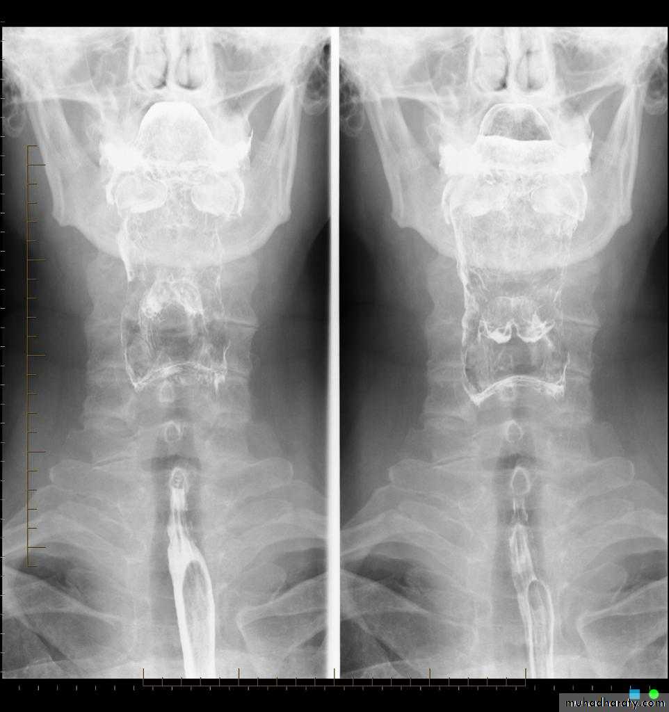
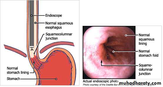
Figure3: OGD of esophagus and cardia
3.Endoscopic ultrasound:
Are transducers at the tip of the OGD and of 2 types:Radial: are rotating transducers giving a circular image.
Linear: for biopsy taking.
4.manometry
Is the measurement of esophageal pressure by the passage of multilumen catheter with many openings at different levels, used to diagnose motility disorder.5. 24 hour pH monitoring:
Is the most useful way to diagnose GERD (pH less than 4 for more than 4% of study time.Foreign bodies in the esophagus
The most common is food bolusThe most dangerous is button batteries in children.
Usually diagnosed by plain radiograph.
Either pass spontaneously or removed by OGD.
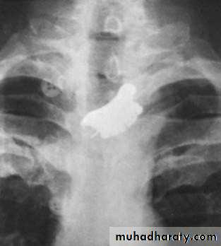
Figure 4: false teeth impacted in the esophagus.
Perforation
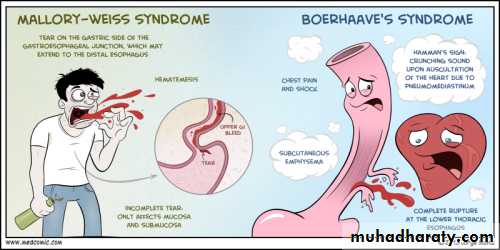
Spontaneous perforation (boerhaave):Occur when a person vomit against closed glottis so the pressure increase rapidly and perforating the esophagus at its weakest point.
Clinical features: 1.chest pain
2. abdominal pain
3. dyspnea
Diagnosis:
• Chest x-ray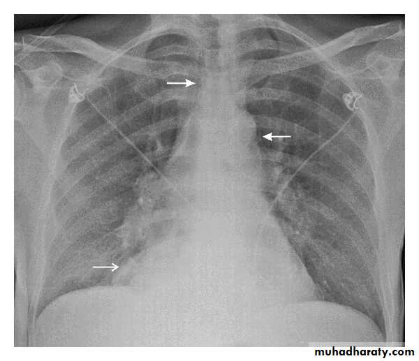
Figure5: CXR showing air in the medistinum
• 2. Water soluble contrast• 3. CT scan
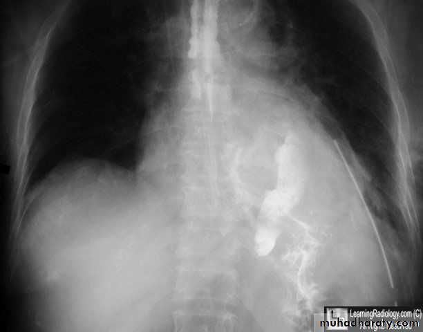
Figure6: contrast material free in the abdomen
Instrumental perforation:Is the most common cause of perforation.
Treatment:
NG tube, antibiotics, hyperalimentation with surgical intervention in deteriorated patients by direct closure or by intercostal muscle flap.
Pathological perforation:
A free perforation of ulcer or tumor and associated with fistula formation.
Mallory- wiess syndrome
A vertical mucosal tear of the cardia caused by forceful vomiting.90% occur in the cardia
Only 10% occur in the esophagus.
Clinical features: hematemisis after large meal and alcoholism.
Treatment: nasogastric tube, antiemetic, OGD, and surgery if condition persist.
Corrosive injury
In attempted suicide or accidental ingestion these injuries may occur by eg: NaOH or sulphuric acid.These agents either acid or alkali.
Alkali are tasteless and colorless so its ingestion is more common.
Alkali cause liquefaction and fibrous scarring.
While acids cause coagulative necrosis and eschar formation.
Acids cause intense pylorospasm with pooling in the antrum so causing more gastric damage.
Treatment:The key is early endoscopy, to determine the extent of damage and subsequent treatment.
If mild so early feeding
If sever with ulceration so feeding jejunostomy.
If there is perforation do esophagectomy.
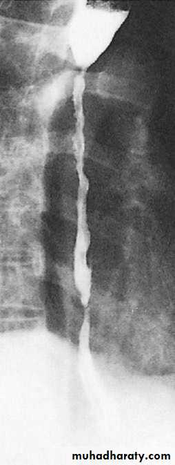
When stricture develop later so treatment is either:Dilatation if small stricture or esophagectomy if large one.
Figure 7: stricture following corrosive injury.
GERD
Normal competence of the gastro- oesophageal junction is maintainedby the LOS. This is influenced by both its physiological
function and its anatomical location relative to the diaphragm
and the oesophageal hiatus. In normal circumstances, the LOS
transiently relaxes as a coordinated part of swallowing, as a
means of allowing vomiting to occur and in response to stretching
of the gastric fundus, particularly after a meal to allow swallowed
air to be vented. Most episodes of physiological reflux
occur during postprandial transient lower oesophageal sphincter
relaxations (TLOSRs). In the early stages of GORD, most
pathological reflux occurs as a result of an increased number of
TLOSRs rather than a persistent fall in overall sphincter pressure.
In more severe GORD, LOS pressure tends to be generallylow, and this loss of sphincter function seems to be made
worse if there is loss of an adequate length of intra-abdominal
oesophagus.
The incidence of GERD is increasing in last 3 decades probably due to obesity.
Clinical features:
A triad of retrosternal burning pain, epigastric pain and regurgitation.
In sever cases there is
• Reflux into the mouth
• Nocturnal reflex
• Odynophagia
• dysphagia
diagnosis
In most cases the diagnosis is assumed rather than proven and treatment is empirical, investigation is required if:• Diagnosis in doubt
• The patient does not respond to PPI.
• If dysphagia develop.
The diagnosis include:
• OGD and biopsy• 24 hour PH monitoring
• Manometry
• Medical treatment
• It include life style change by decreasing weight , smoking, alcohol, tea, coffee, and avoiding large heavy meal at night, and tilting up the head at bed.
2. Antisecretary agents
The most important is PPI
SURGERY
• Endoscopic treatment: it aims to augment the failing LOS and accentuate the angle of His, it include:a. suturing
b. radiofrequency
c. injection of polymers.
Surgical intervention:
All surgical options depend on the creation of intrabdominal segment of esophagus, crural repair and some form of wrap of stomach( fundoplication). They include:• Total fundoplication( nissen) can control reflex effectively but may cause dysphagia.
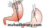
2. Posterior fundoplication( toupet)
3.Anterior fundoplication( Dor,watson)Complication of surgery include shortening or stricture of the esophagus.
Barrett esophagusIs a metaplastic change of the lower esophageal mucosa in response to chronic reflux of gastric content, it occur in 15-20% of patient with GERD.
It may occur in patient mild symptoms.
The hallmark of its epithelium is the presence of mucus secreting goblet cells.
The risk of adenocarcinoma increase 25 folds.
Diagnosis:
By OGD, finding the squamo-columnar junction more proximal, with biopsy taking.
Follow up by every 2 years OGD(no dysplasia)
Treatment:• Of the underlying cause
• Ablation by laser, photodynamic therapy, argon beam and EMR, all used in conjugation with PPIs.