Thymus Thymus is the site of T-Cell differentiation and maturation it is a biolobed gland, situated above heart in the thorax region each lobe is encapsulated and it is dvided into lobules whiche are separated by strands of connective tissue called trabeculae Each lobule contain-lymphocytes& each organized into two compartments 1-Outer cortex 2- Inner medullaFunction:Site of T-cell maturation.Relative size greatest in newbornAbsolute size great at puberty
The cortex contains mostly immature and proliferating thymocytes,Medulla is sparly populated with thymocytes some of which mature and migrate to the medulla in medulla they learn to discriminate between self and non-self during fetal development and for a short time after birth. T-cell leave the medulla to enter the peripheral blood circulation, through which they are transported to the secondary lymphoid organs About 95% of all T cells die in thymus
Beside lymphoid cells it is composed of 1.Epithelial cells(cortical and medullary)2.Macrophage3.Dendritic cells4.Nurse cells5.Hassall,s corpusclesLymphocyte in the thymus are called thymocytes
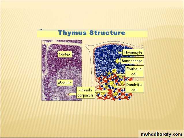
Function of the thymus1-Generate and select T-cells2-Through clonal selection mechanism, thymus cause the death of those T-cell that cannot recognize Ag-MHC-complexes and those that react with self Ag-MHC and stop danger of causing autoimmune disease3- thus about 95% of all T cells die in the thymusNote:Thymectomy is surgical removal of thymusthymectomized mice show decrease in circulating lymphocyte and absence of cell mediated immunity
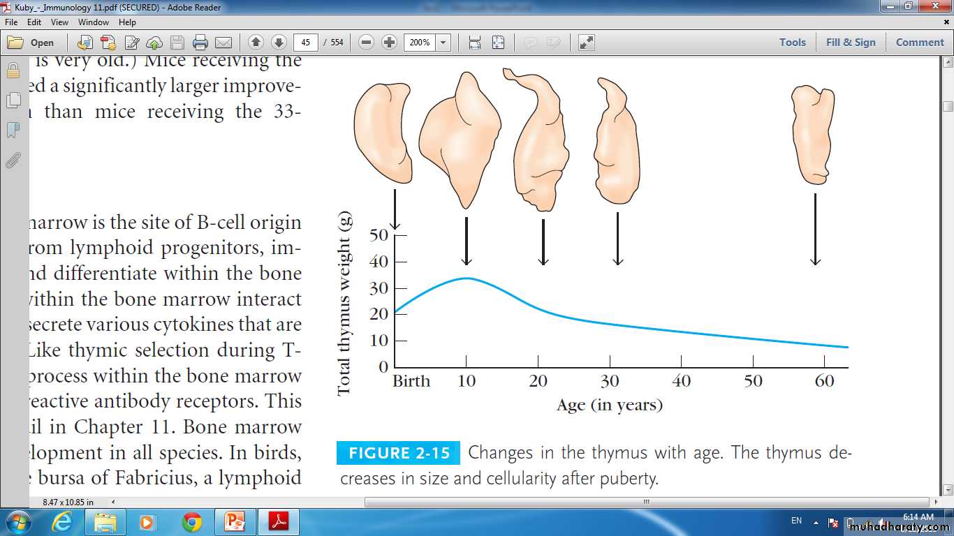
The size of the thymus varies with age
In infants, it is found in the inferior neck and extends into the mediastinum where it partially overlies the heartIt increases in size and is most active during childhood
It stops growing during adolescence and then gradually atrophies
Lymph Nodes
Lymph nodes are the principal lymphoid organs of the bodyNodes are imbedded in connective tissue and clustered along lymphatic vessels
Aggregations of these nodes occur near the body surface in inguinal, axillary, and cervical regions of the body
Chapter 20, Lymphatic System
7
Lymph Nodes
Their two basic functions are:Filtration – macrophages destroy microorganisms and debris
Immune system activation – monitor for antigens and mount an attack against them
Chapter 20, Lymphatic System
8
Structure of a Lymph Node
Nodes are bean shaped and surrounded by a fibrous capsuleTrabeculae extended inward from the capsule and divide the node into compartments
Nodes have two histologically distinct regions: a cortex and a medulla
Chapter 20, Lymphatic System
9
Structure of a Lymph Node
The cortex contains follicles with germinal centers, heavy with dividing B cells
Dendritic cells nearly encapsulate the follicles
The deep cortex houses T cells in transit
T cells circulate continuously among the blood, lymph nodes, and lymphatic stream
Chapter 20, Lymphatic System
10
Structure of a Lymph Node
Medullary cords extend from the cortex and contain B cells, T cells, and plasma cellsThroughout the node are lymph sinuses crisscrossed by reticular fibers
Macrophages reside on these fibers and phagocytize foreign matter
Chapter 20, Lymphatic System
11
Structure of a Lymph Node
Chapter 20, Lymphatic System12
Figure 20.4a, b
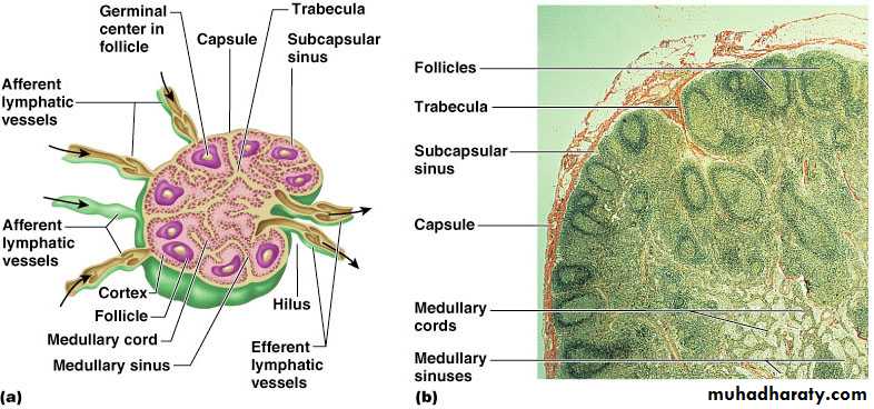
Circulation in the Lymph Nodes
Lymph enters via a number of afferent lymphatic vesselsIt then enters a large subcapsular sinus and travels into a number of smaller sinuses
It meanders through these sinuses and exits the node at the hilus via efferent vessels
Because there are fewer efferent vessels, lymph stagnates somewhat in the node
This allows lymphocytes and macrophages time to carry out their protective functions
Chapter 20, Lymphatic System
13
Other Lymphoid Organs
The spleen, and tonsilsPeyer’s patches and bits of lymphatic tissue scattered in connective tissue
All are composed of reticular connective tissue and all help protect the body
Only lymph nodes filter lymph
Chapter 20, Lymphatic System
14
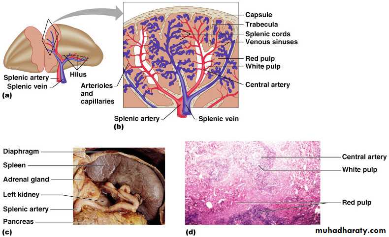
Spleen
Largest lymphoid organ, located on the left side of the abdominal cavity beneath the diaphragmIt extends to curl around the anterior aspect of the stomach
It is served by the splenic artery and vein, which enter and exit at the hilus
Functions
Site of lymphocyte proliferation
Immune surveillance and response
Cleanses the blood by removing old RBC
Chapter 20, Lymphatic System
15
Additional Spleen Functions
Stores breakdown products of RBCs for later reuse
Spleen macrophages salvage and store iron for later use by bone marrow
Site of fetal erythrocyte production (normally ceases after birth)
Stores blood platelets
Chapter 20, Lymphatic System
16
Structure of the Spleen
Surrounded by a fibrous capsule, it has trabeculae that extend inward and contains lymphocytes, macrophages, and huge numbers of erythrocytesTwo distinct areas of the spleen are:
White pulp – area containing mostly lymphocytes suspended on reticular fibers and involved in immune functions
Red pulp – remaining splenic tissue concerned with disposing of worn-out RBCs and bloodborne pathogens
Chapter 20, Lymphatic System
17
Structure of the Spleen
Chapter 20, Lymphatic System18
Figure 20.6a-d
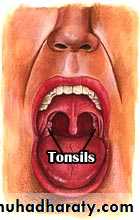
Tonsils
Simplest lymphoid organs; form a ring of lymphatic tissue around the pharynxLocation of the tonsils
Palatine tonsils – either side of the posterior end of the oral cavity
Lingual tonsils – lie at the base of the tongue
Pharyngeal tonsil – posterior wall of the nasopharynx
Tubal tonsils – surround the openings of the auditory tubes into the pharynx
Chapter 20, Lymphatic System
19
Tonsils
Lymphoid tissue of tonsils contains follicles with germinal centersTonsil masses are not fully encapsulated
Epithelial tissue overlying tonsil masses invaginates, forming blind-ended crypts
Crypts trap and destroy bacteria and particulate matter
Chapter 20, Lymphatic System
20
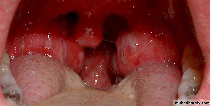
Tonsilitis
Chapter 20, Lymphatic System
21
Peyer’s patches
Isolated clusters of lymphoid tissue, similar to tonsilsFound in the wall of the distal portion of the small intestine
Similar structures are found in the appendix
Peyer’s patches and the appendix:
Destroy bacteria, preventing them from breaching the intestinal wall
Generate “memory” lymphocytes for long-term immunity
Chapter 20, Lymphatic System
22
MALT
Mucosa-Associated Lymphatic Tissue is composed of:Peyer’s patches, tonsils, and the appendix (digestive tract)
Lymphoid nodules in the walls of the bronchi (respiratory tract)
MALT protects the digestive and respiratory systems from foreign matter
Chapter 20, Lymphatic System
23