1
Anatomy
Head and neck
Lec. 1&2
تأليف االستاذ الدكتور
عبدالجبار الحبيطي
--------------------------
تعديل وطباعة الطالب
دي دريد الشيخ نوريَع
ممثل المرحلة
2016
2
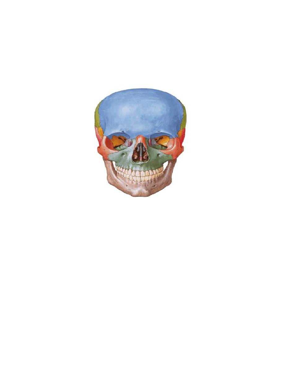
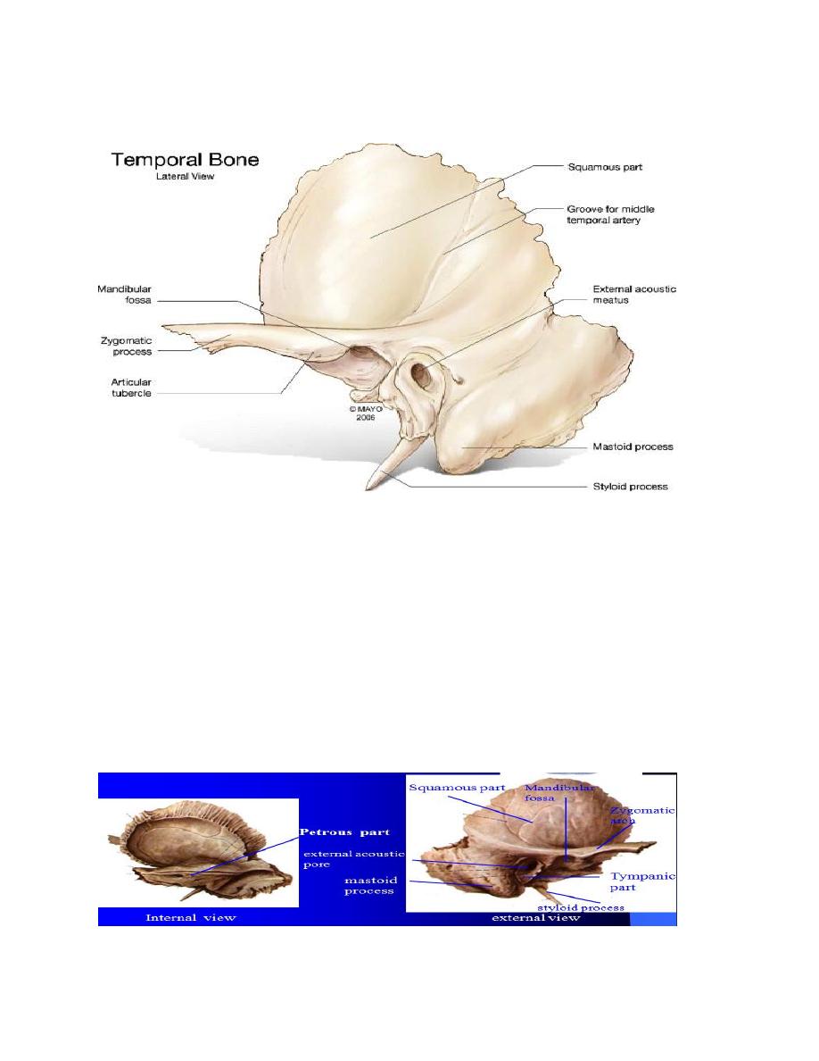
The parts of the temporal bone :
1. Suqamous part , it gives the zygomatic process which forms part of
the zygomatic arch.
2. Petrous part is very hard contains the internal and middle ear, in
addition to the canals for facial nerve and internal carotid artery. The
styloid process projects from its lower surface.
3. Mastoid part having the mastoid process.
4. Tympanic part is a small plate below the external auditory meatus.
3
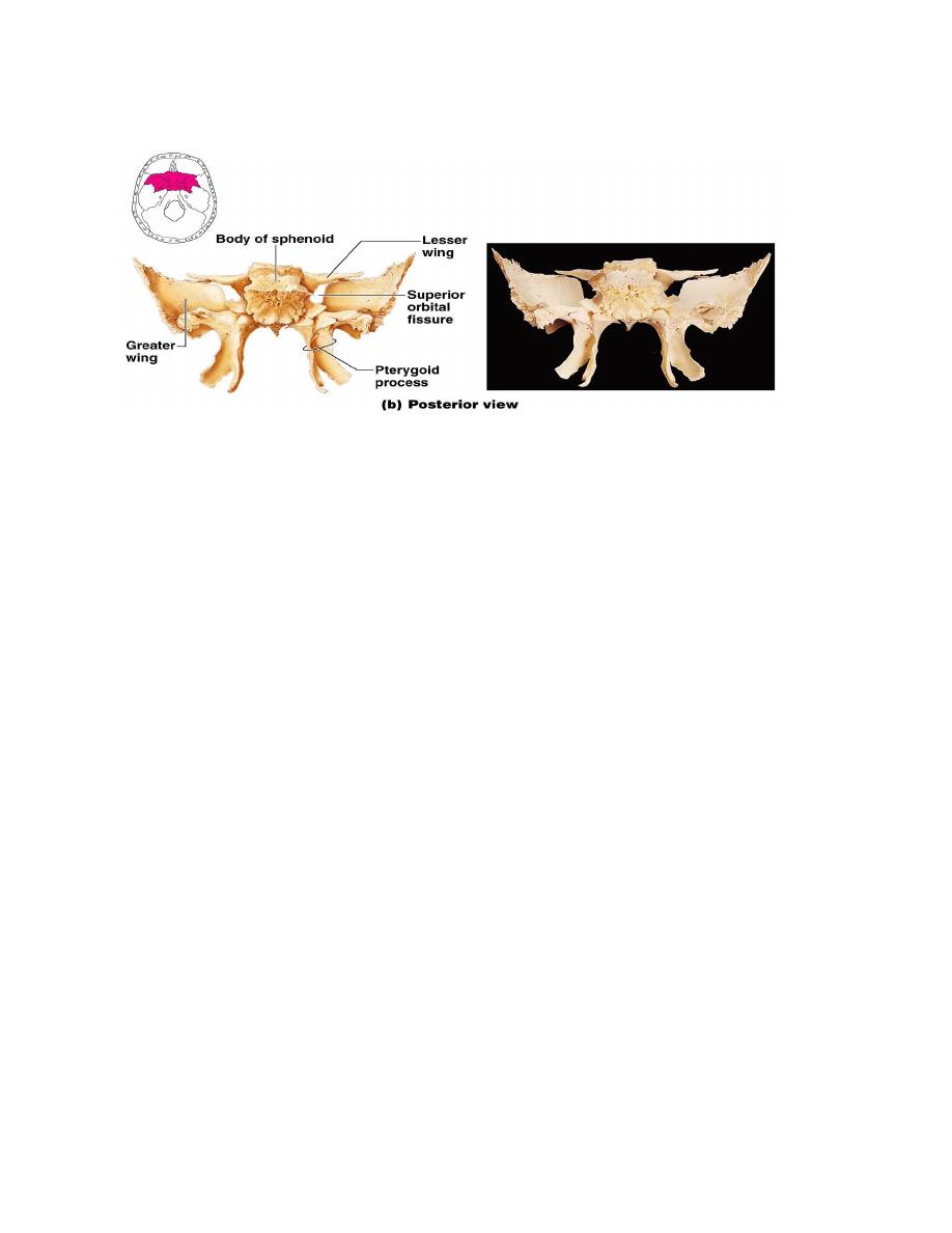
The part of the sphenoid bone :
:
1. Body of the sphenoid, encloses the sphenoid air sinus.
2. Two lesser wings, if s medial end forms the anterior clinoid process,
with the optic canal near to it.
3. Two greater wings forming the floor of middle cranial fossa .it is
separated from the lesser wing by superior orbital fissure.It shows
three ( foramina Rotundum , ovale and Spinosum ).
4. Two pterygoid processes, one on each side. Each pterygoid process
has medial and lateral pterygoid plates with pterygoid fossa between
the two plates.
4
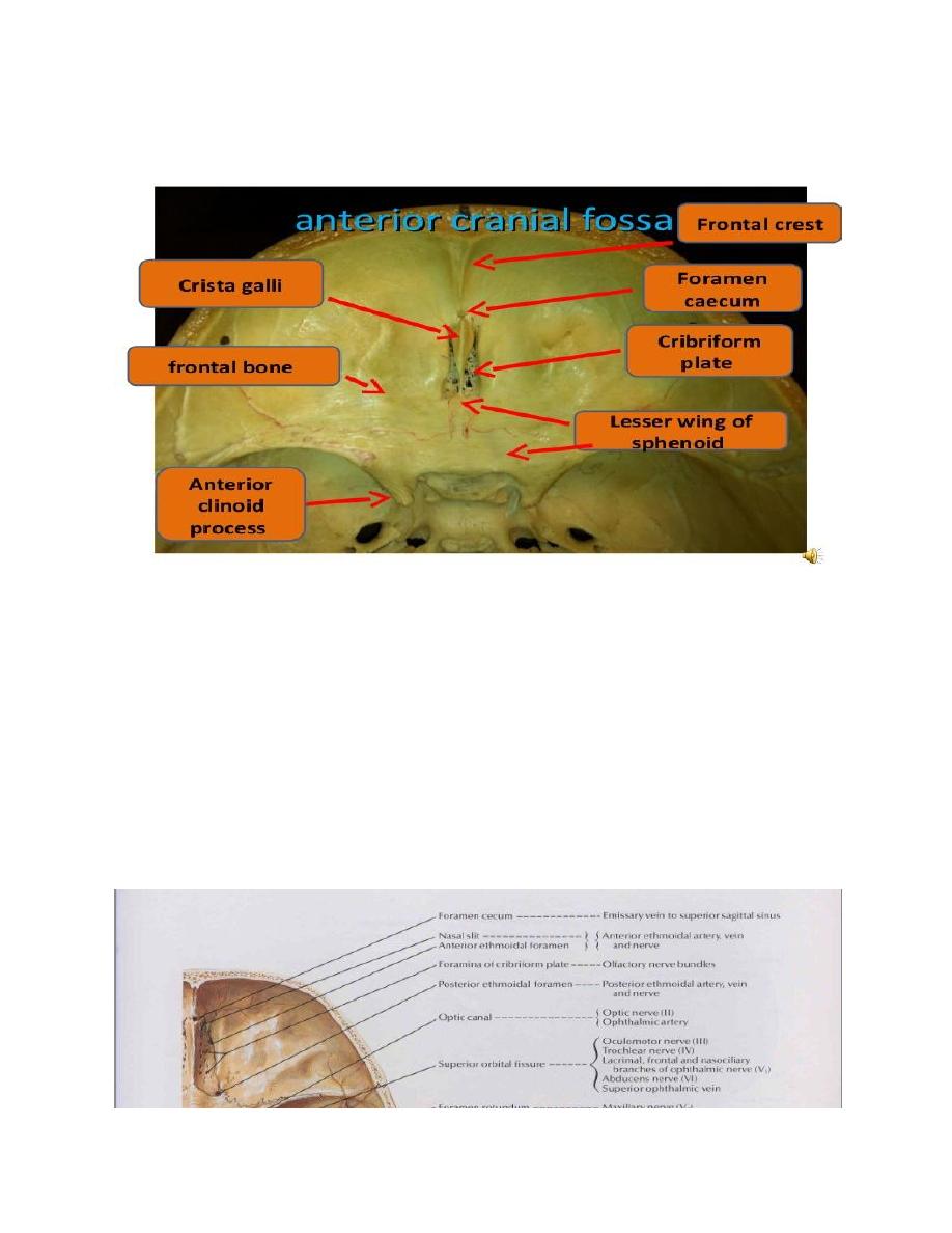
Foramina in the anterior cranial fossa:
1. Foramen caecum transmits a vein connecting the superior sagittal
venous sinus and veins of the nose.
2. Cribriform plate of ethmoid containing olfactory foramina
transmitting nerve bundles of olfactory.
3. Anterior and posterior ethmoidal foramina at the lateral border of
the cribriform plate. They transmits anterior and posterior ethmoidal
arteries and nerves.
5
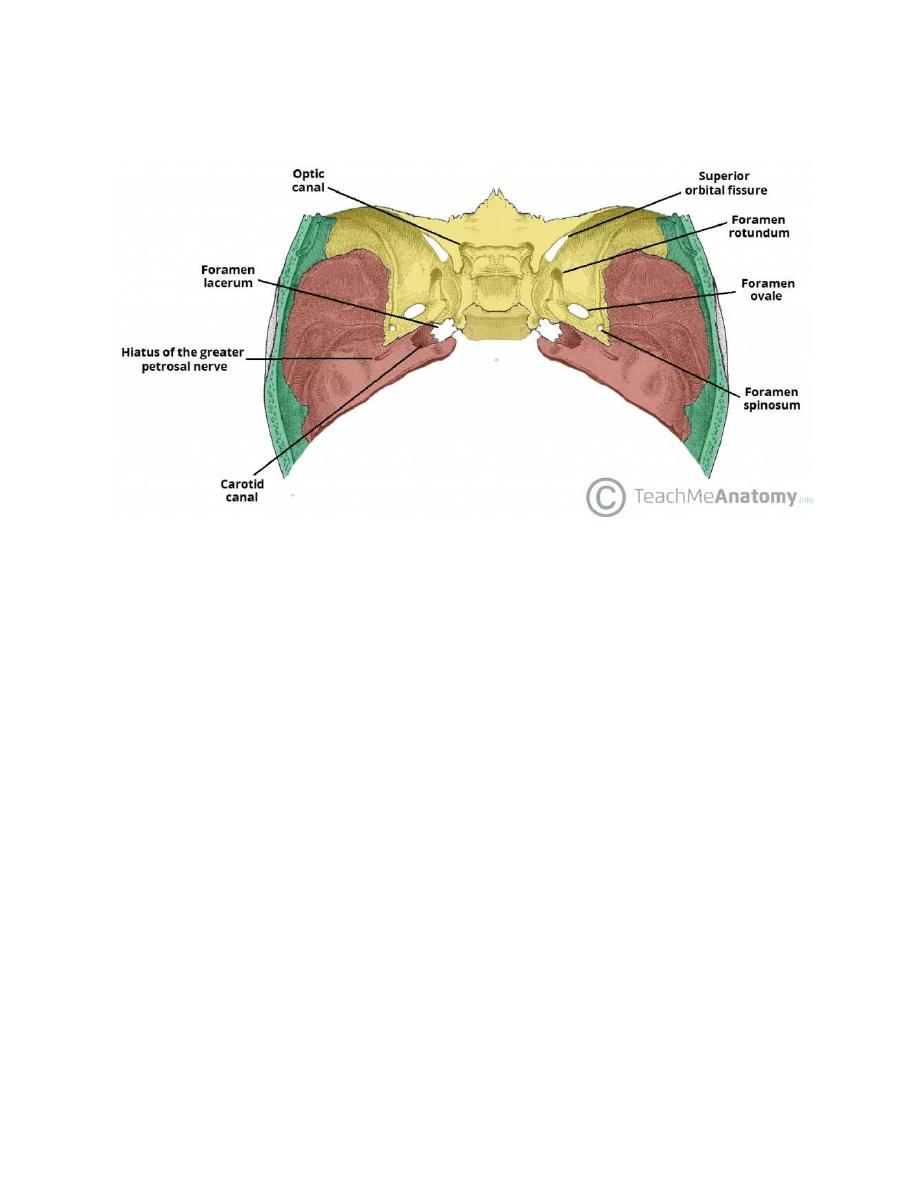
Middle cranial fossa:
1- optic canal transmits:
A. The optic nerve
B. Ophthalmic artery
C. Central vein of retina inside the optic nerve.
2. superior orbital fissure transmits:
a. Occulomotor nerve.
b. Trochlear nerve.
c. Abducent nerve.
d. Ophthalmic division Trigeminal nerve.
e. Ophthalmic veins.
f. Twigs of sympathetic nerve fibers.
6
3. Foramen rotundum transmits Maxillary division of Trigeminal
nerve.
4. Foramen ovale Transmits:
a. Mandibular division of Trigeminal nerve.
b. Lesser petrosal nerve. c. Accessory meningeal artery.
5. Foramen spinosum transmits the middle meningeal artery and
Nervous spinosus from the mandibular nen/e .
6. Foramen lacerum.
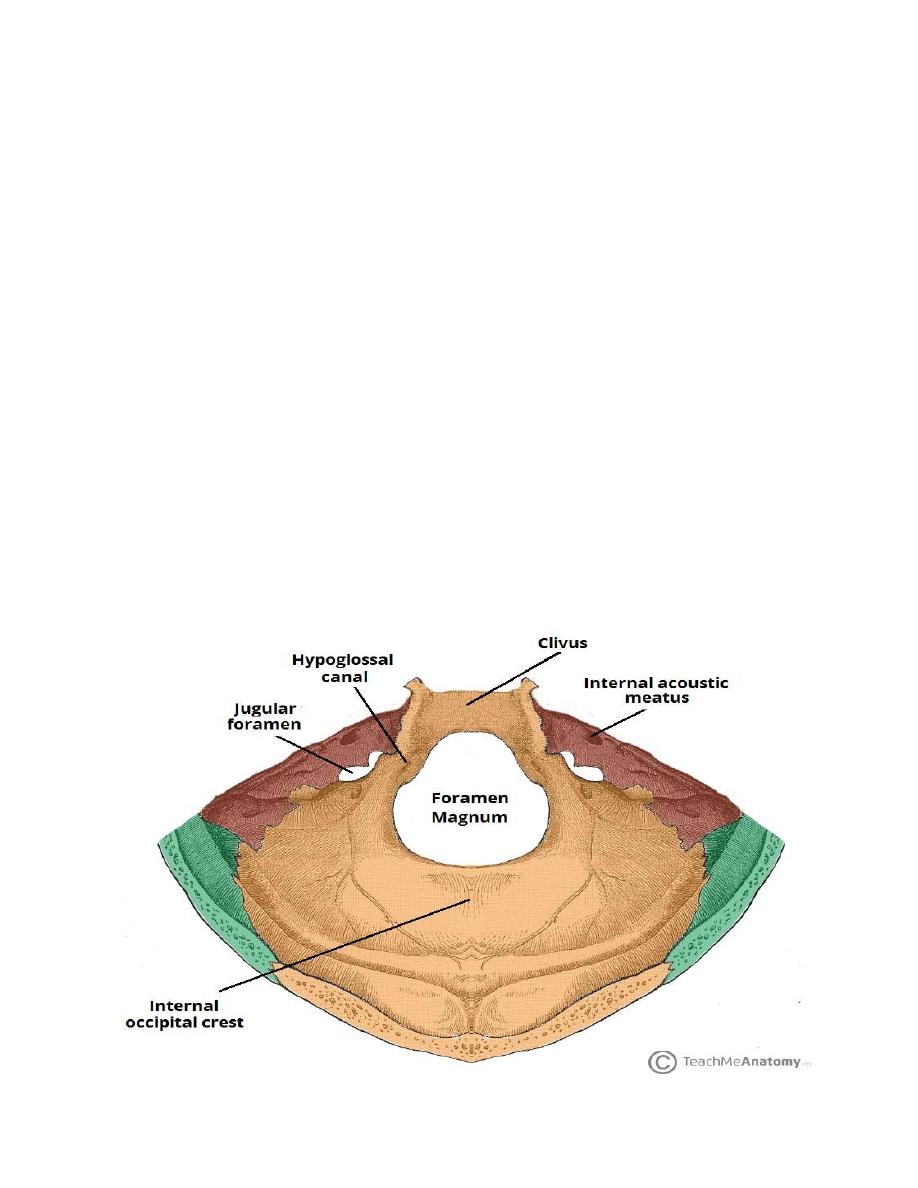
Posterior cranial fossa:

7
1. Internal acoustic meatus transmitting:
a. The facial nerve.
b. Vestibulocochlear nerve.
c. Labyrinthine artery.
2. Jugular foramen transmits:
a. GIossopharyngeal nerve.
b. Vagus nerve
c. Accessory nerve
d. Termination of sigmoid sinus to drain into the bulb of the internal
jugular vein.
e. Inferior petrosat sinus.
3. Hypogfossaf canal for the hypoglossal nerve.
4. Condylar canal if present transmits an emissary vein.
5. Foramen magnum transmits:
a. The lower end of the medulla oblongata to continue as the spinal
cord surrounded by the meninges.
b. The two vertebral arteries.
c. Single anterior spinal artery.
d. A paired posterior spinal artery.==
( C and D from the vertebral )
e. The spinal root of the accessory nerve.

8
Foramina seen from the outside:
1- Incisive foramen for naso-palatine artery and nerve.
2- Greater palatine for grater palatine artery and nerve.
3- Lesser palatine for the lesser palatine artery and nerve.
4- Stylomastoid for facial nerve & stylomastoid artery.
5- Carotic foramen leads to canal that transmit the internal carotid
artery surrounded by sympathetic plexus, emissary veins.
Fissures:
1- Superior orbital fissure.
2- Inferior orbital fissure transmits:
a. Infraorbital nerve (continuity of maxillary nerve) before emerging
from the infraorbital foramen.
b. Infraorbital artery as branch of maxillary artery.
c. Zygomatic nerve as branch of maxillary nerve.
d. Emissary vein joining pterygoid plexus with the ophthalmic veins.
3- Pterygo-maxillary fissure: lies between the back of the maxilla and
the pterygoid process transmits maxillary artery.
4- petrotympanic (squamotympanie) within the mandibular fossa
transmits:
a. Chorda tympani nerve (from facial).
b. Anterior tympanic A (from maxillary).
9
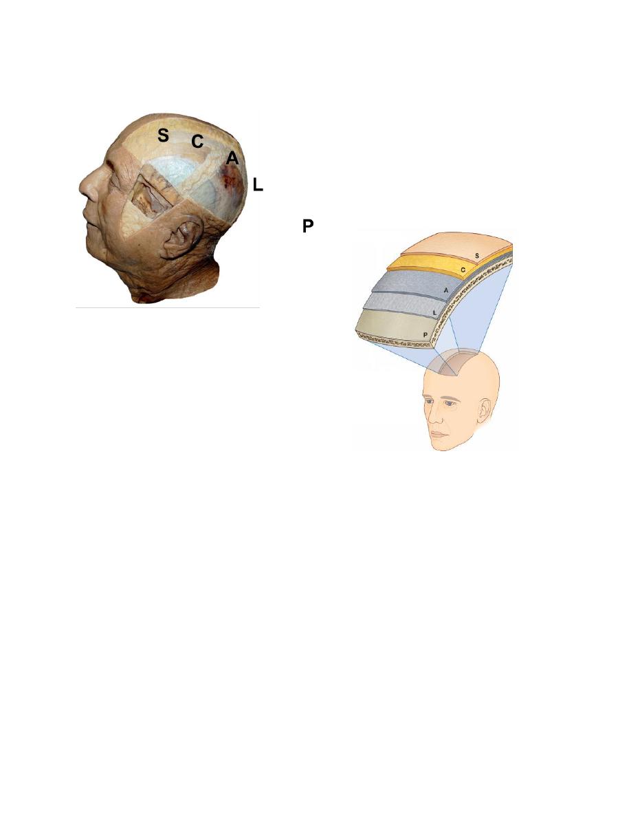
The scalp
Extends from the eye brows to the superior nuchal line (Antero-
posteriorly) and between the two temporal lines (from side to side).
It includes 5 layers:
S= Skin
C= Connective tissue (dense)
A= Aponeurosis of occipitofrontal muscle (epieramic aponeurosis)
L= Loose connective areolar tissue (subaponeurotic)
P= Periosteum of the skull.

10
Surgical Importance
:
1- Bleeding is profuse if an artery in the scalp is cut. (dense fibrous
tissue)
2- If infection in the loose areolar tissue is formed, the infection may
spread to the cranial cavity via emissary vein.
3- Healing if scalp wound is faster than elsewhere in the body because
of rich blood supply thus stitches in the scalp wound can be removed
on
day 5 (other site after 7 days).
The nerve supply of the scalp:
A. Motor to occipitofrontalis muscle. The temporal branch of facial nerve to
frontal belly and the posterior auricular branch of facial nerve to occipital belly.
B. Sensory:
1- Supratrochlear. From ophthalmic division of Trigeminal nerve
2- Supraorbitat. From ophthalmic division of Trigeminal nerve
3- Zygomatico-temporal of Maxillary division.
4- Auriculotemporal of Mandibular division.
5- Great auricular (C2 & C3).
6- Lesser occipital (C2).
7- Greater occipital (C2).
8- Third occipital (C3).

11
Arterial Supply Of The Scalp :
1- Supratrochlear. - from the ophthalmic branch of internal carotid
artery
2- Supraorbital
3- Superficial temporal artery. - Branches of external carotid artery
4- Posterior auricular artery. - Branches of external carotid artery
5- Occipital artery.
Nerves of the face :
A. Motor innervation is by the facial nerve. As it leaves the skull
through
the stylomastoid foramen, it gives off posterior auricular branch (supply
the occipital belly and auricular muscle), it pierces the parotid gland
giving here 5 branches:
1- Temporal to orbicularis oculi and frontal belly.
2- Zygomatic to supply orbicularis oculi.
3- Buccal branch to supply Buccinator muscle.
4- Marginal mandibular to muscles of the lower Iip.
5- Cervical branch to ptatysma.

12
B. Sensory innervations by:
1) From the three division of trigeminal
a. From ophthalmic division via:
- Supratrochtear and supraorbital (of frontal)
- Infratrochlear (from nasodlliary)
- External nasal (from nasocillary)
- Palpebral (from lacrimal nerve)
b. From maxillary division via:
- Nasal , superior labial of inferior palpebral branches (from
infraorbital nerve)
- Zygomatico-facial (from zygomatic nerve)
c. From mandibular division via:
- Buccal nerve to skin over buccinators muscle and the mucous
membrane of cheek
- Mental branch of inferior alveorar.
2) From the cutaneous branches of the cervical plexus via Great
auricular nerve (C2-C3) to supply the skin around ramus of the
mandible.

13
Arterial Supply of the Face
1- Mainly by some branches of the Facial artery (from external carotid artery) via
it's named branches inferior labial, superior labial, lateral nasal and angular.
2- Transverse facial of superficial temporal from external carotid artery .
3- Supraorbital and supratrochlear from the ophthalmic artery of internal carotid.
4- By some branches of infraorbital of the maxillary artery namely, superior
labial, inferior palpebral and external nasal branches.
5- By the mental artery (a branch of inferior alveorar branch of maxillary artery).
The Retromandibular Vein
It is formed by the union of superficial temporal and maxillary vein
behind the neck of the mandible within the substance of the parotid
gland.
It divides near the lower part of the gland in to anterior and posterior
divisions.
The anterior division unites with the facial vein forming the common
facial vein that opens into the I.J.E. as it is formed while the posterior
division unites with the posterior auricular vein to form E.J.V..
The facial vein is formed by the union of supratrochlear and
supraorbital veins at the medial angle of the eye (As angular vein). It
communicates with the superior ophthalmic vein as it starts.

14
Some Important Points :
1- Vertebral levels c3-c4 at this level is the upper border of the thyroid cartilage
and at common carotid artery bifurcates into external and internal .CS - C6 at this
level is the lower limits of both the pharynx (esophagus will start) and the larynx
(the trachea will start).
2- During anasthesia intubation is done through the oral cavity, where as
nasogastric tube for feeding and suction (to access the esophagus and stomach) is
via the nasal cavity.
3- The neck extends from inferior border of the mandible to the upper border of
the manubrium sterni (from the front).lt extends from superior nuchal line to the
disc between C7-T1 vertebral spines (from the back)
4- Among the 12 cranial nerves only 4 of them contains parasympathetic
components these include the occulomotor , facial, glosso pharyngeal and vagus
nerves (3rd-7th-9th and 10th cranial nerves)
5- We have 7 cervical vertebrae and 8 cervical nerves.The ventral rami of
Cl - C4 share in the formation of the cervical plexus while C5 - C8 share in the
formation of the brachial plexus.
6- In airway obstruction (upper) we can do cricothyrotomy i.e incision
through the cricothyroid ligament (very easy and little bleeding).
Tracheostomy is better but more difficult with more bleeding compared
to cricothyrotomy to maintain respiration.