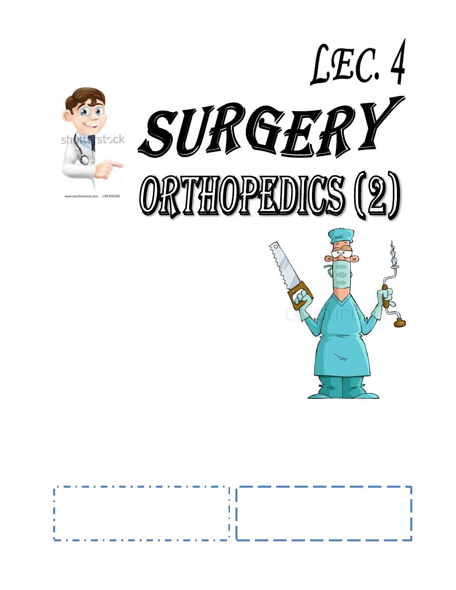
Baghdad College of Medicine / 5
th
grade
Student’s Name :
Dr. Mahmood Al-
Mashhadani
Lec. 2
Post operative complication in
orthopedics
Wed. 28 / 9 / 2016
DONE BY : Mustafa Naser
مكتب اشور لالستنساخ
2016 – 2017
Post. Operative complications Dr. Mahmood
28-9-2016
1
1. Soft tissue swelling;
Its common after limb surgery its aggravated by tight dressing or POP it may affect
wound healing & delay joint movement sometimes its so serious causing vascular
ischemia & compartment syndrome. Prevention is by;
a. Dressing must not be tight.
b. Elevation of the limb.
c. Encourage early joint movement.
d. POP cast must be well padded & not tight.
2. Heamatoma formation;
By post-operative bleeding or ooze sometimes it’s considerable. It’s reduced by using
suction drain.
3. Delayed wound healing;
Usually due to poor circulation.
4. Infection;
It’s seriously disturbing especially in joint replacement surgery.
5. Thromboembolism;
The incidence of DVT is 20%, only 2% gets pulmonary embolism and only
0.2% will die of it.
Highest incidence in hip surgery especially in old debilitated patient.
The commonest type of DVT is that occurring in calf vein which carry better prognosis
than that in proximal thigh vein thrombosis.
—
—
DVT usually silent, if it’s manifested it may cause calf or thigh pain sometimes swelling &
local tenderness.
—
In calf vein thrombosis there will be positive Homan’s sign i.e. increased calf pain on
passive dorsiflexion of the foot. Sudden slight increase in temperature may occur.
—
If pulmonary embolism occurs patient may have shortness in breath, mild chest pain &
sometimes heamoptysis, rarely sudden severe cardiovascular collapse & death.
—
Post. Operative complications Dr. Mahmood
28-9-2016
2
—
—
Doppler U/S can prove the diagnosis. More accurate is ascending veinogreaphy.
—
In pulmonary embolism ECG changes may happen.
—
—
Prevention:
—
•Pre-operative;
by decrease smoking, walking exercises, treatment of systemic diseases
& calf muscle massage with preoperative Heparin 5000 IU twice daily for high risk patients.
Recently we can use low molecular weight heparin.
—
•Per-operative;
avoid trauma to calf veins & frequent massage or use of special elastic
stokes.
• Post-operative;
calf muscle massage, elastic stokes, early active movement &
ambulation, leg elevation together with few days of subcutaneous heparin therapy.
—
—
Calf vein DVT treated by heparin 5000 IU s.c. For 10-14 days then change to oral warfarin
5mg once daily for 2-3 months (all under strict supervision by the doctor with frequent PT &
PTT check).
—
Thigh or proximal vein thrombosis is more serious it needs full heparin therapy of 5000 IU
four times daily intravenously 1-2 weeks followed by 5mg warfarin orally for 3-6 months
(all under strict supervision by the doctor with frequent PT & PTT check).
—
—
It’s used to:
—
1-Reduce pain.
—
2-Increase range of motion.
—
3-Strengthen muscles.
—
4-Restore function.
—
All achieved by;
—
A-Heat; its useful to reduce pain & muscle spasm.
—
B-Cold; like ice packs it decreases pain & reduces swelling.
—
C-Transcutaneous nerve stimulation; also relieves pain.
—
D-Traction; intermittent traction of the spine can relieve neck pain or low back pain.
—
E-Electrical stimulation & muscle exercises; it builds up muscles & improves weakness.
Post. Operative complications Dr. Mahmood
28-9-2016
3
—
Occupational therapy
—
Its used to improve function i.e. teaching the patient to use his skeleton & muscles to
perform useful daily activities as feeding, hygiene, walking. .Etc.
—
—
Functional aids
—
These are used to decrease stresses on musculoskeletal system during weight bearing
like: stick, cane, crutches or frame. During weight bearing these patient uses these to
decrease the mechanical stresses passing through his joints & bones.
—
—
A fracture is an interruption in the bone continuity caused usually by strong trauma in
healthy bones (traumatic fracture).
—
Pathological fracture Stress fr.
—
Simple or closed fr
—
Compound or open
—
complete fr.
—
Incomplete or partial fr.
—
comminuted fr.
—
Segmental fr.
—
Intra-articular fr
—
Direct trauma:
the bone usually breaks at point of impact.
Indirect trauma:
as angulation or rotation or compression of the limb, this can cause
fr. Away from the site of injury.
Multiple or complex
injury of different forces at the same time that may cause
serious complex fracture.
Muscle violence:
sudden strong muscle pull on the localized bony attachment may
cause
(avulsion fr)
of the bony fragment with or without displacement, eg.
Quadriceps pull on the patella cause transverse fr, triceps pull on the olecranon causes
avulsion fr of olecranon.
Post. Operative complications Dr. Mahmood
28-9-2016
4
—
—
Fr may be undisplaced or usually displaced according to the; type of injury and the direction
of ft line also depends on localized muscle action and gravity, displacement described
according to the direction of the distal segment, this can be in form of;
—
Overlap.
—
Side displacement.
—
Angulation.
—
Rotation.
—
Tissue death and heamatoma formation:
—
Local bony necrosis will occur about 1 to 2 mm back from the fracture line and capillary
bleeding will lead to localized fr heamatoma that bridges the fr and later clots.
Inflammation and cellular proliferation:
—
Within 8 hours there will be acute inflammatory cell reaction, within few days there is
chronic inflammatory cell accumulation. This later will lead to local granulation tissue
formation after capillary growth; cellular proliferation will take place as well.
Callus formation:
—
Proliferating cells will show variable chondrogenic and osteogenic activities with
subsequent cartilage and finally bone tissue formation that will later replaces the whole
granulation tissue mass leading to a big irregularly laid fusiform bony mass called the
CALLUS that bridges the fr and locally fix it, the fr now is united but its not healed yet.
Consolidation stage:
—
Here the callus is gradually replaced by physiologically normal solid bone that will cross
through the fr line and fill the gap, the fusiforrn callus replaced by variable osteoclastic
(bone resorbing) and osteoblastic (bone forming) cellular activity, the newly laid solid bone
is of near normal shape and can take normal forces and loading.
Remodeling stage
—
Gradual bone resorbtion and new bone formation will occur where the solid deformed
bone is replaced by new normal bone that takes the original bone shape with reformation
of the medullary canal, this may take months or even years to occur.
—
—
Done BY : Mustafa Naser