(1
st
Lecture (Dentistry 30
th
Sept to 5
th
Oct 2015)
General Histology
Histology:
- Is the science of tissue which is driven from the Greek words histos "tissues"
and the word "logus" which refers to "science" is the study of the microscopic
anatomy of cells and tissues of plants and animals.
- It is commonly performed by examining cells and tissues by sectioning and
staining, followed by examination under a light microscope (LM) or
transmission electron microscope (TEM).
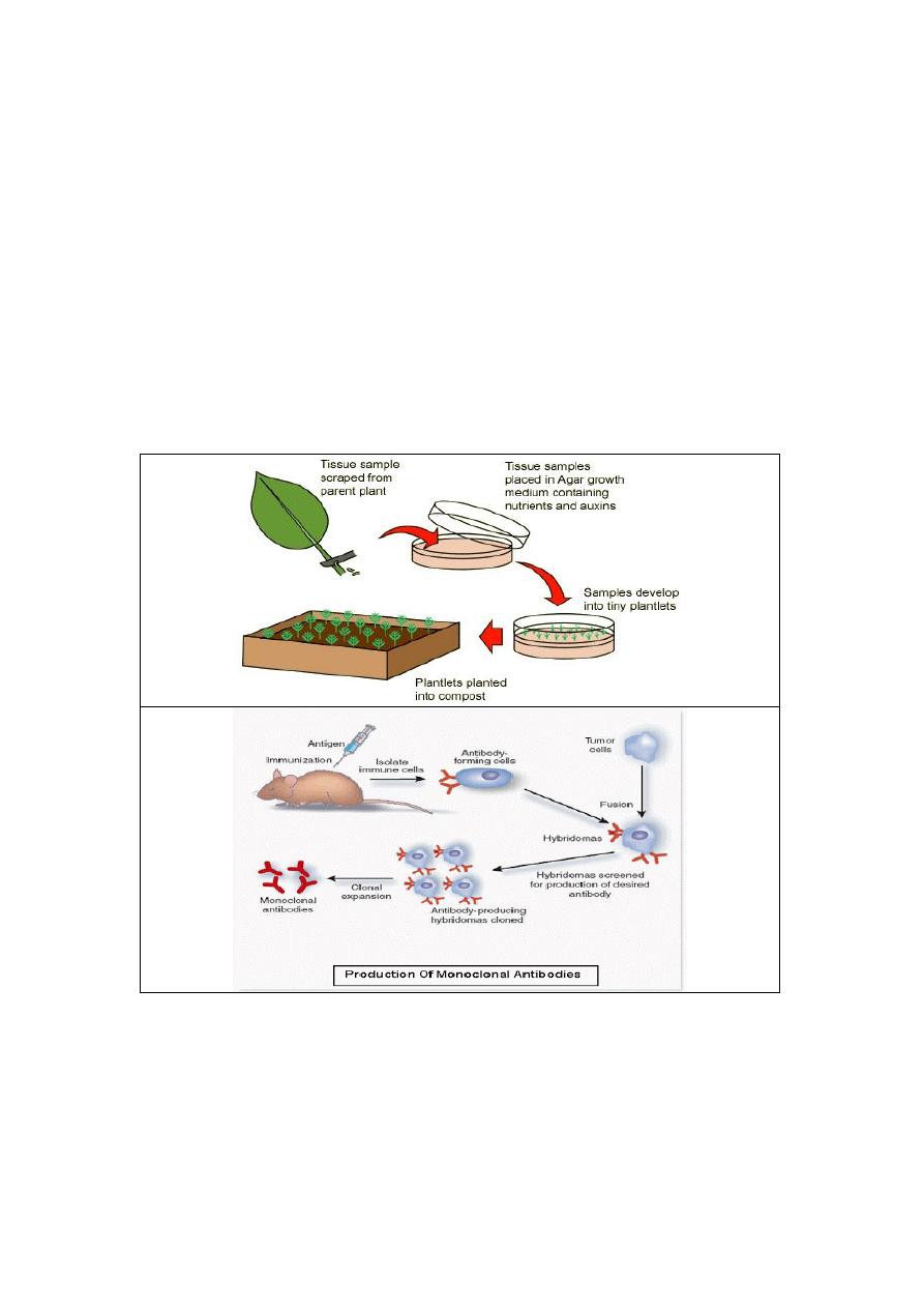
- Histological studies may be conducted via tissue culture (Figure 1,2&3),
where live cells can be isolated and maintained in a proper environment
outside the body for various research projects.
- The ability to visualize or differentially identify microscopic structures is
frequently enhanced through the use of histological stains.
(Figure 1 & 2): "Tissue culture" technique in plant laboratories and immune cell
production in animals where mass production of sister cells can be produced for
agricultural and medical purposes respectively.
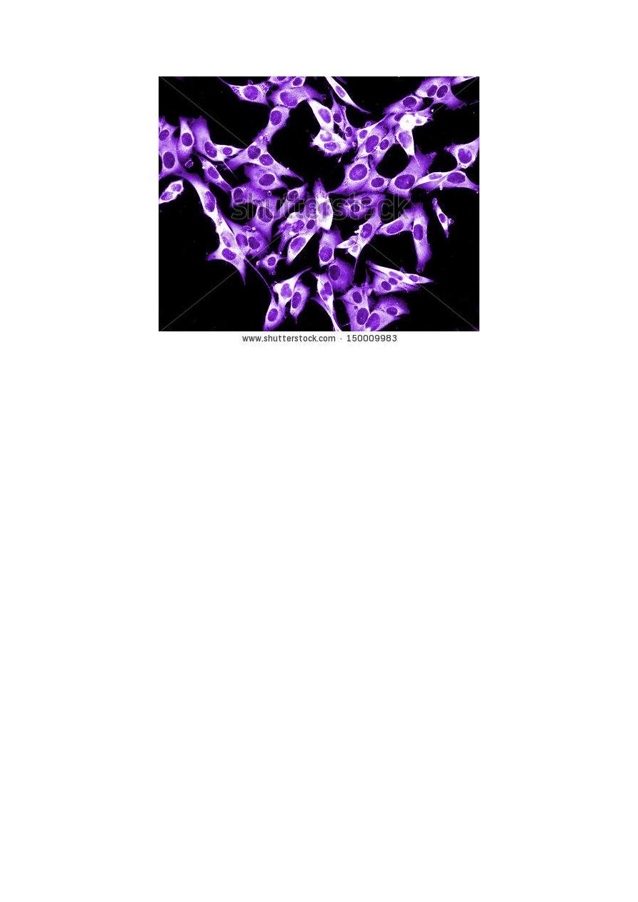
(Fig.3): Sister cells grown in Petri dish using tissue culture technique.
- Histopathology: The microscopic study of diseased tissue is an important tool
in anatomical pathology, since accurate diagnosis of cancer and other diseases
usually requires histopathological examination of samples.
- Histopathologists, are the trained physicians who perform histopathological
examination and provide diagnostic information based on their observations.
- Histotechnicians (HT): Are the trained scientists who perform the preparation
of histological sections in clinical laboratories.
Related sciences:
1). Cell biology: is the study of living cells, their DNA and RNA and the proteins
they express.
2). Anatomy: is the study of organs visible by the naked eye.
3). Morphology: studies entire organisms.
4). Cytology: is the microscopic study of loose cells or clusters obtained from bodily
secretions, aspirations, scrapes, swipes, or washings.
Artifacts: are structures or features in tissue that interfere with normal histological
examination. These are not always present in normal tissue and can come from
outside sources. Artifacts interfere with histology by changing the tissues appearance
and hiding structures. These can be divided into two categories: (1)
:
Pre-histology:
Features and structures that have been introduced prior to the collection of the tissues
e.g. tattoos and freckles (melanin) in skin samples; and (2): Post-histology: Artifacts
can result from tissue processing e.g. shrinkage, washing out of particular cellular
components, color changes in different tissues types and alterations of the structures
in the tissue.
Cells:
Are the basic unit of life. All organisms are made up of cells most are very
small and invisible without using a microscope. Each cells are covered by a cell
membrane and come in many different shapes. The contents of a cell are called the
protoplasm while each contains a nucleus at least except human RBC.
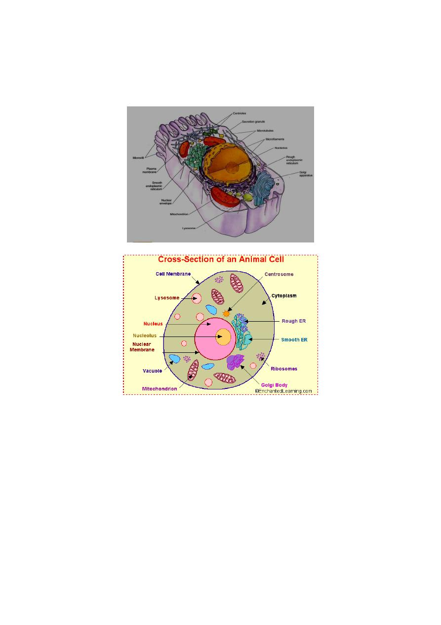
(Fig-4): Schematic diagram of an animal cell shows the cellular organelles.
1. Cell membrane: the thin semi-permeable layer of protein and fat that
surrounds the cell. The cell membrane is, allowing some substances to pass
into the cell and blocking others.
2. Centrosome: (also called the "microtubule organizing center") a small body
located near the nucleus where microtubules are made. During cell division
(mitosis), the centrosome divides and the two parts move to opposite sides of
the dividing cell. The centriole is the dense center of the centrosome.
3. Cytoplasm: the jellylike material outside the cell nucleus in which the
organelles are located.
4. Golgi body: A flattened, layered, sac-like organelle located near the nucleus
which produces lysosomes. The Golgi body packages proteins and
carbohydrates into membrane-bound vesicles for "export" from the cell.
5. Lysosome: Are round organelles surrounded contain digestive enzymes. This
is where the digestion of cell nutrients takes place.
6. Mitochondrion: spherical to rod-shaped organelles with a double membrane
enfolded projections (called cristae) functions as the energy store and converts
glucose into ATP (adenosine triphosphate) for the cell. Mitochondria could be
visualized clearly using TEM.
7. Nuclear membrane: A double layered membrane that surrounds the nucleus
which could only be clearly visible in TEM.
8. Nucleolus: an organelle within the nucleus, where ribosomal RNA is
produced.
9. Nucleus: A spherical body contains many organelles, including the nucleolus.
It controls many functions of the cell e.g. protein synthesis and contains DNA
(in chromosomes). The nucleus is surrounded by the nuclear membrane.
10. Ribosome: small organelles composed of RNA-rich cytoplasmic granules
that are sites of protein synthesis.
11. Rough Endoplasmic Reticulum (RER): located in the cell's cytoplasm
which is continuous with the outer nuclear membrane. RER is covered with
ribosomes that give it a rough appearance. RER transports materials through
the cell and produces proteins in sacks called cisternae sent to the Golgi body.
12. Smooth Endoplasmic Reticulum (SER): Infolded and convoluted tubes that
located in the cytoplasm
13. Vacuole: fluid-filled, membrane-surrounded cavities inside a cell. The vacuole
fills with food being digested and waste material that is on its way out of the
cell (Fig 4).
Cell division:
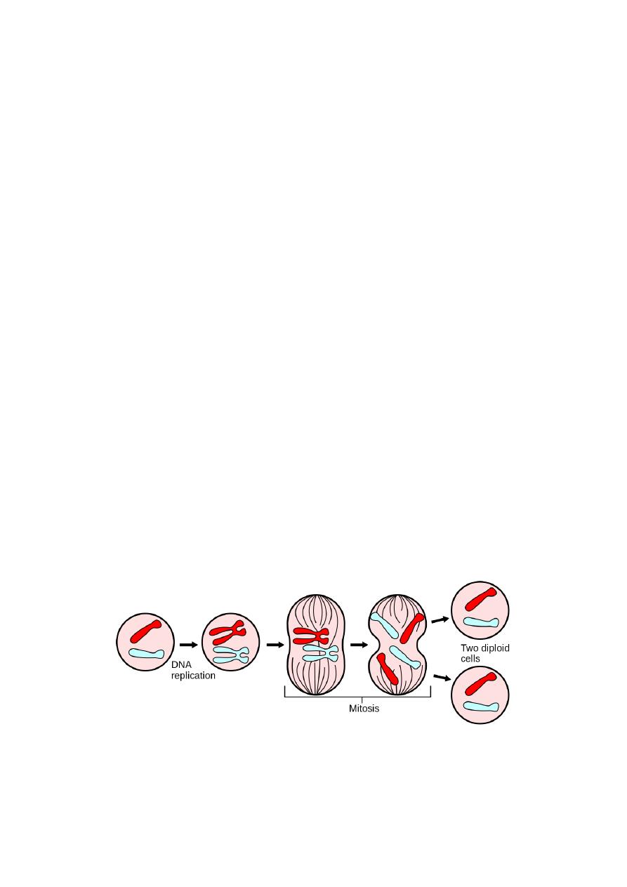
Mitosis is the process, in the cell cycle, by which the chromosomes in the cell nucleus
are separated into two identical sets of chromosomes, each in its own nucleus. Mitosis
occurs only in eukaryoticcells. The process of mitosis is fast and highly complex. Cell
s divide in stages are (1): prophase, (2): prometaphase, (3): metaphase, (4): anaphase,
and (5): telophase. During mitosis, the pairs of chromatids condense and attach to
fibers that pull the sister chromatids to opposite sides of the cell. The cell then divides
in cytokinesis, to produce two daughter cells.
(Fig-5): Mitosis divides the chromosomes in a cell nucleus.
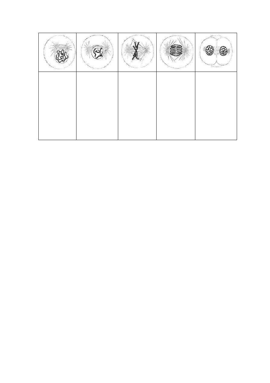
Prophase: The
two round objects
above the nucleus
are the
centrosomes. The
chromatin is
condensing into
chromosomes.
Prometaphase:
The nuclear
membrane
disintegrates and
microtubules have
invaded the nuclear
space. These
microtubules can
attach to
kinetochores or
they can interact
with opposing
microtubules.
Metaphase: The
chromosomes
align at the
metaphase plate.
Anaphase: The
chromosomes
split and the
kinetochore
microtubules
shorten.
Telophase: The
decondensing
chromosomes are
surrounded by
nuclear membranes.
Cytokinesis has
already begun; the
pinched area is
known as the
cleavage furrow.
(Fig-6): Cell division (mitosis) phases in a eukaryotic cell.
Importances of Mitosis:
1). Development and growth: The number of cells within an organism increases by
mitosis e.g.. zygote and also the basis of the growth of a multicellular body.
2). Cell replacement: In skin and digestive tract, cells are constantly sloughed off
and replaced by new ones. New cells are formed by mitosis and so are exact copies of
the cells being replaced. In like manner, RBCs (red blood cells) have short lifespan
(only about 4 months) and new RBCs are formed by mitosis.
3). Regeneration: Some organisms can regenerate body parts. The production of new
cells in such instances is achieved by mitosis e.g. starfish regenerate lost arms through
mitosis.
4). Asexual reproduction:Some organisms produce genetically similar offspring
through asexual reproduction e.g. the hydra reproduces asexually by budding.
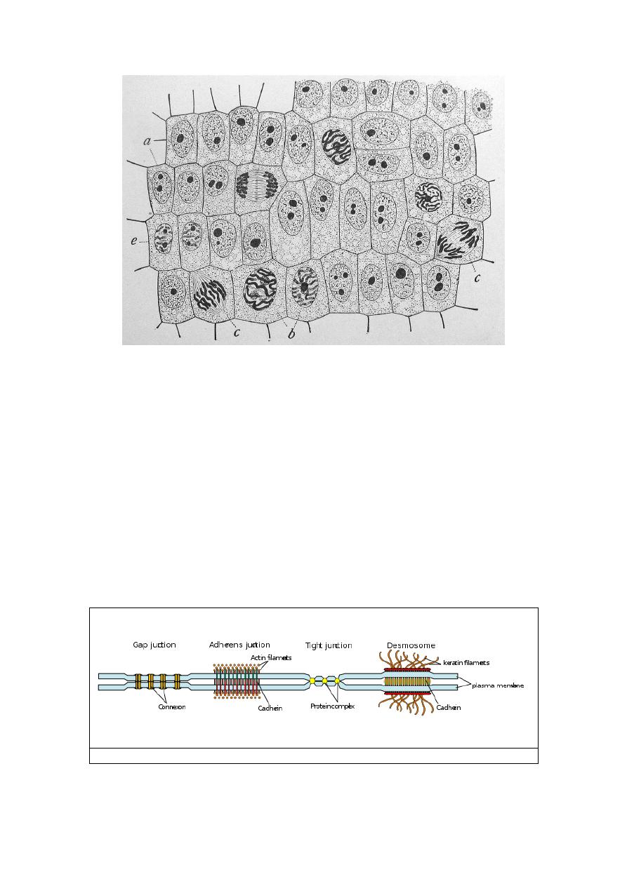
(Fig.7): Onion (Allium) cells in different phases of the cell cycle enlarged 800
diameters.
a. non-dividing cells (Interphase)
b. nuclei preparing for division (Prophase).
c. dividing cells showing mitotic figures (Metaphase).
e. pair of daughter-cells shortly after division (Sister cells).
Cellular Junctions: or intercellular bridge join cells to each other by proteins where
the point of connection between two cells is called a junction. Junctions can be found
in epithelial tissues which bind cells together. Some kinds of junctions prevent the
passage of molecules between cells. They consist of multi-protein complexes that
provide contact between neighboring cells or between a cell and the extracellular
matrix. They also enable communication between neighboring cells. There are 4
major types of cell junctions (1): Anchoring junctions (Adherens junctions); (2):
desmosomes and hemidesmosomes; 3): Gap junctions (communicating junction) and
4): Tight junctions (occluding junctions).
(Fig: 8): various types of cell junctions.
.
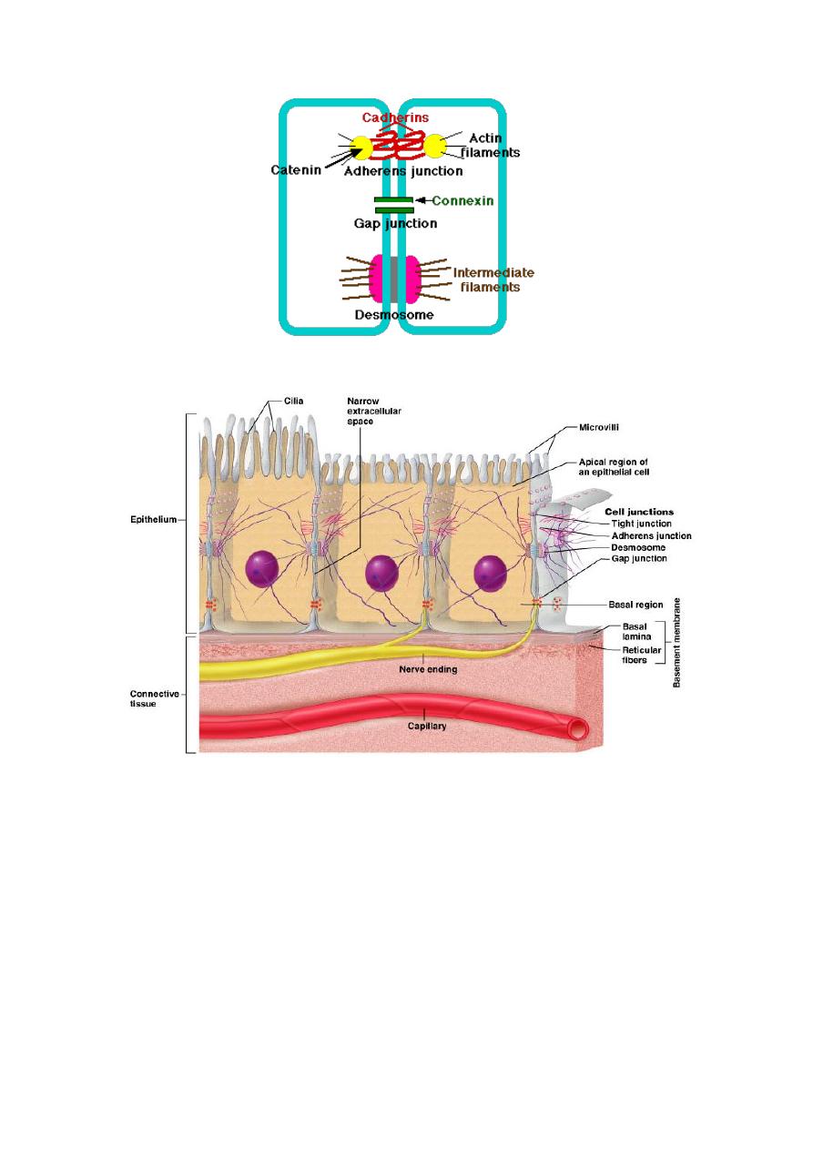
(Fig 9): Schematic diagram of various types of cell junctions.
(Fig. 10): Cellular Junctions.
Levels of Organization:
Tissues:
A tissue is composed of cells that function together in a specialized
activity. Multicellular (large) organisms function more efficiently if cells become
specialized for specific functions. There are four types of tissues found in animals:
(1): Epithelial, (2): connective; (3):Nervous and (4): Muscle tissue.
Organs
:
Organs are composed of two or more tissues which function together to
perform a common task. For example, the heart contains all 4 types of tissues.

Organ systems:
An organ system consists of two or more organs which perform a
specific task. Some organ systems are: the integumentary, nervous, sensory,
endocrine, skeletal, muscular, circulatory, immune, digestive, respiratory, excretory,
and reproductive systems.
Embryonic Tissues:
This part will be studied in details next course within "Oral
Histology" or "Embryology". Ectoderm, mesoderm, and endoderm are embryonic
tissues that give rise to all of the tissues, organs, and organ systems in the body.
Ectoderm forms the outer layer of skin and nervous system; Mesoderm forms the
muscles, connective tissues, skeleton, kidneys, and circulatory and reproductive
organs and Endoderm forms the lining of the gut, respiratory tract, and urinary
bladder. It also forms the glands associated with the gut and respiratory tract.
Epithelial Tissues:
Covers a body surface or lines a body cavity
Forms most glands
Functions of epithelium
Protection
Absorption, secretion, and ion transport
Filtration
Forms slippery surfaces
Special Characteristics of Epithelia
Cellularity
cells are in close contact with each other with little or no intercellular
space between them
Specialized contacts
may have junctions for both attachment and communication
Polarity
epithelial tissues always have an apical and basal surface
Support by connective tissue
at the basal surface, both the epithelial tissue and the connective tissue
contribute to the basement membrane
Avascular
nutrients must diffuse
Innervated
Regeneration
epithelial tissues have a high capacity for regeneration
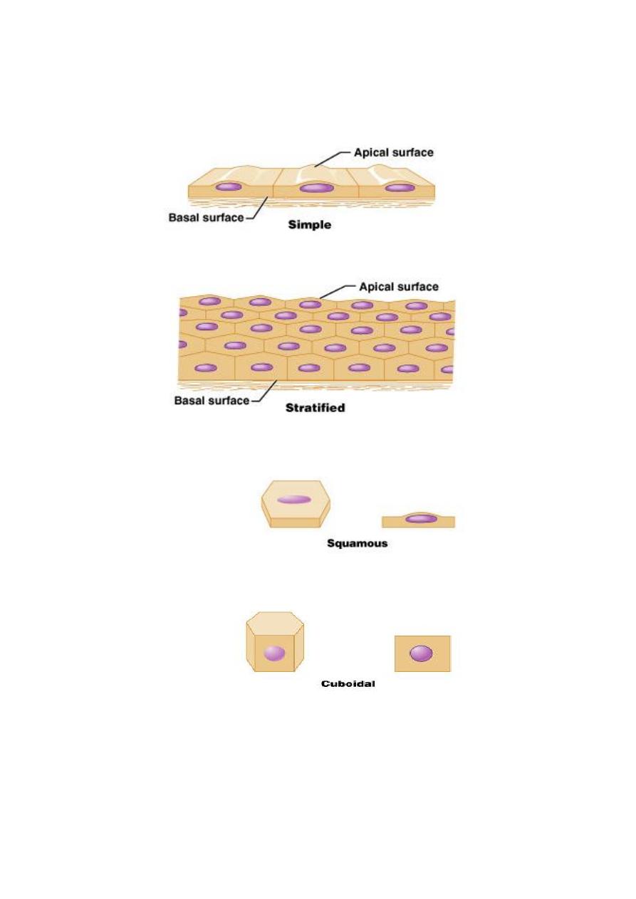
Classifications & Naming of Epithelia:
First name of tissue indicates number of layers
Simple – one layer of cells:
Stratified – more than one layer of cell:
Last name of tissue describes shape of cells
Squamous – cells wider than tall (plate or “scale” like):
Cuboidal – cells are as wide as tall, as in cubes.
Columnar – cells are taller than they are wide, like
columns
Naming Epitheli
Naming the epithelia includes both the layers (first) and the shape of the cells
(second)
i.e. stratified cuboidal epithelium
The name may also include any accessory structures
Goblet cells
Cilia
Keratin
Special epithelial tissues (don’t follow naming convention)
Psuedostratified
Transitional
Simple Squamous Epithelial Tissue:
Description
single layer of flat cells with disc-shaped nuclei
Special types
Endothelium (inner covering)
slick lining of hollow organs
Mesothelium (middle covering)
Lines peritoneal, pleural, and pericardial cavities
Covers visceral organs of those cavities
Function
Passage of materials by passive diffusion and filtration
Secretes lubricating substances in serosae
Location
Renal corpuscles
Alveoli of lungs
Lining of heart, blood and lymphatic vessels
Lining of ventral body cavity (serosae)
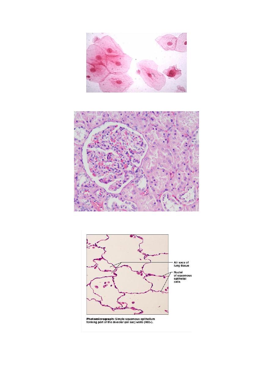
Fig: 11A: macerated Simple Squamous Epiteilal Tissue.
Fig. 11B: Bowman capsules in the kidney
.
Fig. 11C: Pulmonary Tissues (Lung tissue).
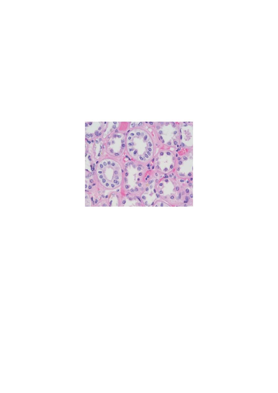
Simple Cuboidal Epithelial Tissue:
Description
single layer of cube-like cells with large, spherical central
nuclei
Function
secretion and absorption
Location
kidney tubules, secretory
portions of small glands,
ovary & thyroid follicles.
(Fig. 12): Simple Cuboidal Tissue, in Proximal and distal tubules.
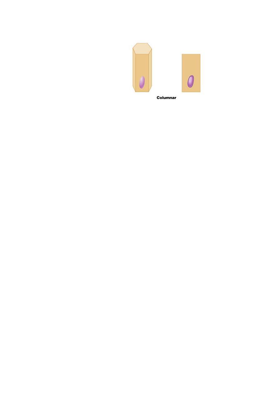
Simple Columnar Epitheila Tissue:
Description
single layer of column-shaped (rectangular) cells with oval
nuclei
Some bear cilia at their apical surface
May contain goblet cells
Function
Absorption; secretion of mucus, enzymes, and other
substances
Ciliated type propels mucus or reproductive cells by ciliary
action.
Location
Non-ciliated form
Lines digestive tract,
gallbladder, ducts of
some glands
Ciliated form
Lines small bronchi,
uterine tubes, uterus
(Fig. 13): Lining of the Digestive system.
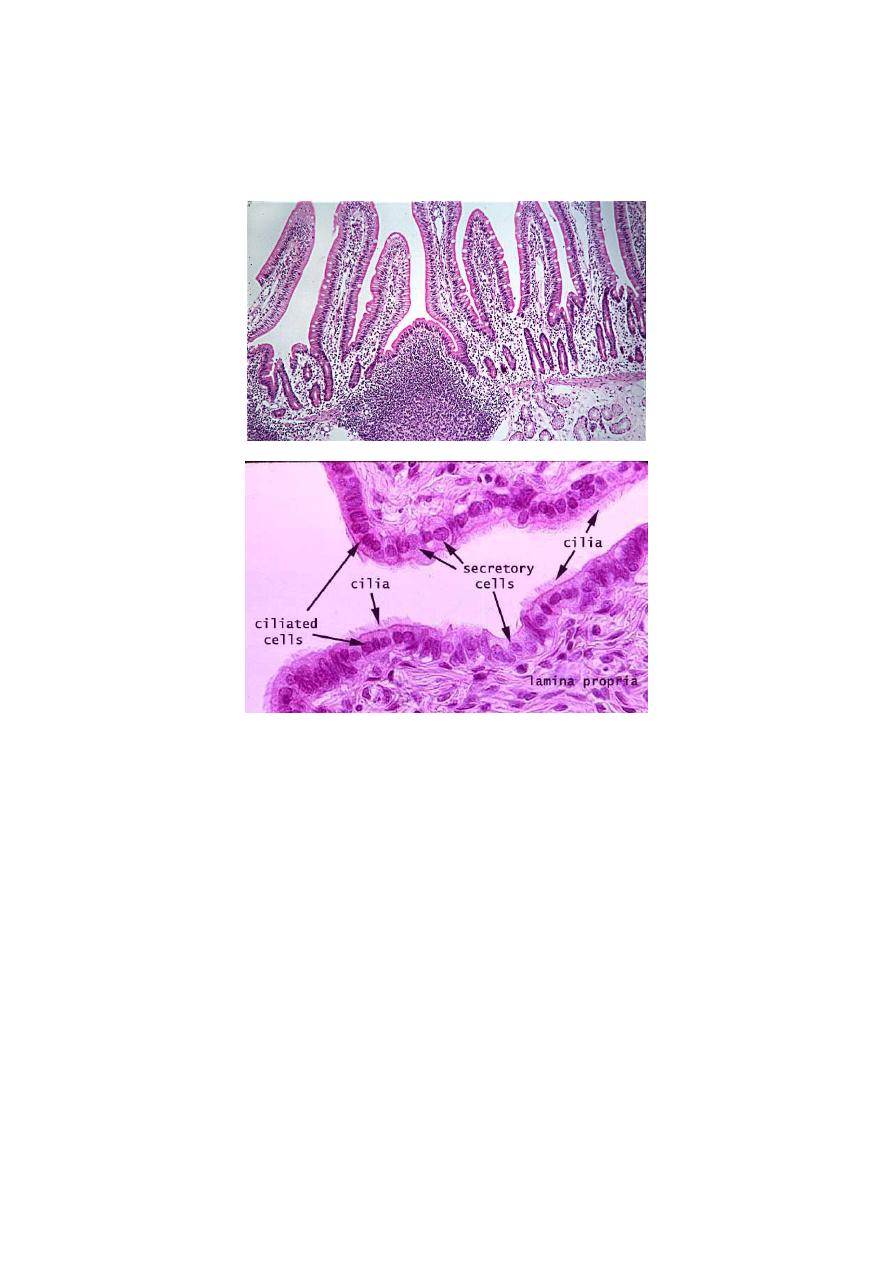
Pseudostratified Columnar Epithelial Tissue:
Description
All cells originate at basement membrane
Only tall cells reach the apical surface
May contain goblet cells and bear cilia
Nuclei lie at varying heights within cells
Gives false impression of stratification
Function
secretion of mucus; propulsion of mucus by cilia
Locations
Non-ciliated type
Ducts of male reproductive tubes
Ducts of large glands
Ciliated variety
Lines trachea and most of upper respiratory tract.
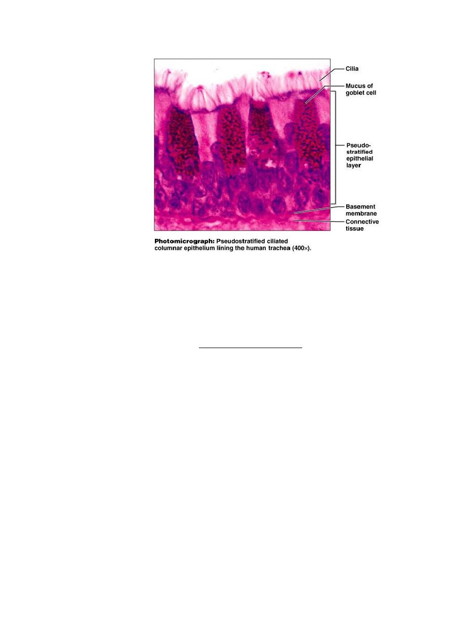
(Fig. 14: Pseudostratified Epithelial Tissue in trachea.
Stratified Epithelial Tissue:
Contain two or more layers of cells
Regenerate from below
Major role is protection
Are named according to the shape of cells at apical layer.
Stratified Squamous Epithelial Tissue.
Description
Many layers of cells – squamous in shape
Deeper layers of cells appear cuboidal or columnar
Thickest epithelial tissue – adapted for protection.
Specific types
Keratinized – contain the protective protein keratin
Surface cells are dead and full of keratin
Non-keratinized – forms moist lining of body openings
Function
Protects underlying tissues in
areas subject to abrasion
Location
Keratinized – forms epidermis
Non-keratinized – forms lining of
esophagus, mouth, and vagina.
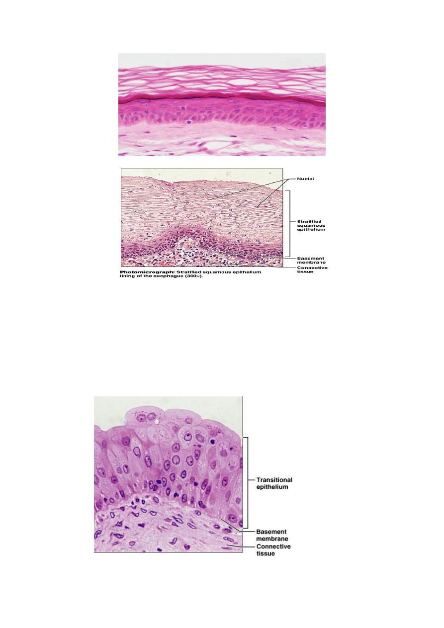
Transitional Epithelium:
Description
Basal cells usually cuboidal or columnar
Superficial cells dome-shaped or squamous
Function
stretches and permits distension of urinary bladder
Location
Lines ureters, urinary bladders and part of urethra.