SUPERFICIAL MYCOSES
By
Dr. Mohammed H. Mushrif
Lecturer of Medical Mycology
1- Pityriasis versicolor Skin
2- Tinea nigra Skin
3- Black piedra Hair
4- White piedra Hair
Superficial mycoses

Causative agent:
o Malassezia furfur is the causative agent.
o Malasseszia furfur is a lipophilic fungus living on the
skin as part of the normal flora.
o So, it is cultivated on media containing fatty
substances like olive oil.
Pityriasis versicolor
Diseases caused by Malassezia furfur:
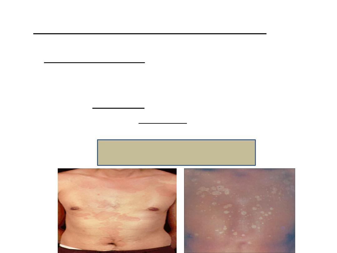
o Pityriasis versicolor is associated with hyperpigmented
or hypopigmented macules usually on the chest and
upper back of the patients.
o Pityriasis folliculitis.
o Catheter acquired fungemia in patients receiving total
parenteral nutrition containing lipid emulsion.
Pityriasis versicolor

Laboratory diagnosis:
Wood’s lamp:
o
Wood’s light is ultraviolet light at wave length 365 nm.
o
The lamp is held four or five inches from the affected skin. If
pityriasis versicolor is present on the skin, the affected skin
will appear yellowish green in color.
Specimen:
o
Skin scrapings from the lesions.
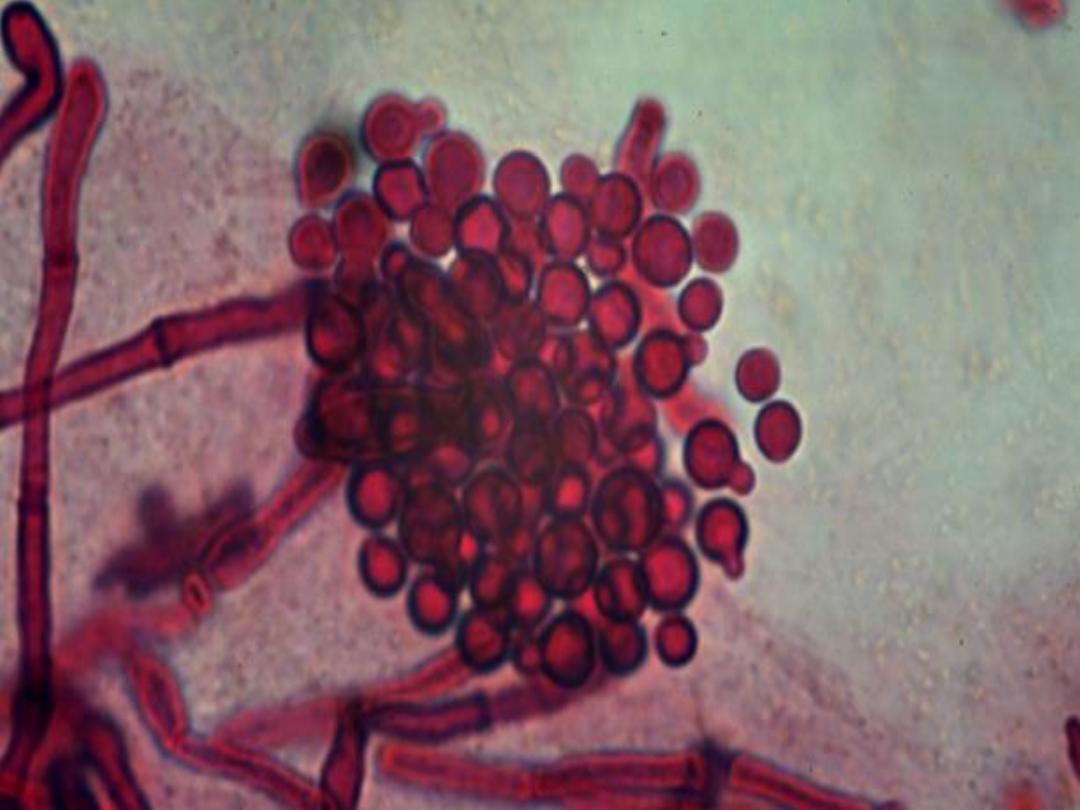
Direct Microscopy:
o
The skin scrapings are mounted in 10-20% KOH.
o
It will show spherical budding cells and short unbranched
angular septate hyphae (spaghetti and meat balls).
o
These microscopic features are diagnostic for Malassezia
furfur and culture is not necessary.

Treatment:
Daily application of selenium sulfide.
Topical or oral azoles are very effective.

Causative agent:
o Exophiala (Hortae) werneckii is the causative agent.
o It is a saprophytic fungus which occurs in the soil.
o It is a dematicaeous fungus (contains melanin in its cell
wall and appear dark in color under the microscope).
Tinea nigra
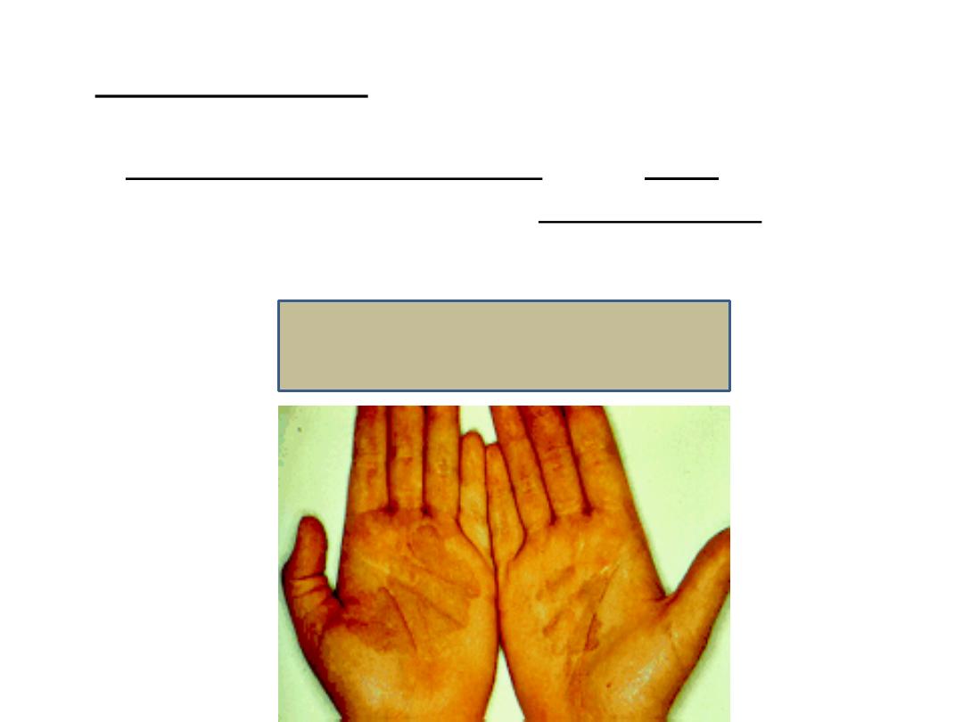
Clinical picture:
o Brown to black discoloration of the palm of the hand.
o So, Tinea nigra is also called Tinea palmaris.
o But, it may affect the sole of the foot.
Tinea nigra
Laboratory diagnosis:
Specimen:
o
Skin scrapings from the lesions.
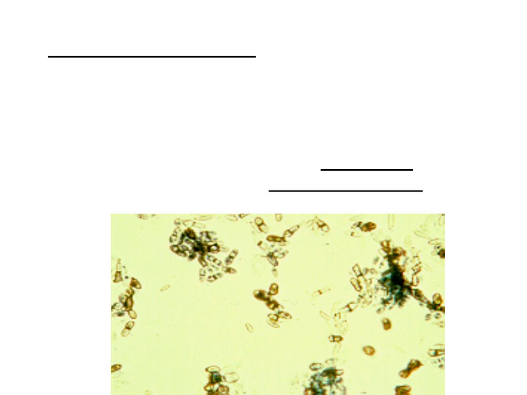
Direct Microscopy:
o
The skin scrapings are mounted in 10-20% KOH.
o
It will show brown to black two celled yeast cells + branched
septate hyphae.

Treatment:
Keratolytic solutions like salicylic acid.
Azoles are also effective.

o Piedraia hortai is the causative agent.
o It is a dematicaeous mold.
o It affects the hair like beard, moustache and scalp &
pubic hair.
o It causes brown to black hard nodules around the hair
shaft.
o It is treated by removal of infected hairs + topical
antifungal drugs.
Black piedra

o Trichosporon beigelii is the causative agent.
o It is a mold.
o It affects the hair.
o It causes large, white to yellow soft nodules around
the hair shafts.
o It is treated similar to black piedra.
White piedra