11/28/2010
1
Pectoral Region &
Breast
Lab Session 5
Dr. Hayder Jalil Al‐Assam
MBChB (Iraq), Mres Anatomy (UK)
: dr_hayder_anatomy@yahoo.com
The Breast
•
Specialized accessory gland that
secretes milk
•
Breast base extends
1‐ From 2
nd
to 6
th
rib.
2 F
l
l
b d
id
2‐ From lateral sternum border to mid‐
axillary line.
•
In Male & immature female, it consists
of nipple, duct system embedded in
connective tissue that does not extend
beyond the areola
•
In Female, consist of elongated duct
system embedded in fat tissue that
forms 15‐20 lobes.
The Breast
•
Breast mainly in superficial
fascia except the tail that
extend deep to the Pectoralis
major muscle fascia.
•
The lobular fibrous septa =
suspensary ligaments
•
Behind the breast there is a
space filled with loose
connective tissue = retro
mammary space.
The Breast
•
Blood Supply branches from
1‐ internal thoracic artery
2‐ intercostal arteries
3‐ Axillary artery by lateral thoracic
artery and thoraco‐acromial artery
y
y
•
Lymph drainage
1‐ medial quadrants – internal
thoracic group
2‐ Lateral quadrants – anterior axillary
group
3‐ few pass to the other side &
abdomen
4‐ few pass to the posterior
intercostal
Breast Cancer
•
60% in lateral upper quadrant
•
Lymph drainage is important way of spread
and Axillary LNs are commonly involved.
ibl
li i l f
•
Possible clinical features:
1‐ Skin tethering /Dimbling
2‐ Nipple retraction / destruction
3‐ Peau d’orange (orange peel like)
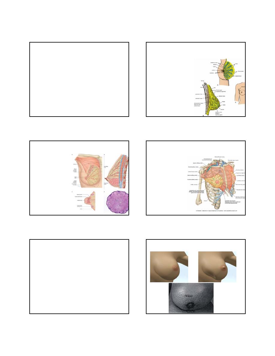
Breast Cancer
Nipple Retraction
Skin tethering
Peau d’Orange sign
11/28/2010
2
Axilla
•
Is a pyramidal space between the arm and
the chest.
•
It provide a passage for blood vessels,
lymphatics and nerves to the upper limb
lymphatics and nerves to the upper limb.
•
The pyramid has apex and base
1‐ Apex – directed towards the root of the neck
2‐ Base – the floor of the axilla
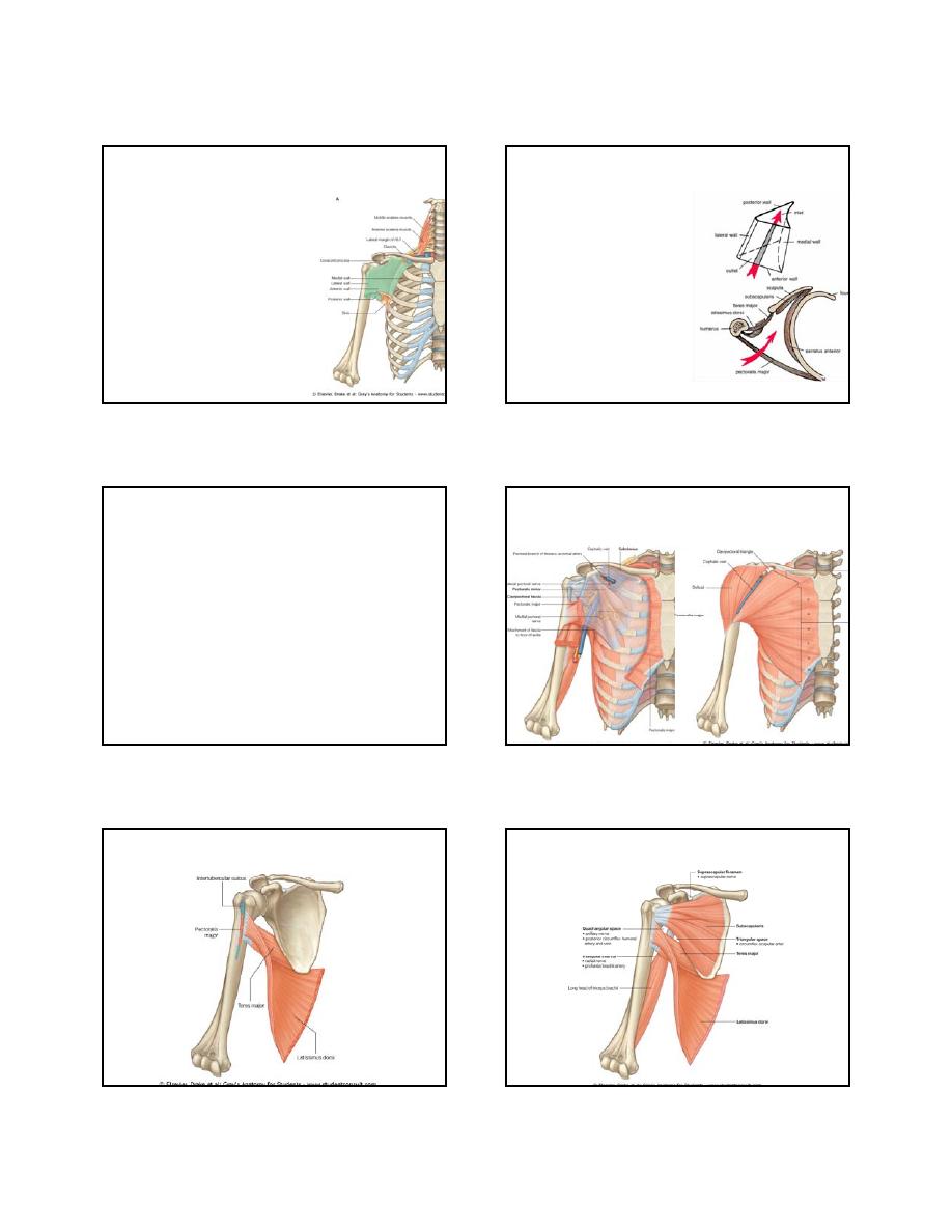
Axilla
•
APEX
Bounded anteriorly by clavicle,
posteriorly by scapula and
medially by first rib
•
BASE
Bounded anteriorly by anterior
axillary fold (Pectoralis Major
muscle), posteriorly by posterior
axillary fold (Latissmus dori &
Teres major Muscles) and
medially by the chest wall.
Axilla
•
Walls of the axilla
Anterior wall – pectoralis major, pectoralis minor &
subclavius.
Poterior wall – Subscapularis, Teres major & latissmus
Poterior wall Subscapularis, Teres major & latissmus
dorsi muscles.
Medial wall – upper 4‐5 ribs, intercostal spaces &
serratus anterior muscle.
Lateral wall – humerus, coracobrachialis and biceps
muscles.
Base – Skin of the axilla.
Anterior Axillary wall
Lateral Axillary wall
Posterior Axillary wall
11/28/2010
3
Medial Axillary wall
Pectoral region muscles
Muscles of the pectoral region
•
4 Muscles that connect the UL to thoracic wall
(Pectoralis Major, Pectorlais Minor, Serratus Anterior &
Sub‐scapularis)
Sub scapularis)
•
5 Muscles that connect the UL to vertebral column
(Trapizus, Latissmus dorsi, Levator scapulae, Rhomboid
Major & Rhomboid Minor)
•
6 Muscles that connect the scapula to the Humerus
(Subscapularis, Supraspinatus, infraspinatus, deltoid,
teres major & teres minor)
The End
The End