
490
CHAPTER 10
to absorb shocks, such as in jumping.
shape to uneven surfaces. It also serves as a resilient spring
structed in the form of arches, which enable it to adapt its
age for walking and running. It is unique in that it is con
The foot supports the body weight and provides lever
(see Fig. 10.48).
The fat and the large tendo calcaneus lie behind the ankle
Structures That Lie Directly behind the Ankle
rate sheaths.
beneath the inferior peroneal retinaculum, they have sepa
10.59) share a common synovial sheath. Lower down,
The peroneus longus and brevis tendons (Figs. 10.48 and
Malleolus beneath the Superior Peroneal
Structures That Pass behind the Lateral
Small saphenous vein (see Fig. 10.48)
The sural nerve
Malleolus Superficial to the Superior Peroneal
Structures That Pass behind the Lateral
lum, it is surrounded by a synovial sheath.
As each of these tendons passes beneath the flexor retinacu
Flexor hallucis longus (Figs. 10.48 and 10.49)
Tibial nerve
Posterior tibial artery with venae comitantes
Flexor digitorum longus
Tibialis posterior tendon
From Medial to Lateral
Malleolus beneath the Flexor Retinaculum
Structures That Pass behind the Medial
Saphenous nerve (Figs. 10.48 and 10.51)
Great saphenous vein
Structures That Pass in Front of the Medial
oneus tertius share a common synovial sheath.
The tendons of extensor digitorum longus and the per
extensor retinacula, it is surrounded by a synovial sheath.
As each of the above tendons passes beneath or through the
Peroneus tertius (Fig. 10.48)
Extensor digitorum longus tendons
Deep peroneal nerve
Anterior tibial artery with venae comitantes
Extensor hallucis longus tendon
Tibialis anterior tendon
Extensor Retinacula from Medial to Lateral
Structures That Pass Beneath or Through the
(Fig. 10.48)
Superficial peroneal nerve (medial and lateral branches)
the medial malleolus)
(in front of
Saphenous nerve and great saphenous vein
Retinacula from Medial to Lateral
Structures That Pass Anterior to the Extensor
identify as many of the structures as possible.
to lateral. At the same time, examine your own ankle and
in Figure 10.48; on it, identify the structures from medial
A transverse section through the ankle joint is shown
site for fractures, sprains, and dislocations.
joint. From the clinical standpoint, the ankle is a common
the tendons, arteries, and nerves in the region of the ankle
a student have a sound knowledge of the arrangement of
Before learning the anatomy of the foot, it is essential that
See pages 496 and
Medial and lateral plantar nerves:
to the ankle joint.
Articular branch
skin over the medial surface of the heel (Fig. 10.49).
supplies the
medial calcaneal branch
The
Cutaneous:
gus, flexor hallucis longus, and tibialis posterior.
to the soleus, flexor digitorum lon
Muscular branches
Branches in the Leg (Below the Popliteal Fossa)
The Lower Limb
■
■
-
■
■
■
■
■
■
497.
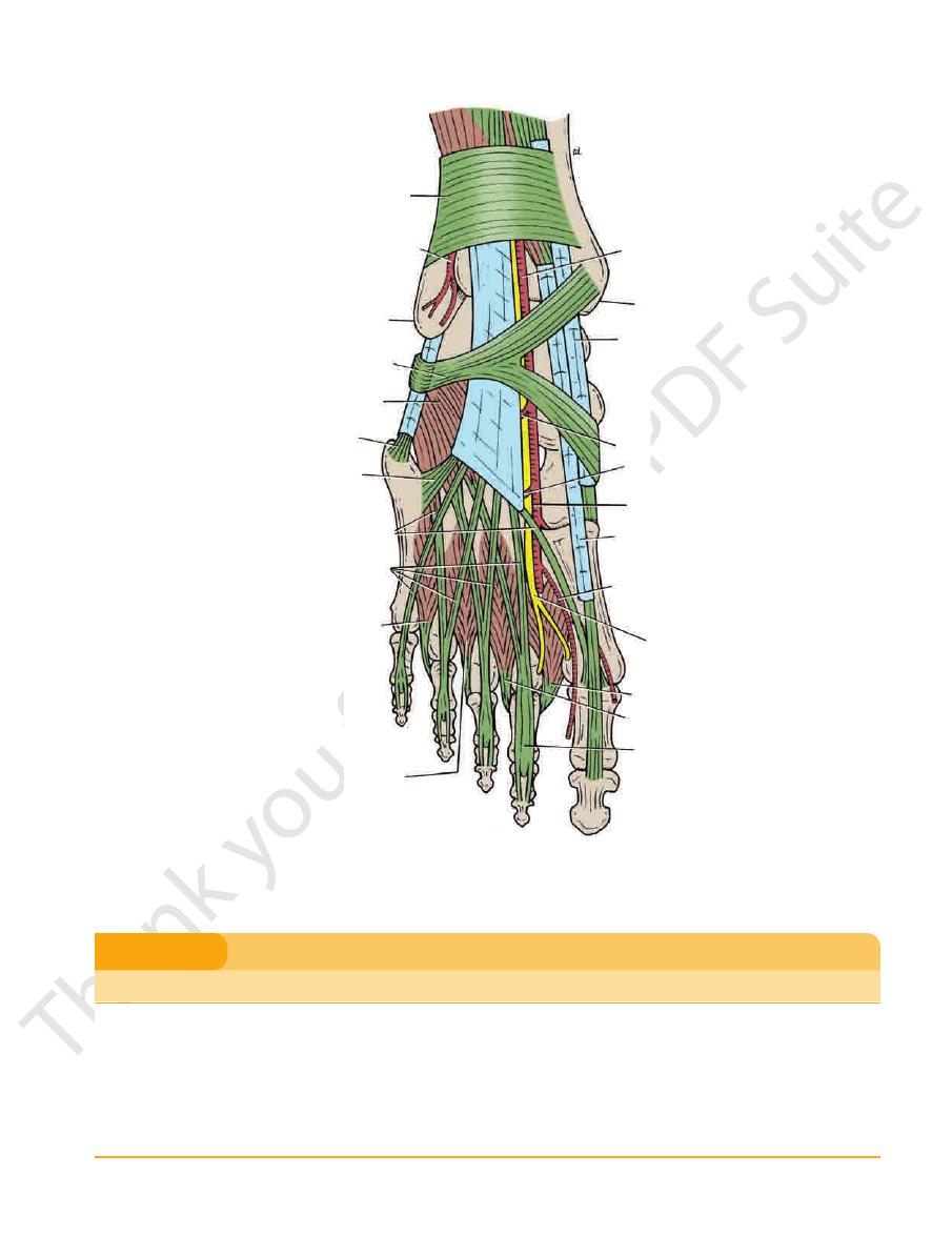
The Region of the Ankle
Anterior Aspect of the Ankle
■
■
■
■
■
■
■
■
■
■
■
■
■
■
■
■
-
Malleolus
■
■
■
■
Posterior Aspect of the Ankle
■
■
■
■
■
■
■
■
■
■
-
Retinaculum
■
■
■
■
Retinaculum
-
The Foot
-
-
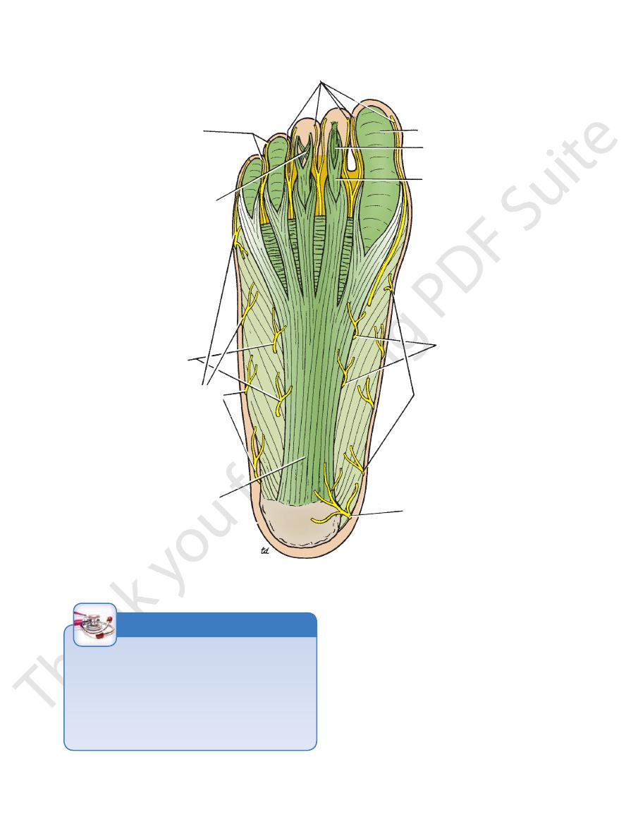
plantar aponeurosis
The
Deep Fascia
the sole (Figs. 10.1 and 10.54).
which innervate the lateral third of
lateral plantar nerve,
the medial two thirds of the sole; and branches from the
which innervate
medial plantar nerve,
branches from the
tibial nerve, which innervates the medial side of the heel;
of the
medial calcaneal branch
foot is derived from the
to the skin of the sole of the
sensory nerve supply
The
large numbers.
the sites of skin movement. Sweat glands are present in
ous fibrous bands. The skin shows a few flexure creases at
firmly bound down to the underlying deep fascia by numer
The skin of the sole of the foot is thick and hairless. It is
The Sole of the Foot
Skin
-
is a triangular thickening of the
into the toes.
base of the aponeurosis divides into five slips that pass
the medial and lateral tubercles of the calcaneum. The
vessels, and muscles (Fig. 10.54). Its apex is attached to
deep fascia that protects the underlying nerves, blood
Basic Anatomy
491
digital branches of medial plantar nerve
digital branches
of lateral plantar nerve
decussating fibers of
flexor digitorum brevis
branches of lateral
plantar nerve
branches of sural nerve
plantar aponeurosis
medial calcaneal nerve
branches of saphenous nerve
branches of medial
plantar nerve
tendon of flexor
digitorum brevis
tendon of flexor
digitorum longus
fibrous flexor sheath
FIGURE 10.54
Plantar aponeurosis and cutaneous nerves of the sole of the right foot.
Plantar Fasciitis
tion in the posterior attachment of the aponeurosis, forming
Plantar fasciitis, which occurs in individuals who do a great
deal of standing or walking, causes pain and tenderness of the
sole of the foot. It is believed to be caused by repeated minor
trauma. Repeated attacks of this condition induce ossifica-
a calcaneal spur.
C L I N I C A L N O T E S
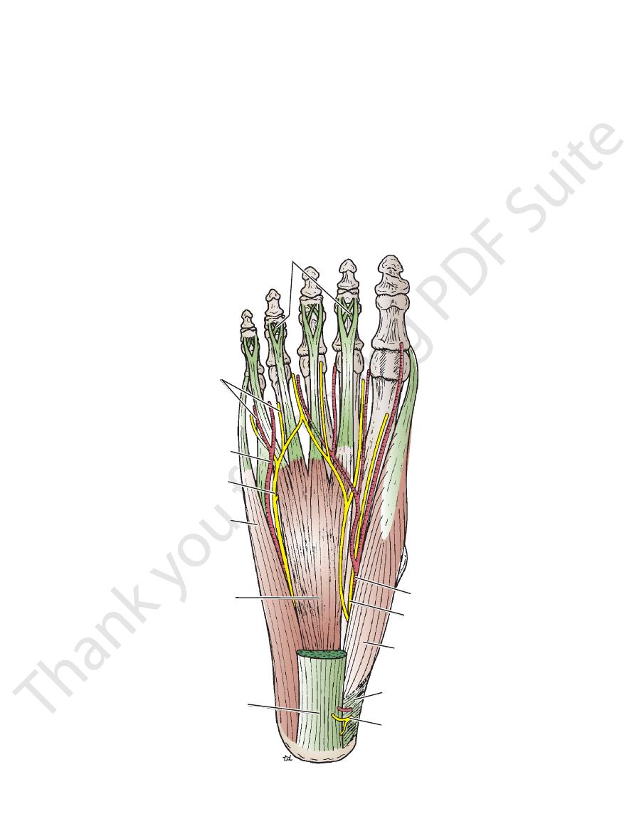
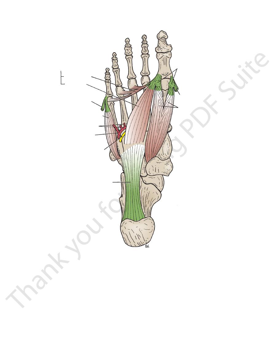
Muscles of the Sole of the Foot
alis posterior tendon
Interossei, peroneus longus tendon, tibi
Fourth layer:
flexor digiti minimi brevis
Flexor hallucis brevis, adductor hallucis,
Third layer:
tendon
digitorum longus tendon, flexor hallucis longus
Quadratus plantae, lumbricals, flexor
Second layer:
abductor digiti minimi
Abductor hallucis, flexor digitorum brevis,
First layer:
layers from the inferior layer superiorly.
The muscles of the sole are conveniently described in four
■
■
■
■
■
■
■
■
-
492
CHAPTER 10
passing behind the medial malleolus beneath the flexor
The flexor digitorum longus tendon enters the sole by
Flexor Digitorum Longus Tendon
Long Tendons of the Sole of the Foot
10.59 and are described in Table 10.8.
The muscles of the sole are seen in Figures 10.55 through
used in most people.
suggest control of individual toes, this function is rarely
porting the arches of the foot. Although their names would
few delicate functions and are chiefly concerned with sup
Unlike the small muscles of the hand, the sole muscles have
The Lower Limb
-
retinaculum (Figs. 10.47 and 10.56). It passes forward across
dons then enter the fibrous sheaths of the lateral four toes
forward, giving origin to the lumbrical muscles. The ten
now divides into its four tendons of insertion, which pass
the insertion of the quadratus plantae muscle. The tendon
a strong slip. It is here that it receives on its lateral border
the tendon of flexor hallucis longus, from which it receives
the medial surface of the sustentaculum tali and then crosses
-
(Fig. 10.54). Each tendon perforates the corresponding ten
flexor digitorum profundus in the hand (see page 400).
the method of insertion is similar to that found for the
into the base of the distal phalanx. It should be noted that
don of flexor digitorum brevis and passes on to be inserted
-
decussating fibers of flexor digitorum brevis
medial plantar arte
flexor retinaculum
flexor digitorum brevis
lateral plantar artery
digital nerves and arteries
lateral plantar nerve
abductor digiti minimi
plantar aponeurosis
medial calcaneal nerve
abductor hallucis
medial plantar nerve
ry
FIGURE 10.55
First layer of the plantar muscles of the right foot. Medial and lateral plantar arteries and nerves are also shown.

Muscles of the Sole of the Foot
T A B L E 1 0 . 8
transverse head from
metatarsal bones;
2nd, 3rd, and 4th
Oblique head bases of
into lateral side of base of
of big toe; lateral tendon
of base of proximal phalanx
Medial tendon into medial side
See Table 10.7
See Table 10.7
remainder: lateral
plantar nerve;
First lumbrical: medial
Tendons of flexor digito
longus in flexing
Assists flexor digitorum
Tendon of flexor digitorum
perforated by those of
of middle phalanx; tendons
toes—inserted into borders
Four tendons to four lateral
toe; braces medial
Flexes and abducts big
Muscle
Origin
Insertion
Nerve Supply
Nerve Root
a
Action
First Layer
Abductor hallucis
Medial tuberosity of
calcaneum and flexor
retinaculum
Base of proximal phalanx of
big toe
Medial plantar nerve
S2, 3
longitudinal arch
Flexor digitorum
brevis
Medial tubercle of
calcaneum
flexor digitorum longus
Medial plantar nerve
S2, 3
Flexes lateral four toes;
braces medial and
lateral longitudinal
arches
Abductor digiti
minimi
Medial and lateral tuber-
cles of calcaneum
Base of proximal phalanx of
fifth toe
Lateral plantar nerve
S2, 3
Flexes and abducts fifth
toe; braces lateral
longitudinal arch
Second Layer
Quadratus plantae
Medial and lateral sides
of calcaneum
longus
Lateral plantar nerve
S2, 3
lateral four toes
Lumbricals (4)
-
rum longus
Dorsal extensor expansion;
bases of proximal phalan-
ges of lateral four toes
plantar nerve
S2, 3
Extends toes at inter-
phalangeal joints
Flexor digitorum
longus tendon
Flexor hallucis
longus tendon
Third Layer
Flexor hallucis
brevis
Cuboid, lateral cunei-
form, tibialis posterior
insertion
proximal phalanx of big toe
Medial plantar nerve
S2, 3
Flexes metatar-
sophalangeal joint
of big toe; supports
medial longitudinal
arch
Adductor hallucis
plantar ligaments
Lateral side of base of proxi-
mal phalanx of big toe
Deep branch lateral
plantar nerve
S2, 3
Flexes metatar-
sophalangeal joint
of big toe; holds
together metatarsal
bones
Flexor digiti minimi
brevis
Base of 5th metatarsal
bone
Lateral side of base of proxi-
mal phalanx of little toe
See Table 10.7
Tibialis posterior
See Table 10.6
joints and extends
metatarsophalangeal
Adduction of toes; flexes
Lateral plantar nerve
S2, 3
Flexes metatar-
sophalangeal joint of
little toe
Fourth Layer
Interossei
Dorsal (4)
Adjacent sides of meta-
tarsal bones
Bases of proximal phalan-
ges—first: medial side of
second toe; remainder:
lateral sides of second,
third, and fourth toes—also
dorsal extensor expansion
Lateral plantar nerve
S2, 3
Abduction of toes;
flexes metatar-
sophalangeal joints
and extends inter-
phalangeal joints
Plantar (3)
Inferior surfaces of
3rd, 4th, and 5th
metatarsal bones
Medial side of bases of proxi-
mal phalanges of lateral
three toes
Lateral plantar nerve
S2, 3
interphalangeal joints
Peroneus longus
tendon
tendon
a
The predominant nerve root supply is indicated by boldface type.
494
CHAPTER 10
The Lower Limb
first lumbrical
flexor hallucis longus
flexor digitorum longus
quadratus plantae
lateral plantar
lateral plantar ne
plantar arch
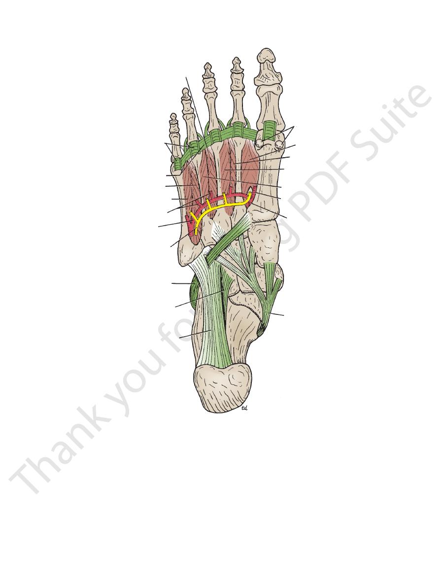
th lumbrical
third lumbrical
four
digital nerve
deep branch of
lateral plantar nerve
rve
artery
medial plantar artery
medial plantar nerve
digital nerves
second lumbrical
FIGURE 10.56
oot. Medial and lateral plantar arteries and nerves are also
Second layer of the plantar muscles of the right f
from behind the lateral malleolus and runs obliquely across
The peroneus longus tendon (Fig. 10.59) enters the foot
Peroneus Longus Tendon
by synovial sheaths (Figs. 10.49 and 10.57).
cis longus and the flexor digitorum longus are surrounded
The tendons of the flexor hallu
Synovial Flexor Sheaths
blind tunnel in which lie the flexor tendons of the toe (Fig.
of the phalanges and the interphalangeal joints, forms a
398). The fibrous sheath, together with the inferior surfaces
arrangement is similar to that found in the fingers (see page
is attached to the sides of the phalanges (Fig. 10.54). The
tal phalanx, is provided with a strong fibrous sheath, which
from the head of the metatarsal bone to the base of the dis
The inferior surface of each toe,
Fibrous Flexor Sheaths
the distal phalanx.
fibrous sheath of the big toe and is inserted into the base of
tendon, to which it gives a strong slip. It then enters the
lum tali and crosses deep to the flexor digitorum longus
flexor retinaculum. It runs forward below the sustentacu
sole by passing behind the medial malleolus beneath the
The flexor hallucis longus tendon (Fig. 10.56) enters the
Flexor Hallucis Longus Tendon
shown.
-
-
10.57).
-
Basic Anatomy
495
fibrous flexor sheath of second toe
digital synovial sheaths
flexor digitorum longus
synovial sheath
of peroneus brevis
synovial sheath
of peroneus longus
synovial sheath
of flexor hallucis longus
tibialis posterior
synovial sheath
of flexor digitorum
longus
flexor hallucis
longus
FIGURE 10.57
Synovial sheaths of the tendons seen on the sole of the right foot.
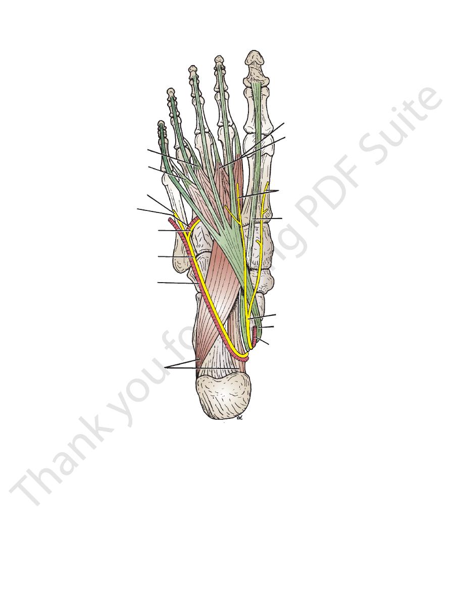
arises beneath the flexor retinaculum and passes forward
branches of the posterior tibial artery (see page 488). It
The lateral plantar artery is the larger of the terminal
Lateral Plantar Artery
articular branches.
its course, it gives off numerous muscular, cutaneous, and
supplying the medial side of the big toe (Fig. 10.55). During
deep to the abductor hallucis muscle (Fig. 10.49). It ends by
arises beneath the flexor retinaculum and passes forward
branches of the posterior tibial artery (see page 488). It
The medial plantar artery is the smaller of the terminal
Medial Plantar Artery
Arteries of the Sole of the Foot
a synovial sheath.
third, and fourth metatarsals. The tendon is surrounded by
cuboid and the cuneiforms and to the bases of the second,
osity of the navicular. Small tendinous slips pass to the
the sustentaculum tali to be inserted mainly into the tuber
flexor retinaculum and runs downward and forward above
from behind the medial malleolus. It passes beneath the
The tibialis posterior tendon (Fig. 10.59) enters the foot
Tibialis Posterior Tendon
and is surrounded by a synovial sheath (Fig. 10.57).
where it is held in position by the long plantar ligament
The tendon grooves the inferior surface of the cuboid
sal bone and the adjacent part of the medial cuneiform.
the sole to be inserted into the base of the first metatar-
-
496
CHAPTER 10
The Lower Limb
adductor hallucis
transverse head
oblique head
tendon of abductor
digiti minimi
flexor digiti minimi brevis
metatarsal arteries
plantar arch
deep branch of
lateral plantar nerve
long plantar ligament
flexor hallucis brevis
tendon of abductor hallucis
sesamoid bones
FIGURE 10.58
Third layer of the plantar muscles of the right foot. The deep branch of the lateral plantar nerve and the plantar
deep to the abductor hallucis and the flexor digitorum
arterial arch are also shown.
brevis (Figs. 10.49, 10.55, and 10.56). On reaching the base
of
one, the artery curves medially to
the 5th metatarsal b
nerve (see page 489). It arises beneath the flexor retinacu
The medial plantar nerve is a terminal branch of the tibial
Nerves of the Sole of the Foot
olus to form the posterior tibial venae comitantes.
sponding arteries, and they unite behind the medial malle
accompany the corre
lateral plantar veins
Medial
Veins of the Sole of the Foot
plies the cleft between the big and second toes.
The first plantar metatarsal artery, which sup
Branches
ately joins the lateral plantar artery (Fig. 10.59).
sal interosseous muscle, the dorsalis pedis artery immedi
On entering the sole between the two heads of the first dor
of the Foot)
he Dorsal Artery
Dorsalis Pedis Artery (
arch gives off plantar digital arteries to the toes.
muscular, cutaneous, and articular branches. The plantar
artery (Fig. 10.59). During its course, it gives off numerous
of the first intermetatarsal space joins the dorsalis pedis
(Fig. 10.58) and at the proximal end
plantar arch
form the
T
-
-
-
and
-
-
Medial Plantar Nerve
-
lum (Fig. 10.49) and runs forward deep to the abductor
Basic Anatomy
497
third dorsal interosseous
plantar ligaments of
metatarsophalangeal joints
deep transverse ligaments
fourth dorsal interosseous
third plantar interosseous
metatarsal arteries
plantar arch
deep branch of
lateral plantar nerve
peroneus longus
short plantar ligament
long plantar ligament
tibialis posterior
dorsalis pedis artery
first plantar metatarsal artery
second plantar interosseous
first plantar interosseous
second dorsal interosseous
first dorsal interosseous
sesamoid bones
FIGURE 10.59
Fourth layer of the plantar muscles of the right foot. The deep branch of the lateral plantar nerve and the plan
tar arterial arch are also shown. Note the deep transverse ligaments.
-
hallucis, with the medial plantar artery (Fig. 10.55). It
deep branches (Fig. 10.56).
of the fifth metatarsal bone, it divides into superficial and
the lateral plantar artery (Fig. 10.56). On reaching the base
hallucis and the flexor digitorum brevis, in company with
lum (Fig. 10.49) and runs forward deep to the abductor
nerve (see page 489). It arises beneath the flexor retinacu
The lateral plantar nerve is a terminal branch of the tibial
palm of the hand.
Compare with the distribution of the median nerve in the
and the tips of the toes.
nerves extend onto the dorsum and supply the nail beds
sides of the medial three and a half toes (Fig. 10.54). The
run to the
Cutaneous branches: Plantar digital nerves
lumbrical muscle.
digitorum brevis, the flexor hallucis brevis, and the first
to the abductor hallucis, the flexor
Muscular branches
Branches
and the flexor digitorum brevis.
comes to lie in the interval between the abductor hallucis
■
■
■
■
Lateral Plantar Nerve
-

498
CHAPTER 10
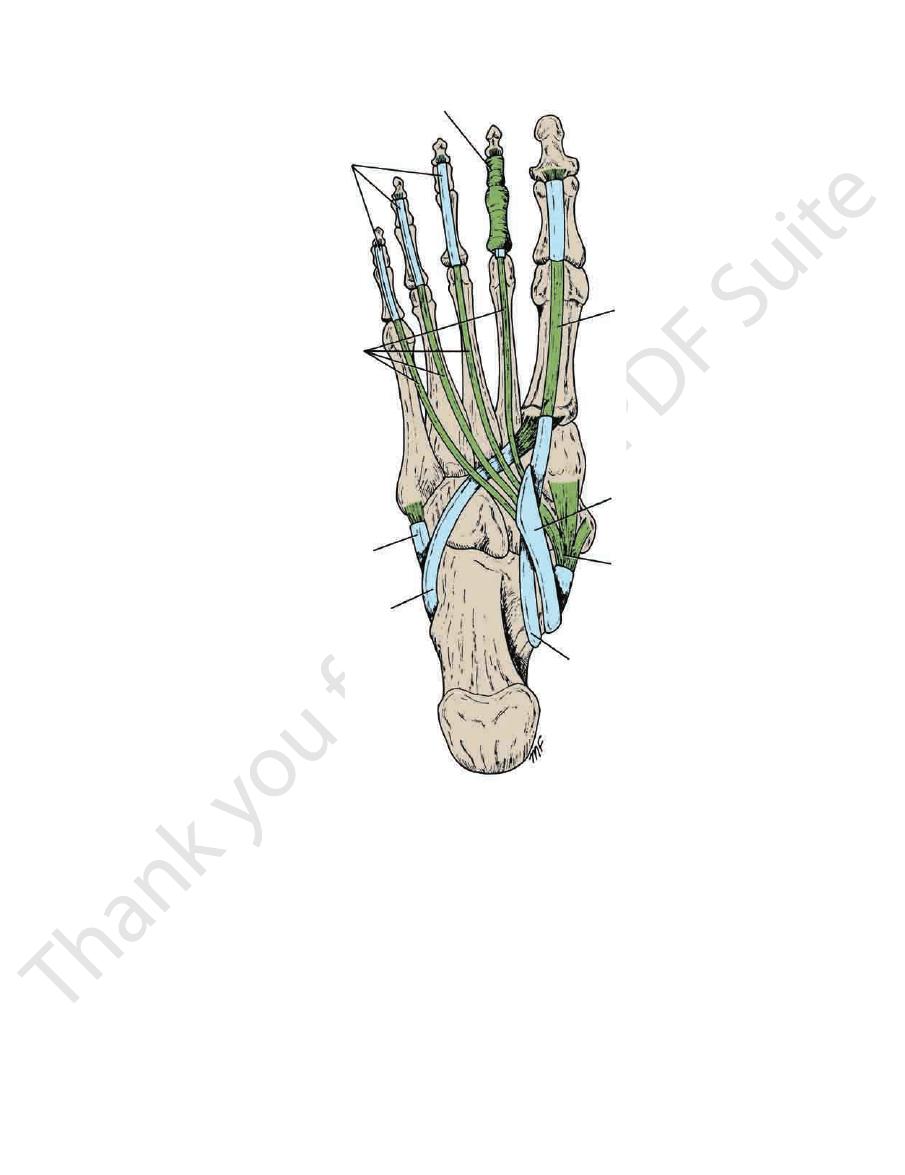
longus tendons. On the medial side lies the tendon of extensor
nal part of the deep peroneal nerve and the extensor digitorum
digitorum brevis (Fig. 10.60). On its lateral side lie the termi
inferior extensor retinaculum and the first tendon of extensor
(Fig. 10.59). It is superficial in position and is crossed by the
joins the lateral plantar artery and completes the plantar arch
two heads of the first dorsal interosseous muscle, where it
It terminates by passing downward into the sole between the
a continuation of the anterior tibial artery (see page 485).
The dorsalis pedis artery begins in front of the ankle joint as
of the Foot)
Dorsalis Pedis Artery (the Dorsal Artery
Artery of the Dorsum of the Foot
atarsal bone.
malleoli and distally to the level of the base of the fifth met
sheath extends proximally for a short distance above the
they pass beneath the extensor retinacula (Fig. 10.60). The
dons are surrounded by a common synovial sheath as
The extensor digitorum longus and peroneus tertius ten
Digitorum Longus
Synovial Sheath of the Tendon of Extensor
dons of insertion of the interosseous and lumbrical muscles.
The dorsal expansion, as in the fingers, receives the ten
distal phalanx (Fig. 10.60).
eral parts, which converge to be inserted into the base of the
inserted into the base of the middle phalanx, and two lat
expansion splits into three parts: a central part, which is
Near the proximal interphalangeal joint, the extensor
sion.
extensor expan
joins the fascial expansion called the
On the dorsal surface of each toe, the extensor tendon
side by a tendon of extensor digitorum brevis (Fig. 10.60).
third, and fourth toes, each tendon is joined on its lateral
toes. Opposite the metatarsophalangeal joints of the second,
out over the dorsum of the foot and pass to the lateral four
muscle (Fig. 10.60). The tendon divides into four, which fan
extensor retinaculum, in company with the peroneus tertius
the superior extensor retinaculum and through the inferior
The tendon of extensor digitorum longus passes beneath
The Insertion of the Long Extensor Tendons
Table 10.9.
The muscle is seen in Figure 10.60 and described in
Muscles of the Dorsum of the Foot
pass through the interosseous spaces.
digital veins and communicating veins from the sole, which
of the blood from the whole foot drains into the arch via
back of the leg is described on page 487. The greater part
into the leg behind the lateral malleolus. Its course in the
is described on page 451. The small saphenous vein ascends
the leg in front of the medial malleolus. Its further course
nous vein leaves the dorsum of the foot by ascending into
into the small saphenous vein (Fig. 10.19). The great saphe
side into the great saphenous vein and on the lateral side
the heads of the metatarsal bones and drains on the medial
The dorsal venous arch lies in the subcutaneous tissue over
Dorsal Venous Arch (or Network)
lateral plantar nerves (see above).
of the terminal phalanges are supplied by the medial and
The nail beds and the skin covering the dorsal surfaces
margin of the foot and the lateral side of the little toe.
lateral malleolus and supplies the skin along the lateral
(Fig. 10.1) enters the foot behind the
sural nerve
The
the head of the first metatarsal bone.
the skin along the medial side of the foot as far forward as
foot in front of the medial malleolus (Fig. 10.2). It supplies
passes onto the dorsum of the
saphenous nerve
The
cent sides of the big and second toes (Fig. 10.2).
supplies the skin of the adja
deep peroneal nerve
The
fourth, and fifth toes.
of the big toe; and the adjacent sides of the second, third,
supply the skin on the dorsum of the foot; the medial side
divides into medial and lateral cutaneous branches that
muscle in the lower part of the leg (see page 486). It now
the peroneus brevis and the extensor digitorum longus
emerges from between
superficial peroneal nerve
The
nerves.
nerve, assisted by the deep peroneal, saphenous, and sural
dorsum of the foot is derived from the superficial peroneal
(Fig. 10.2) to the skin on the
sensory nerve supply
The
mobile on the underlying tendons and bones.
The skin on the dorsum of the foot is thin, hairy, and freely
palm of the hand.
Compare with the distribution of the ulnar nerve in the
branch above).
those in the fourth intermetatarsal space (see superficial
and fourth lumbricals; and all the interossei, except
and supplies the adductor hallucis; the second, third,
branch curves medially with the lateral plantar artery
(Fig. 10.59). This
From the deep terminal branch
the dorsum and supply the nail beds and tips of the toes.
of the lateral one and a half toes. The nerves extend onto
metatarsal space. Plantar digital branches pass to the sides
minimi and the interosseous muscles of the fourth inter
to the flexor digiti
From the superficial terminal branch
of the lateral part of the sole.
abductor digiti minimi; cutaneous branches to the skin
to the quadratus plantae and
From the main trunk
Branches
The Lower Limb
■
■
■
■
-
■
■
The Dorsum of the Foot
Skin
-
-
Extensor Digitorum Brevis
-
-
-
-
-
-
sor tendons opposite the bases of the metatarsal bones
which runs laterally under the exten
Arcuate artery,
foot just below the ankle joint (Fig. 10.60).
which crosses the dorsum of the
Lateral tarsal artery,
Branches
Its pulsations can easily be felt.
hallucis longus (Fig. 10.60).
■
■
■
■
-
(Fig. 10.60). It gives off metatarsal branches to the toes.
passing deep to the extensor retinacula on the lateral side
The deep peroneal nerve enters the dorsum of the foot by
Deep Peroneal Nerve
Nerve Supply of the Dorsum of the Foot
sides of the big toe (Fig. 10.60).
which supplies both
First dorsal metatarsal artery,
■
■
Basic Anatomy
499
superior extensor retinaculum
perforating branch of peroneal artery
lateral malleolus
inferior extensor retinaculum
extensor digitorum brevis
peroneus brevis
peroneus tertius
extensor digitorum brevis tendons
extensor digitorum longus tendons
fourth dorsal interosseous
third dorsal interosseous
extensor expansion
second dorsal interosseous
first dorsal interosseous
medial terminal branch
of deep peroneal nerve
first dorsal metatarsal artery
extensor hallucis longus
dorsalis pedis artery
arcuate artery
lateral tarsal artery
tibialis anterior
medial malleolus
anterior tibial artery
FIGURE 10.60
Structures in the dorsal aspect of the right foot.
Muscle of the Dorsum of the Foot
T A B L E 1 0 . 9
Muscle
Origin
Insertion
Nerve Supply
Nerve Root
Action
Extensor digitorum
brevis
Anterior part of
upper surface of
the calcaneum
and from the
inferior extensor
retinaculum
By four tendons
into the proximal
phalanx of big
toe and long
extensor ten-
dons to second,
third, and fourth
toes
Deep peroneal
nerve
S1, S2
Extends toes

500
CHAPTER 10
the condyles of the tibia and their cartilaginous menisci
Above are the rounded condyles of the femur; below are
Articulation
Note that the fibula is not directly involved in the joint.
between the patella and the patellar surface of the femur.
the corresponding condyles of the tibia, and a gliding joint,
between the medial and lateral condyles of the femur and
in the body. Basically, it consists of two condylar joints
The knee joint is the largest and most complicated joint
The hip joint is fully described on page 467.
articular branches to the joints of the foot.
sor digitorum brevis muscle. Both terminal branches give
ond toes (Fig. 10.60). The lateral branch supplies the exten
supplies the skin of the adjacent sides of the big and sec
terminal, medial, and lateral branches. The medial branch
of the dorsalis pedis artery (see page 485). It divides into
The Lower Limb
-
-
Joints of the Lower Limb
Knee Joint
(Fig. 10.35); in front is the articulation between the lower
tellar bursa
suprapa
beneath the quadriceps tendon, forming the
permitting the synovial membrane to pouch upward
the joint. On the front of the joint, the capsule is absent,
surfaces and surrounds the sides and posterior aspect of
The capsule is attached to the margins of the articular
vial joint of the plane gliding variety.
possible. The joint between the patella and femur is a syno
the hinge variety, but some degree of rotatory movement is
The joint between the femur and tibia is a synovial joint of
Type
tibial plateaus
referred to clinically as the medial and lateral
faces of the medial and lateral condyles of the tibia are often
covered with hyaline cartilage. Note that the articular sur
The articular surfaces of the femur, tibia, and patella are
end of the femur and the patella.
-
.
-
Capsule
-
(Fig.
h side of the patella, the
10.35). On eac
above to the lateral condyle of the femur and below to the
is cordlike and is attached
lateral collateral ligament
The
the common tendon of the quadriceps femoris muscle.
10.35). It is, in fact, a continuation of the central portion of
der of the patella and below to the tuberosity of the tibia (Fig.
is attached above to the lower bor
ligamentum patellae
The
Extracapsular Ligaments
capsule and those that lie within the capsule.
The ligaments may be divided into those that lie outside the
Ligaments
permits the tendon of the popliteus to emerge (Fig. 10.35).
An opening in the capsule behind the lateral tibial condyle
(Fig. 10.35).
oblique popliteal ligament
muscle called the
is strengthened by an expansion of the semimembranous
vastus lateralis and medialis. Behind the joint, the capsule
capsule is strengthened by expansions from the tendons of
-
head of the fibula (Fig. 10.35). The tendon of the popliteus
and below to the medial surface of the shaft of the tibia
attached above to the medial condyle of the femur
medial collateral ligament
The
meniscus (Fig. 10.61).
muscle intervenes between the ligament and the lateral
is a flat band and is
(Fig. 10.35).
popliteal bursa
popliteus, forming the
longed downward on the deep surface of the tendon of the
At the back of the joint, the synovial membrane is pro
muscle (Fig. 10.35).
articularis genus
called the
ment of a small portion of the vastus intermedius muscle,
. This is held in position by the attach
suprapatellar bursa
cle for three fingerbreadths above the patella, forming the
which extends up beneath the quadriceps femoris mus
10.61). On the front and above the joint, it forms a pouch,
to the margins of the articular surfaces (Figs. 10.35 and
The synovial membrane lines the capsule and is attached
Synovial Membrane
it is relatively immobile.
meniscus is also attached to the medial collateral ligament,
tibia by anterior and posterior horns. Because the medial
Each meniscus is attached to the upper surface of the
cushions between the two bones.
to receive the convex femoral condyles; they also serve as
tion is to deepen the articular surfaces of the tibial condyles
surfaces are in contact with the tibial condyles. Their func
faces are in contact with the femoral condyles. The lower
forms a free edge (Figs. 10.35 and 10.61). The upper sur
capsule, and the inner border is thin and concave and
tilage. The peripheral border is thick and attached to the
The menisci are C-shaped sheets of fibrocar
Menisci
being pulled posteriorly.
With the knee joint flexed, the PCL prevents the tibia from
prevents anterior displacement of the femur on the tibia.
medial femoral condyle (Figs. 10.35 and 10.61). The PCL
be attached to the anterior part of the lateral surface of the
of the tibia and passes upward, forward, and medially to
ament (PCL) is attached to the posterior intercondylar area
The posterior cruciate lig
Posterior Cruciate Ligament
from being pulled anteriorly.
tibia. With the knee joint flexed, the ACL prevents the tibia
ACL prevents posterior displacement of the femur on the
the lateral femoral condyle (Figs. 10.35 and 10.61). The
be attached to the posterior part of the medial surface of
of the tibia and passes upward, backward, and laterally, to
ment (ACL) is attached to the anterior intercondylar area
The anterior cruciate liga
Anterior Cruciate Ligament
tibia throughout the joint’s range of movement.
ligaments are the main bond between the femur and the
to their tibial attachments (Fig. 10.61). These important
10.35). They are named anterior and posterior, according
ments that cross each other within the joint cavity (Fig.
are two strong intracapsular liga
cruciate ligaments
The
Intracapsular Ligaments
strengthens the posterior aspect of the capsule (Fig. 10.35).
sion derived from the semimembranosus muscle. It
is a tendinous expan
oblique popliteal ligament
The
(Fig. 10.61).
meniscus
It is firmly attached to the edge of the medial
-
-
-
-
-
-
-
-
-
-
. A bursa is
osed
interp