
Basic Anatomy
tal ends of the anterior borders of the fibula and the tibia
The superior extensor retinaculum is attached to the dis
Superior Extensor Retinaculum
481
-
(Figs. 10.46, 10.47, and 10.50).
The inferior extensor retinaculum is a Y-shaped band
Inferior Extensor Retinaculum
located in front of the ankle joint (Figs. 10.44, 10.46, and
The superior peroneal retinaculum connects the lateral
Superior Peroneal Retinaculum
lined by a synovial sheath.
tendons lie in compartments (Fig. 10.48), each of which is
medial malleolus as they pass forward to enter the sole. The
the deep muscles of the back of the leg to the back of the
face of the calcaneum (Fig. 10.49). It binds the tendons of
downward and backward to be attached to the medial sur
The flexor retinaculum extends from the medial malleolus
Flexor Retinaculum
synovial sheath.
ments (Figs. 10.48 and 10.50), each of which is lined by a
10.47). Fibrous bands separate the tendons into compart
-
-
malleolus to the lateral surface of the calcaneum (Fig. 10.49).
big
toe
peroneus
longus
anterior border
of tibia
tibialis anterior
extensor hallucis longus
superficial peroneal nerve
extensor digitorum longus
superior extensor
retinaculum
medial malleolus
great saphenous vein
extensor digitorum longus
dorsalis pedis artery
peroneus
brevis
superficial
peroneal nerve
inferior extensor
retinaculum
lateral
malleolus
peroneus longus
and brevis tendons
extensor digitorum
brevis
peroneus
tertius
dorsal
venous
network
extensor
hallucis
longus
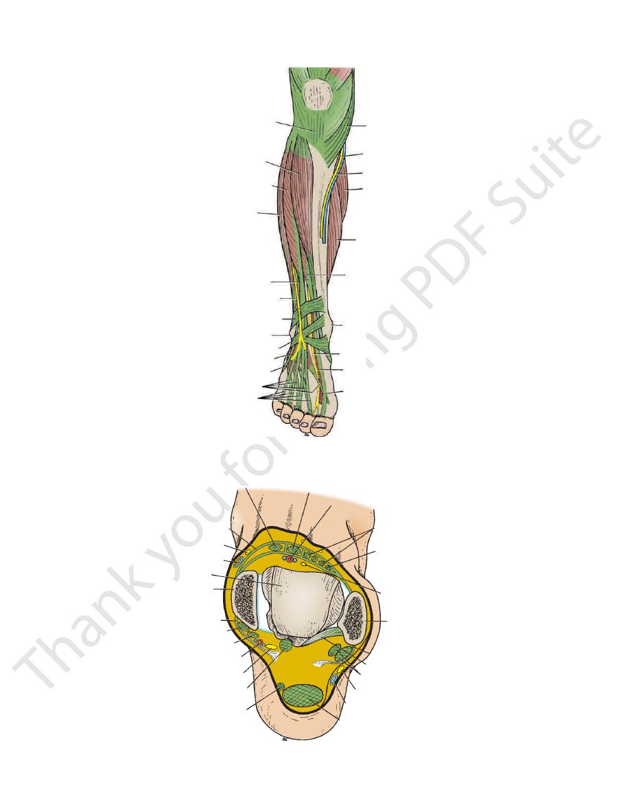
FIGURE 10.46
Dissection of the front of the right leg and
mon peroneal nerve (see page 479), supplies the skin of the
a branch of the com
superficial peroneal nerve,
The
on the upper part of the lateral surface of the leg (Fig. 10.1).
common peroneal nerve (see page 479), supplies the skin
a branch of the
lateral cutaneous nerve of the calf,
The
Cutaneous Nerves
retinacula is described on pages 490.
The arrangement of the tendons beneath the different
mon sheath.
synovial sheath, which is continuous above with the com
of the calcaneum (Fig. 10.49). The tendons each possess a
the peroneus longus and brevis muscles to the lateral side
The inferior peroneal retinaculum binds the tendons of
Inferior Peroneal Retinaculum
with a common synovial sheath.
the back of the lateral malleolus. The tendons are provided
It binds the tendons of the peroneus longus and brevis to
dorsum of the foot.
-
The Front of the Leg
Skin
-
lower part of the anterolateral surface of the leg (Fig. 10.2).
joint along with the other muscles in this compartment
The peroneus tertius muscle extends the foot at the ankle
of the foot away from the ground.
Extension, or dorsiflexion of the ankle, is the movement
Note the following:
10.48, and 10.49 and are described in Table 10.5.
The muscles are seen in Figures 10.44, 10.45, 10.46, 10.47,
Muscles of the Anterior Fascial Compartment
Deep peroneal nerve
Nerve supply:
Anterior tibial artery
Blood supply:
gus, peroneus tertius, and extensor hallucis longus
The tibialis anterior, extensor digitorum lon
Muscles:
(Fig. 10.4).
the small saphenous vein and drain into the popliteal nodes
of the front of the leg may pass via vessels that accompany
10.4). A small amount of lymph from the upper lateral part
the vertical group of superficial inguinal lymph nodes (Fig.
in vessels that follow the great saphenous vein, to end in
fascia on the front of the leg drains upward and medially
The greater part of the lymph from the skin and superficial
Lymph Vessels
(Fig. 10.51).
the leg and ultimately drain into the great saphenous vein
Numerous small veins curve around the medial aspect of
Superficial Veins
face of the leg (Fig. 10.2).
(see page 463), supplies the skin on the anteromedial sur
a branch of the femoral nerve
saphenous nerve,
The
-
Contents of the Anterior Fascial
Compartment of the Leg
■
■
-
■
■
■
■
of the Leg
■
■
■
■
482
CHAPTER 10
The Lower Limb
ligamentum patellae
tibialis anterior
extensor digitorum longus
peroneus longus
peroneus brevis
superficial peroneal nerve
superior extensor retinaculum
lateral malleolus
inferior extensor retinaculum
extensor digitorum brevis
extensor digitorum longus
extensor hallucis longus
dorsalis pedis artery
deep peroneal nerve
medial malleolus
extensor hallucis longus
soleus
gastrocnemius
great saphenous vein
saphenous nerve
sartorius
peroneus tertius
extensor digitorum brevis
FIGURE 10.47
Structures in the anterior and lateral aspects of the right leg and the dorsum of the foot.
tibialis anterior
inferior extensor retinaculum
saphenous nerve
great saphenous vein
talus
medial malleolus
flexor retinaculum
tibialis posterior
flexor digitorum longus
posterior tibial artery
tibial nerve
flexor hallucis longus
plantaris tendon
tendo calcaneus
small saphenous vein
sural nerve
peroneus brevis and longus
posterior talofibular ligament
superior peroneal retinaculum
lateral malleolus
deep peroneal nerve
peroneus tertius
extensor digitorum longus
dorsalis pedis artery
extensor hallucis longus
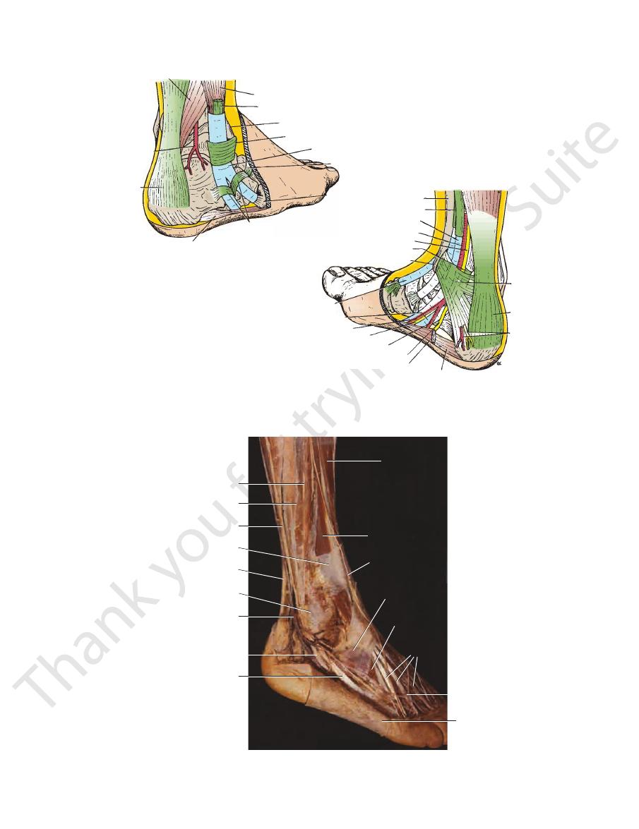
FIGURE 10.48
Relations of the right ankle joint.
Basic Anatomy
483
flexor hallucis longus
peroneal
artery
tendo
calcaneus
abductor digiti minimi
inferior peroneal retinaculum
fifth metarsal
bone
inferior extensor retinaculum
synovial sheath
superior peroneal retinaculum
lateral malleolus
peroneus longus
peroneus brevis
tibia
tibialis posterior
flexor digitorum longus
posterior tibial artery
tibial nerve
flexor hallucis longus
medial malleolus
tibialis anterior
flexor hallucis longus
medial plantar nerve
medial plantar artery
lateral plantar artery
lateral plantar nerve
abductor hallucis
flexor digitorum brevis
medial
calcaneal
nerve and
artery
tendo
calcaneus
flexor
retinaculum
A
B
FIGURE 10.49
Structures passing behind the lateral malleolus
dons are shown in
. Synovial sheaths of the ten
(A) and the medial malleolus (B)
-
blue. Note the positions of the retinacula.
peroneus brevis
peroneus longus
sural nerve
superior extensor retinaculum
tendo calcaneus
lateral malleolus
small saphenous vein
peroneus longus
peroneus brevis
extensor digitorum
longus
superficial peroneal
nerve
inferior extensor
retinaculum
extensor
digitorum brevis
extensor
digitorum
longus
dorsal venous arch
reflected skin
tibialis anterior
FIGURE 10.50
Dissection of the right ankle region showing the structures passing behind the lateral malleolus. Note the posi-
tion of the retinacula.
484
CHAPTER 10
The Lower Limb
dorsal venous network
big toe
medial malleolus of tibia
great saphenous vein
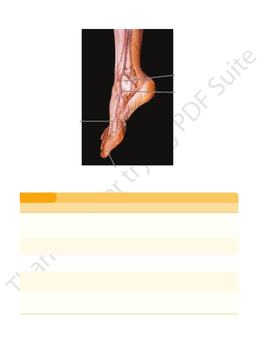
FIGURE 10.51
Dissection of the right ankle region showing the origin of the great saphenous vein from the dorsal venous
arch. Note that the great saphenous vein ascends in front of the medial malleolus of the tibia.
Muscles of the Anterior Fascial Compartment of the Leg
T A B L E 1 0 . 5
and long extensor tendons to
proximal phalanx of big toe
By four tendons into the
foot at subtalar and
foot at ankle joint; inverts
Extends big toe; extends
everts foot at subtalar and
Extends foot at ankle joint;
Tibialis
Muscle
Origin
Insertion
Nerve Supply
Nerve Root
a
Action
anterior
Lateral surface of
shaft of tibia and
interosseous
membrane
Medial cuneiform and base of
1st metatarsal bone
Deep peroneal
nerve
L4, 5
Extends
b
foot at ankle joint;
inverts foot at subtalar
and transverse tarsal
joints; holds up medial
longitudinal arch of foot
Extensor
digitorum
longus
Anterior surface of
shaft of fibula
Extensor expansion of lateral
four toes
Deep peroneal
nerve
L5; S1
Extends toes; extends foot
at ankle joint
Peroneus
tertius
Anterior surface of
shaft of fibula
Base of 5th metatarsal bone
Deep peroneal
nerve
L5; S1
transverse tarsal joints
Extensor
hallucis
longus
Anterior surface of
shaft of fibula
Base of distal phalanx of
great toe
Deep peroneal
nerve
L5; S1
transverse tarsal joints
Extensor
digitorum
brevis
Calcaneum
second, third, and fourth toes
Deep peroneal
nerve
S1, 2
Extends toes
a
The predominant nerve root supply is indicated by boldface type.
b
Extension, or dorsiflexion, of the ankle is the movement of the foot away from the ground.
Basic Anatomy
485
B
medial
malleolus
tendon of
tibialis
anterior
lateral
malleolus
tendons of
extensor
digitorum
longus
tendons of
extensor
digitorum
longus
tendon of
tibialis
anterior
great saphenous
vein
medial
malleolus
lateral
malleolus
sites for
palpation
of dorsalis
pedis artery
A
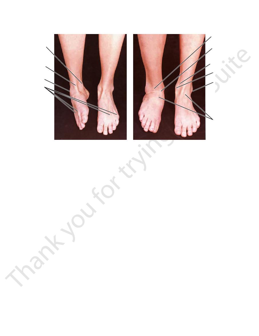
FIGURE 10.52
-old woman showing inversion
Anterior view of the ankles and feet of a 29-year
sor digitorum longus on its lateral side. It is here that its
and the deep peroneal nerve and the tendons of exten
tendon of the extensor hallucis longus on its medial side
passed behind the superior extensor retinaculum, it has the
the lower end of the tibia (Figs. 10.44 and 10.47). Having
the lower part of its course, it lies superficial in front of
it lies deep beneath the muscles of the compartment. In
peroneal nerve (Fig. 10.44). In the upper part of its course,
of the interosseous membrane, accompanied by the deep
membrane (Fig. 10.42). It descends on the anterior surface
through an opening in the upper part of the interosseous
passes forward into the anterior compartment of the leg
lower border of the popliteus muscle (see page 477) and
branches of the popliteal artery. It arises at the level of the
The anterior tibial artery is the smaller of the terminal
Anterior Tibial Artery
Artery of the Anterior Fascial Compartment
with the insertion of extensor digitorum in the hand.)
inserted into the base of the distal phalanx. (Compare
middle phalanx, and the two lateral parts converge to be
part of the expansion is inserted into the base of the
. The central
extensor expansion
expansion called the
surface of each toe become incorporated into a fascial
The extensor digitorum longus tendons on the dorsal
peroneal nerve.
cles but receives no innervation from the superficial
joints along with the peroneus longus and brevis mus
also everts the foot at the subtalar and transverse tarsal
and is supplied by the deep peroneal nerve. The muscle
of the right foot.
and eversion
(A)
(B)
-
■
■
of the Leg
-
pulsations can easily be felt in the living subject. In front of
to the ankle joint
Articular branch
hallucis longus
digitorum longus, the peroneus tertius, and the extensor
to the tibialis anterior, the extensor
Muscular branches
Branches
the foot is described on page 498.
passes behind the extensor retinacula. Its further course in
lateral to the anterior tibial artery (Fig. 10.44). The nerve
longus muscle, first lying lateral, then anterior, and finally
septum. It then descends deep to the extensor digitorum
the anterior compartment by piercing the anterior fascial
side of the neck of the fibula (Fig. 10.44). The nerve enters
the substance of the peroneus longus muscle on the lateral
of the common peroneal nerve (see page 479). It arises in
The deep peroneal nerve is one of the terminal branches
Deep Peroneal Nerve
Compartment of the Leg
Nerve Supply of the Anterior Fascial
form the popliteal vein.
of the posterior tibial artery in the popliteal fossa to
of the anterior tibial artery join those
Venae comitantes
of other arteries around the knee and ankle joints
that anastomose with branches
Anastomotic branches
to neighboring muscles
Muscular branches
Branches
(see page 498).
the ankle joint, the artery becomes the dorsalis pedis artery
■
■
■
■
■
■
■
■
■
■

486
CHAPTER 10
The Lower Limb
Anterior Compartment of the Leg Syndrome
deep fascia and thus decompress the area and prevent anoxic
ment of the leg by making a longitudinal incision through the
peroneal nerve—that is, the skin cleft between the first and
Loss of sensation is limited to the area supplied by the deep
gus, and the extensor hallucis longus muscles are paralyzed.
disappears. The tibialis anterior, the extensor digitorum lon
cut off by compression, and the dorsalis pedis arterial pulse
in pressure. In severe cases, the arterial supply is eventually
the venous return is diminished, thus producing a further rise
of the ankle also increases the pain. As the pressure rises,
that pass through the compartment by passive plantar flexion
increases the severity of the pain. Stretching of the muscles
can become severe. Dorsiflexion of the foot at the ankle joint
compartment of the leg that is characteristic of this syndrome
diagnosis is critical. The deep, aching pain in the anterior
associated with bone fractures is a common cause, and early
from an increased production of tissue fluid. Soft tissue injury
increase in the intracompartmental pressure that results
The anterior compartment syndrome is produced by an
-
second toes. The surgeon can open the anterior compart-
necrosis of the muscles.
C L I N I C A L N O T E S
Contents of the Lateral Fascial
It arises in the substance of the peroneus longus muscle on
branches of the common peroneal nerve (see page 479).
The superficial peroneal nerve is one of the terminal
Superficial Peroneal Nerve
Nerve of the Lateral Fascial Compartment
muscles.
pierce the posterior fascial septum, and supply the peroneal
488), which lies in the posterior compartment of the leg,
Numerous branches from the peroneal artery (see page
Artery of the Lateral Fascial Compartment
tie to the transverse arch of the foot.
foot. In addition, the peroneus longus tendon serves as a
role in holding up the lateral longitudinal arch in the
and transverse tarsal joints. They also play an important
foot at the ankle joint and evert the foot at the subtalar
Both the peroneus longus and brevis muscles flex the
Note the following:
10.48, 10.49, and 10.50 and described in Table 10.6.
The muscles are seen in Figures 10.44, 10.45, 10.46, 10.47,
Muscles of the Lateral Fascial Compartment
Superficial peroneal nerve
Nerve supply:
Branches from the peroneal artery
Blood supply:
Peroneus longus and peroneus brevis
Muscles:
Compartment of the Leg
■
■
■
■
■
■
of the Leg
■
■
of the Leg
of the Leg
Muscles of the Lateral Fascial Compartment of the Leg
T A B L E 1 0 . 6
Muscle
Origin
Insertion
Nerve Supply
Nerve Root
a
Action
Peroneus
longus
Lateral surface of
shaft of fibula
Base of 1st
metatarsal
and the medial
cuneiform
Superficial
peroneal nerve
L5; S1, 2
Plantar flexes foot at ankle joint;
everts foot at subtalar and
transverse tarsal joints; supports
lateral longitudinal and transverse
arches of foot
Peroneus
brevis
Lateral surface of
shaft of fibula
Base of 5th
metatarsal bone
Superficial
peroneal nerve
L5; S1, 2
Plantar flexes foot at ankle joint;
everts foot at subtalar and
transverse tarsal joint; supports
lateral longitudinal arch of foot
a
The predominant nerve root supply is indicated by boldface type.
Tenosynovitis and Dislocation of the Peroneus
retinaculum must be torn. It usually occurs in older children
malleolus. For this condition to occur, the superior peroneal
Tendon dislocation can occur when the tendons of peroneus
Treatment consists of immobilization, heat, and physiotherapy.
Tenosynovitis (inflammation of the synovial sheaths) can
Longus and Brevis Tendons
affect the tendon sheaths of the peroneus longus and bre-
vis muscles as they pass posterior to the lateral malleolus.
longus and brevis dislocate forward from behind the lateral
and is caused by trauma.
C L I N I C A L N O T E S

Basic Anatomy
the ground.
ning by using the foot as a lever and raising the heel off
the main forward propulsive force in walking and run
powerful plantar flexors of the ankle joint. They provide
Together, the soleus, gastrocnemius, and plantaris act as
Note the following:
described in Table 10.7.
The muscles are seen in Figures 10.45 and 10.53 and are
of the Leg: Superficial Group
Muscles of the Posterior Fascial Compartment
Tibial nerve
Nerve supply:
Posterior tibial artery
Blood supply:
gus, flexor hallucis longus, and tibialis posterior
Popliteus, flexor digitorum lon
Deep group of muscles:
and soleus
Gastrocnemius, plantaris,
Superficial group of muscles:
superficial and deep groups (see Fig. 10.45).
divides the muscles of the posterior compartment into
of the leg is a septum that
deep transverse fascia
The
liteal nodes (Fig. 10.4).
group of superficial inguinal nodes or drain into the pop
around the medial side of the leg to end in the vertical
the back of the leg drain upward and either pass forward
Lymph vessels from the skin and superficial fascia on
Lymph Vessels
great saphenous vein.
division joining the popliteal and the other joining the
join the great saphenous vein; or it may split in two, one
subject to variation: It may join the popliteal vein; it may
The mode of termination of the small saphenous vein is
medially and join the great saphenous vein (Fig. 10.19)
that run upward and
anastomotic branches
Important
with the deep veins of the foot
Communicating veins
from the back of the leg
small veins
Numerous
Tributaries
small saphenous vein has numerous valves along its course.
and 10.40); it ends in the popliteal vein (see page 478). The
muscle in the lower part of the popliteal fossa (Figs. 10.19
fascia and passes between the two heads of the gastrocnemius
up the middle of the back of the leg. The vein pierces the deep
lows the lateral border of the tendo calcaneus and then runs
the lateral malleolus in company with the sural nerve. It fol
behind
dorsal venous arch of the foot (Fig. 10.19). It ascends
arises from the lateral part of the
small saphenous vein
The
Superficial Veins
the posteromedial surface of the leg (Fig. 10.1).
(see page 463), gives off branches that supply the skin on
a branch of the femoral nerve
saphenous nerve,
The
eral surface of the leg (Fig. 10.1).
479), supplies the skin on the lower part of the posterolat
a branch of the tibial nerve (see page
sural nerve,
The
(Fig. 10.1).
on the upper part of the posterolateral surface of the leg
common peroneal nerve (see page 479), supplies the skin
a branch of the
lateral cutaneous nerve of the calf,
The
of the back of the leg (Fig. 10.1).
supplies the skin over the popliteal fossa and the upper part
the back of the thigh (see page 465). In the popliteal fossa, it
of the thigh descends on
posterior cutaneous nerve
The
Cutaneous Nerves
of the little toe (see page 498).
cent sides of the first and second toes and the lateral side
dorsal surfaces of the skin of all the toes, except the adja
the dorsum of the foot. In addition, branches supply the
to the skin on the lower part of the front of the leg and
Medial and lateral branches are distributed
Cutaneous:
(Fig. 10.44)
branches to the peroneus longus and brevis
Muscular
Branches
cutaneous (Figs. 10.47 and 10.50).
brevis muscles, and in the lower part of the leg it becomes
and 10.50). It descends between the peroneus longus and
the lateral side of the neck of the fibula (Figs. 10.44, 10.46,
487
■
■
■
■
-
The Back of the Leg
Skin
-
-
■
■
■
■
■
■
-
Contents of the Posterior Fascial
Compartment of the Leg
■
■
■
■
-
■
■
■
■
■
■
-
fibers of the soleus or partial tearing of the tendo calcaneus is
gastrocnemius and soleus muscles retract proximally, leaving
tion. A sudden, sharp pain is felt, with immediate disability. The
Tearing of the gastrocnemius or soleus muscles will produce
Gastrocnemius and Soleus Muscle Tears
severe localized pain over the damaged muscle. Swelling may
be present.
Ruptured Tendo Calcaneus
Rupture of the tendo calcaneus is common in middle-aged
men and frequently occurs in tennis players. The rupture
occurs at its narrowest part, about 2 in. (5 cm) above its inser-
a palpable gap in the tendon. It is impossible for the patient to
actively plantar flex the foot. The tendon should be sutured as
soon as possible and the leg immobilized with the ankle joint
plantar flexed and the knee joint flexed.
Rupture of the Plantaris Tendon
Rupture of the plantaris tendon is rare, although tearing of the
frequently diagnosed as such a rupture.
Plantaris Tendon and Autografts
The plantaris muscle, which is often missing, can be used for
tendon autografts in repairing severed flexor tendons to the
fingers; the tendon of the palmaris longus muscle can also be
used for this purpose.
C L I N I C A L N O T E S