
Chapter 11
Muscular
System
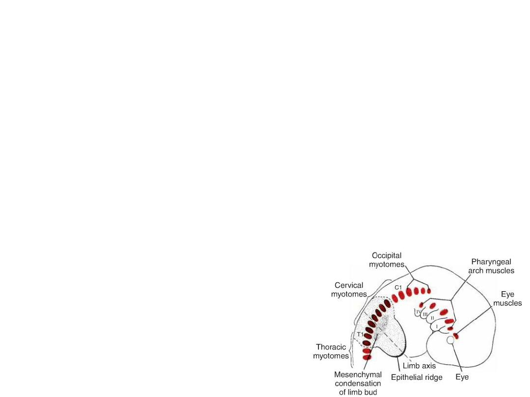
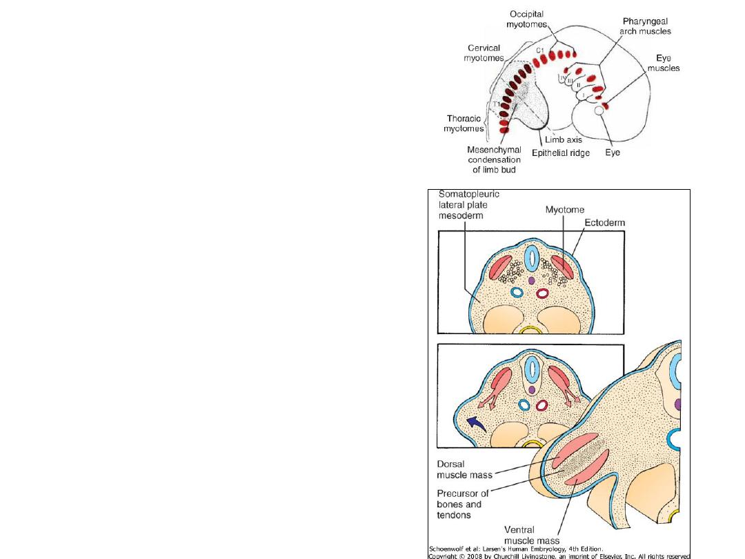
Drawing showing musculature in the
head and neck derived from
somitomeres and myotomes that form
from the occipital region caudally in a 7-
week embryo.
STRIATED SKELETAL MUSCULATURE
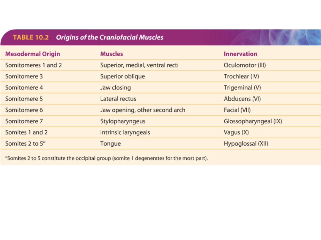
Head musculature: Derived from seven
somitomeres
Musculature of the a
xial skeleton, body wall,
and limbs: derived from somites
ORIGIN
• Skeletal muscles: paraxial mesoderm
(Somites & somitomeres)
• Smooth muscles:
Splanchnic mesoderm surrounding gut tube & its derivatives
Ectoderm: pupillary muscle, mammary gland & sweat glands
• Cardiac muscles: splanchnic mesoderm surrounding heart tube
Ch 11 / Muscular System (Smooth – Skeletal – Cardiac)
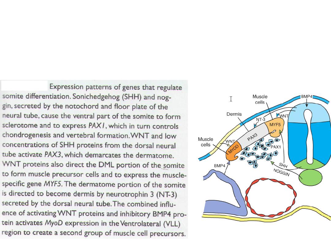
Sclerotome: vertebrae & ribs
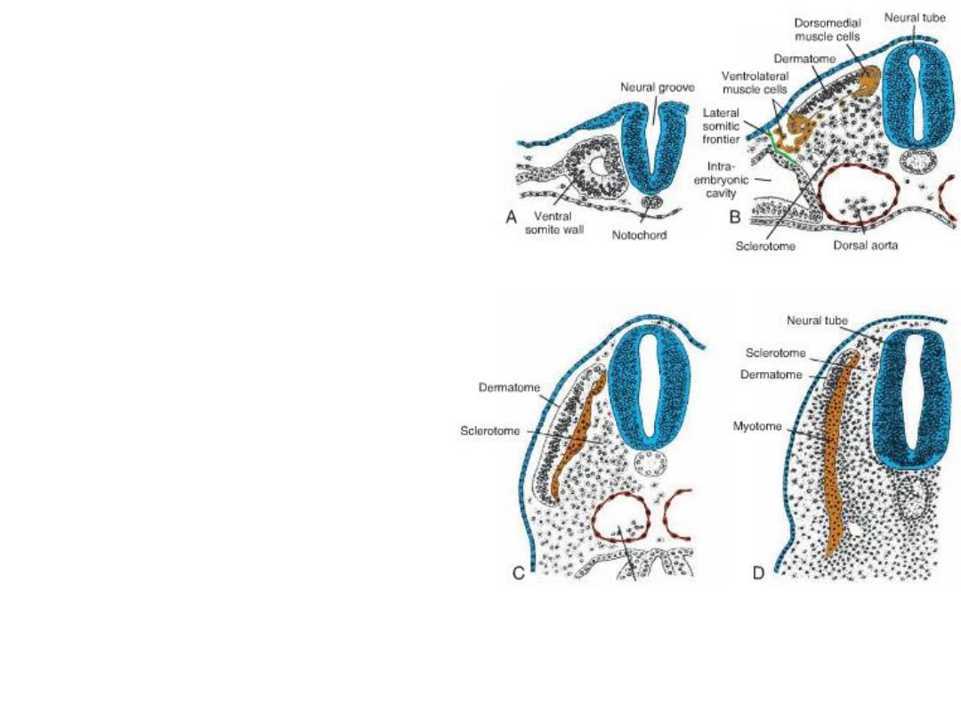
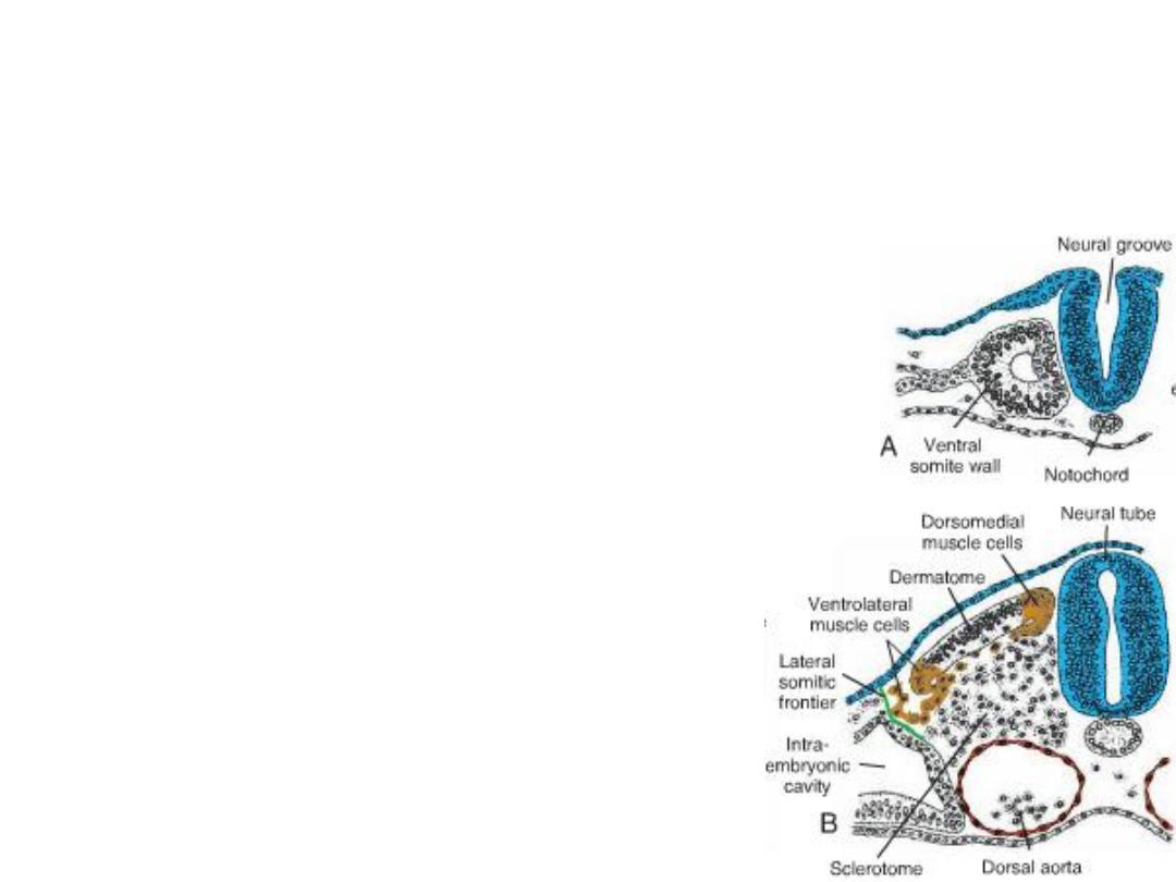
•Cross-sectional drawings showing
the stages of development in a
somite.
•A. Mesoderm cells become epithelial
and are arranged around a small
lumen.
•B. Cells in the ventral and medial
walls of the somite lose their
epithelial characteristics and migrate
around the neural tube and
notochord, and some move into the
parietal layer of lateral plate
mesoderm. Collectively, these cells
constitute the sclerotome.
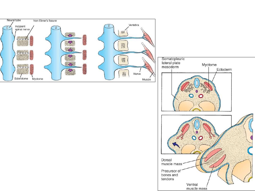
Cells at the dorsomedial (DML) and
ventrolateral (VLL) regions of the somite
form muscle cell precursors.
Dermomyotome
Cells from both regions migrate ventral to the dermatome to form the dermomyotome.
Some cells from the VLL region also migrate into the adjacent parietal layer of lateral
plate mesoderm across the lateral somitic frontier(greenline): infrahyoid, abdominal
wall (Rec abd., Int obl. Ext.Obl. & Transv. Abd.) and limb muscles.
The remaining cells in the myotome form muscles of the back, shoulder girdle and
intercostal muscles
The lateral somitic frontier
The lateral somitic frontier separates
between two mesodermal domains:
1.
The primaxial domain: region around
neural tube, contains only somite-derived
(paraxial mesoderm) cells.
2.
The abaxial domain: consists of parietal
layer of lateral plate mesoderm with somite
cells that migrated across the lateral somitic
frontier.
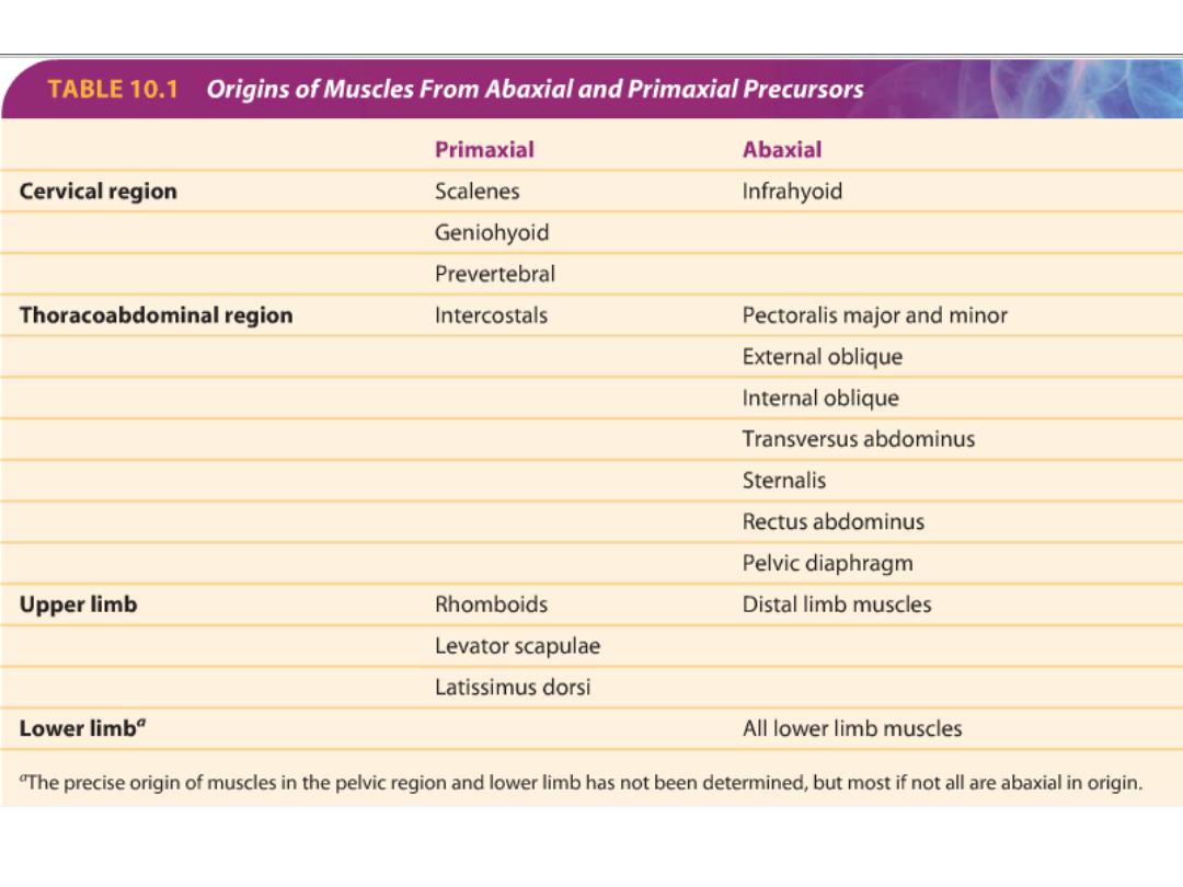
• Abaxial muscle cell precursors (from VLL edge of myotome) cross the frontier
and receive signals for differentiation from lateral plate mesoderm.
• The primaxial muscle cell precursors: that remain in the paraxial mesoderm
and do not cross the frontier (the remaining VLL cells and all of the DML cells)
receive their development signals from the neural tube & notochord.
• Regardless of their domain, each myotome receives its innervation from
spinal nerves derived from the same segment as the muscle cells.
The lateral somitic frontier
• The lateral somitic frontier also defines the
border of dermis derived from dermatomes in
the back and dermis derived from lateral plate
mesoderm in the body wall.
• It also defines a border for rib development with
the bony components of each rib derived from
primaxial sclerotome cells and the cartilaginous
parts of those ribs that attach to the sternum
derived from sclerotome cells that migrate
across the lateral somitic frontier (abaxial cells).
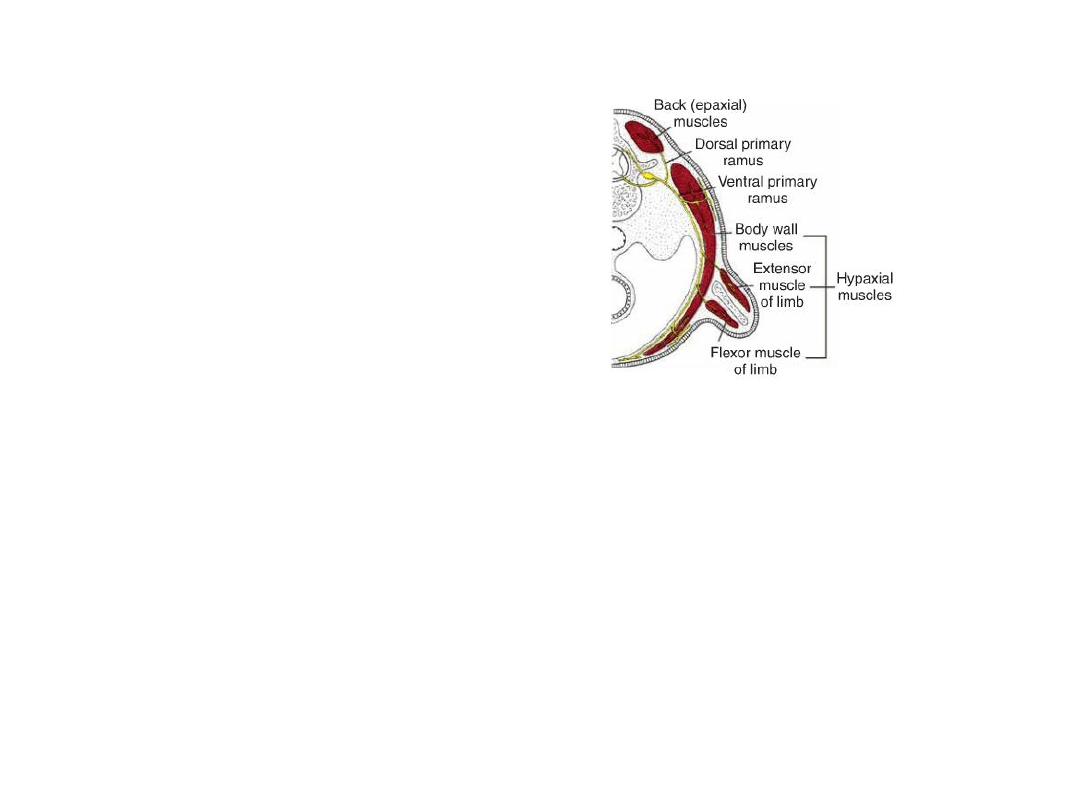
• Epaxial (true back muscles) are
innervated by dorsal (posterior)
primary rami.
• Hypaxial muscles (limb and body
wall) are innervated by ventral
(anterior) primary rami.
Innervation of axial skeletal muscles
Skeletal muscle &tendon
• During differentiation, precursor cells, the myoblasts, fuse and form long,
multinucleated muscle fibers.
• Myofibrils soon appear in the cytoplasm, and by the end of the third
month, cross-striations, typical of skeletal muscle, appear.
• Tendons: for the attachment of muscles to bones are derived from
sclerotome cells lying adjacent to myotomes at the anterior and posterior
borders of somites.
• The transcription factor SCLERAXIS regulates development of tendons.

Patterning of muscles
• Patterns of muscle formation are controlled by connective tissue into
which myoblasts migrate.
• Origin of connective tissue:
• Neural crest cells: in head region
• Somitic mesoderm: in cervical and occipital regions
• The parietal layer of lateral plate mesoderm: in body wall and limbs
Head musculature
• All voluntary muscles of the head region are derived from paraxial
mesoderm (somitomeres and somites), including:
– Musculature of the tongue,
– Eye muscles (except that of the iris, which is derived from optic cup
ectoderm), and
– Muscles associated with the pharyngeal (visceral) arches
• Patterns of muscle formation in the head are directed by connective tissue
elements derived from
neural crest cells
.
The first indication of limb
musculature is observed in the
seventh week of development as a
condensation of mesenchyme near
the base of the limb buds.
The mesenchyme is derived from
VLL cells of the somites that
migrate into the limb bud to form the
muscles.
As in other regions, connective
tissue dictates the pattern of muscle
formation, and this tissue is derived
from the parietal layer of lateral
plate mesoderm, which also gives
rise to the bones of the limb.
LIMB MUSCULATURE

CARDIAC MUSCLE
• Cardiac muscle develops from splanchnic mesoderm surrounding the
endothelial heart tube.
• myoblasts do not fuse.
SMOOTH MUSCLE
• Smooth muscle for the dorsal aorta and large arteries is derived from
lateral plate mesoderm and neural crest cells.
• In the coronary arteries, smooth muscle originates from proepicardial cells
and
neural crest cells
(proximal segments).
• Smooth muscle in the wall of the gut and gut derivatives is derived from
the
splanchnic layer of lateral plate mesoderm
that surrounds these
structures.
• Only the
sphincter and dilator muscles of the pupil
and muscle tissue in
the
mammary
and
sweat glands
are derived from ectoderm.

Poland sequence.
The pectoralis minor and part of the
pectoralis major muscles are missing on
the patient's left side. Note displacement
of the nipple and areola.
Prune belly syndrome
Partial or complete absence of
abdominal musculature. A
distended abdomen from atrophy of
abdominal wall musculature.