
Chapter 6
Third to Eighth
weeks:
Embryonic Period

EMBRYONIC PERIOD
3
rd
– 8
th
weeks
Period of Organogenesis
• Derivatives of the Ectodermal Germ Layer
• Clinical Correlates
• Derivatives of the Mesodermal Germ Layer
• Clinical Correlates
• Derivatives of the Endodermal Germ Layer
• Patterning of the Anteroposterior Axis:
• Regulation by Homeobox Genes
• External Appearance During the Second Month
• Clinical Correlates
The embryonic period
• Period of organogenesis
• From 3
rd
to 8
th
weeks of development
• Germ layers: ectoderm, mesoderm & endoderm tissues & organs
• By end of embryonic period (end of 2
nd
month):
main organ systems
are
established
recognizable body form
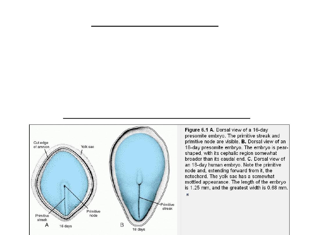
Derivatives of ectodermal germ layer
NEURULATION
• Notochord and prechordal mesoderm induce the overlying ectoderm to
thicken and form the
neural plate
.
• Cells of the plate make up the
neuroectoderm
, and their induction
represents the initial event in the process of
neurulation
.

Molecular regulation of neural induction
BMP4 ( bone morphogenic protein4)
•
Inactivated (absent) BMP4
Ectoderm becomes Neuralized neural plate
Mesoderm becomes Dorsalized notochord & paraxial mesoderm
Inactivation of BMP4 by secretion of:
o noggin, chordin & follistatin (from organizer, notochord &
prechordal mesoderm)
Which induce formation of: Forebrain & midbrain
o WNT3 & FGF
Which induce formation of hindbrain & spinal cord
In addition, retinoic acid (RA) appears to play a role in organizing the
cranial-to-caudal axis
• Presence of BMP4
Ectoderm epidermis
Ventralize Mesoderm intermediate & lateral plate mesoderm
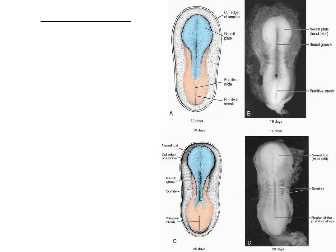
NEURULATION
• Dorsal view of a late presomite
embryo (approximately 19 days).
The amnion has been removed, and
the neural plate is clearly visible.
• Dorsal view of an embryo at
approximately 20 days showing
somites and formation of the neural
groove and neural folds.
• The process whereby the neural plate
forms the neural tube.
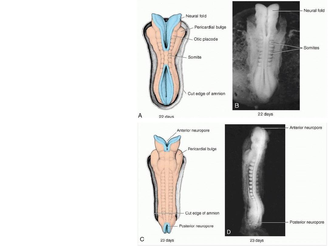
Dorsal view of an embryo at
approximately day
22.
Seven
distinct somites are visible on
each side of the neural tube.
Dorsal view of an embryo at
approximately day
23
. Note the
pericardial bulge on each side of
the midline in the cephalic part of
the embryo.
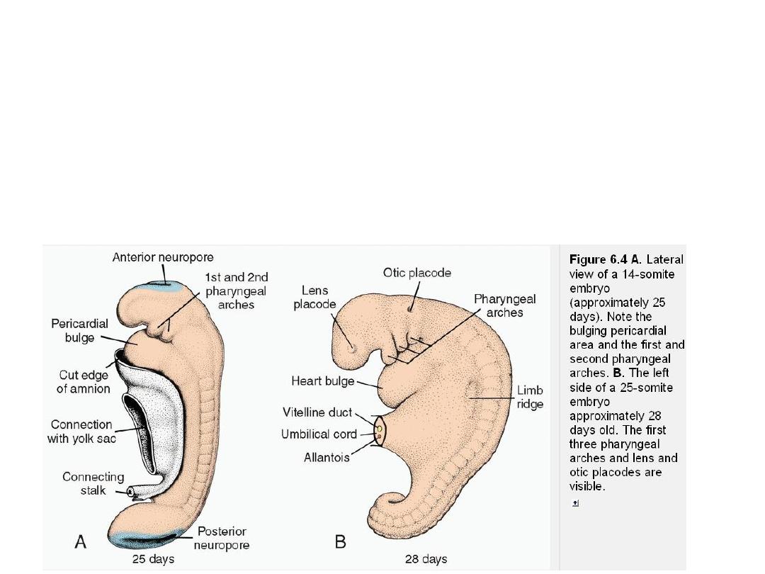
CLOSURE OF ANT NEUROPORE
- DAY 25
14-somite
embryo
Note the bulging pericardial area
and the first and second
pharyngeal arches .
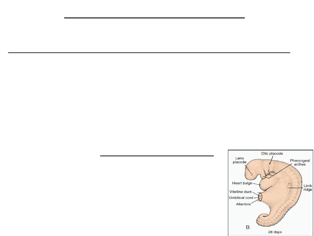
Closure of post neuropore- day
28
25
-somite embryo
The first three pharyngeal
arches and lens and otic
placodes are visible
.
Completion of neurulation
• By closure of ant & post neuropores the central nervous system is formed:
brain vesicles & spinal cord.
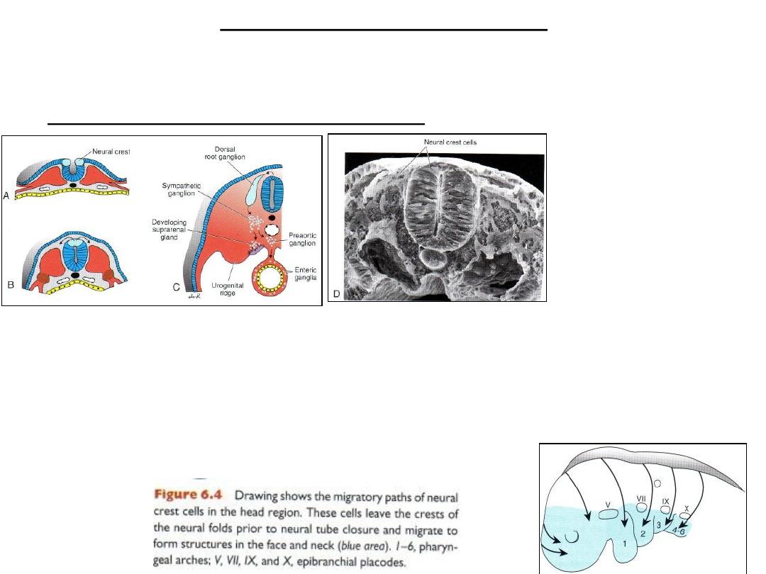
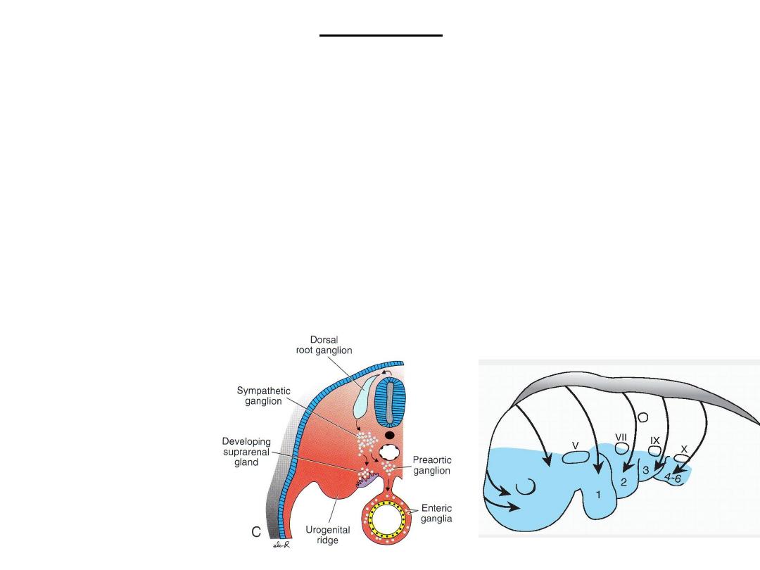
Neural crest formation & migration
Figure 6.5 Formation and migration of neural crest cells in the spinal cord. A,B. Crest cells form at the tips
of neural
folds and do not migrate away from this region until neural tube closure is complete. C. After migration,
crest cells
contribute to a heterogeneous array of structures, including dorsal root ganglia, sympathetic chain
ganglia, adrenal medulla,
and other tissues (Table 6.1, p. 69). D. In a scanning electron micrograph, crest cells at the top of the
closed neural tube can
be seen migrating away from this area.
Crest cells
• Crest cells form at the tips of neural folds and do not migrate away from
this region until neural tube closure is complete.
Neural crest formation & migration
epithelial-t0-mesenchymal transition
In trunk region
• DORSAL PATHWAY:
– Melanocytes in skin
• VENTRAL PATHWAY:
– sensory ganglia,
– Sympathetic
– enteric neurons
– Schwann cells
– Adrenal medulla
Neural crest formation &
migration
epithelial-t0-mesenchymal
transition
In head region
craniofacial skeleton, cranial ganglia
neurons, glial cells, melanocytes
Induction of crest cells
• Intermedite levels of bmp4.
The fate of the entire ectodermal germ layer depends on BMP concentrations:
• High levels induce
epidermis
formation;
• intermediate levels, at the border of the neural plate and surface ectoderm,
induce the
neural crest
;
• and very low concentrations cause formation of
neural ectoderm.
• BMPs also regulate neural crest cell migration, proliferation, and differentiation,
• and abnormal concentrations of the proteins have been associated with neural
crest defects in the craniofacial region of laboratory animals
Otic placodes and the lens placodes
• By the time the neural tube is closed, 2 bilateral ectodermal
thickenings, the otic placodes and the lens placodes,
• The otic placodes invaginate and form the otic vesicles
– Structures for hearing and maintenance of equilibrium
• the lens placodes the lenses of the eyes

Ectodermal derivatives
contact with outside
•
CNS, PNS
•
Sensory epith.: ear, nose, eyes
•
Epidermis: hair, nails
•
Subcutaneous glands, mammary gl, pituitary, enamel of teeth.
TABLE 6.1 - Neural Crest Derivatives
•
Connective tissue and bones of the face and skull
•
Cranial nerve ganglia
•
Cells of the thyroid gland
•
Conotruncal septum in the heart
•
Odontoblasts
•
Dermis in face and neck
•
Spinal (dorsal root) ganglia
•
Sympathetic chain and preaortic ganglia
•
Parasympathetic ganglia of the gastrointestinal tract
•
Adrenal medulla
•
Schwann cells
•
Glial cells
•
Meninges (forebrain)
•
Melanocytes
•
Smooth muscle cells to blood vessels of the face and forebrain
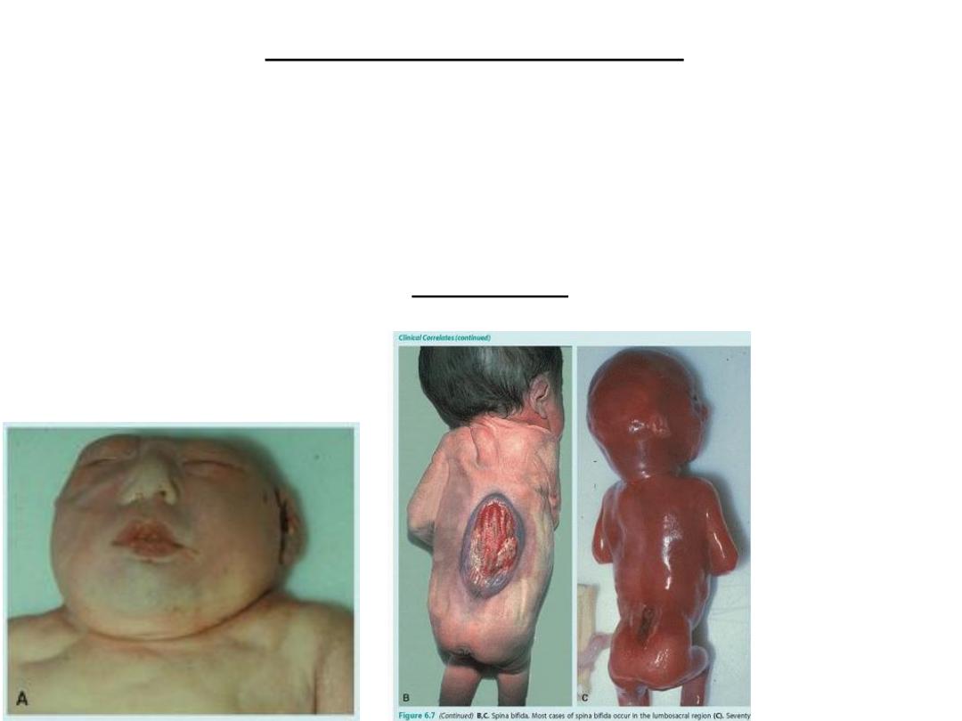
Neural tube defects (NTDs)
• result when neural tube closure fails to occur.
• If the neural tube fails to close in the cranial region, then most of the brain
fails to form, and the defect is called anencephaly
• If closure fails anywhere from the cervical region caudally, then the defect
is called spina bifida
Examples of neural tube defects
(NTDs), Anencephaly.
Spina bifida
• Most cases of
spina bifida
occur in the
lumbosacral
region
• Seventy
percent of all
of these NTDs
can be
prevented by
the vitamin
folic acid.
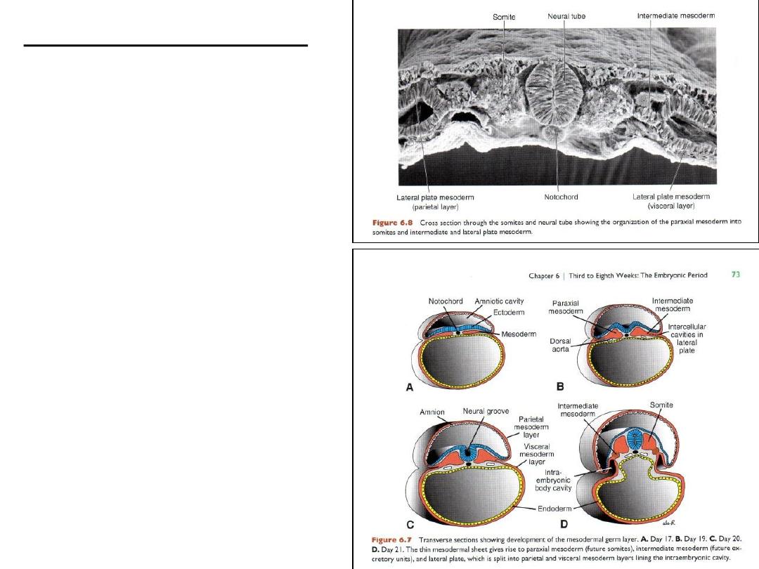
Mesoderm derivatives
• Paraxial mesoderm
• Intermediate mesoderm
• Lateral plate mesoderm

Paraxial mesoderm
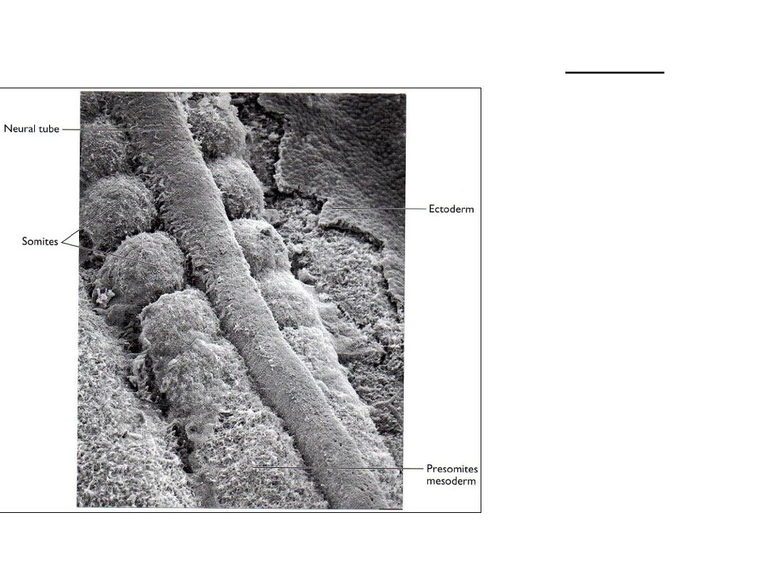
Paraxial mesoderm
Somites forming along the neural tube
Somites
• Head region: neuromeres-
mesenchyme of head
• Occipital---caudal region:
somites ...axial skeleton
– 1
st
pair: occipital region:
20
th
day
– 3pairs/day
– End of 5
th
week: 42-44
somites
• 4occiptal pairs
• 12thoracic pairs
• 5lumbar pairs
• 5sacral pairs
• 8-10 coccygeal pairs
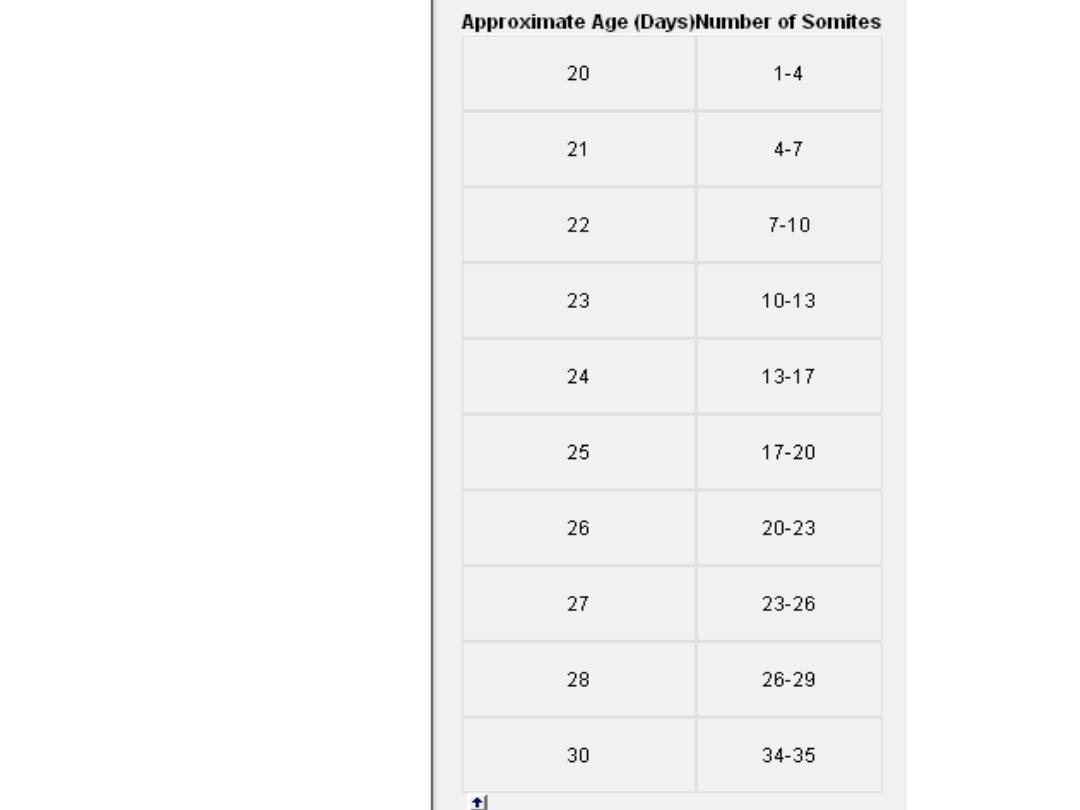
Age of embryo
in days &
Number of
somites
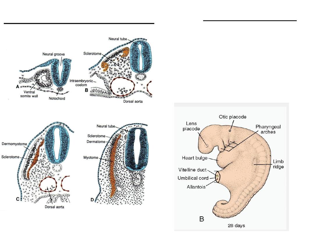
Stages in development of a somite
Somite derivatives
• Each somite: own…
Sclerotome: tendon, cartilage & bone
Myotome: segmental muscles
Dermatome: dermis of back
Myotome+dermatome: segmental nerve
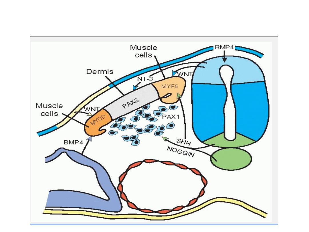
EXPRESSION PATTERNS OF GENES
THAT REGULATE SOMITE DIFFERENTIATION
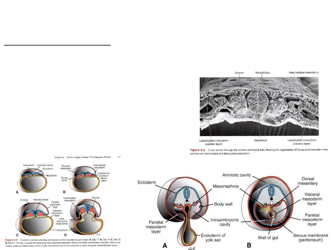
Intermediate Mesoderm - urogenital structures
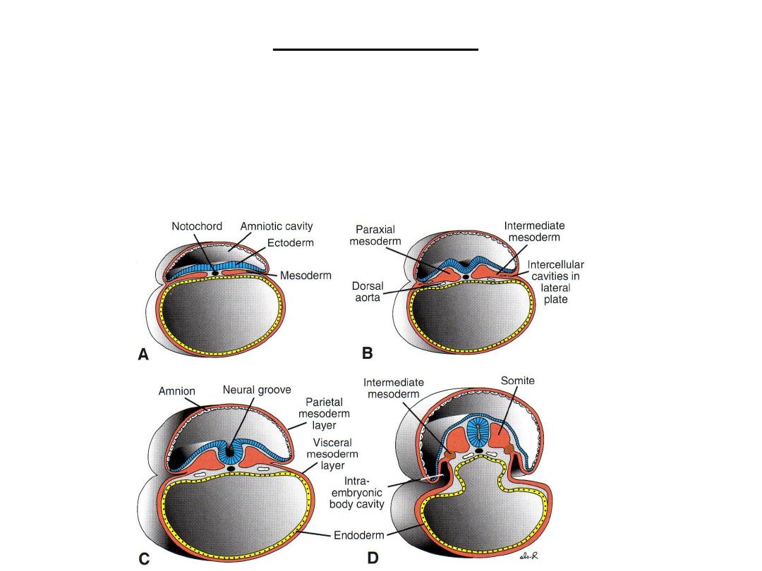
LATERAL PLATE MESODERM
Lateral plate mesoderm splits into parietal
(somatic) and visceral (splanchnic) layers,
Line the intraembryonic cavity and surround the
organs
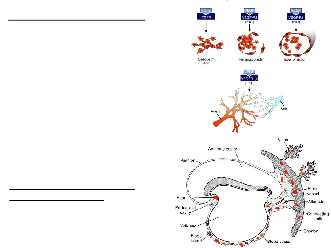
VASCULOGENESIS
ANGIOGENESIS
BLOOD & BLOOD VESSELS
Blood cells and blood vessels also arise
from mesoderm
EXTRAEMBRYONIC BLOOD
VESSEL FORMATION
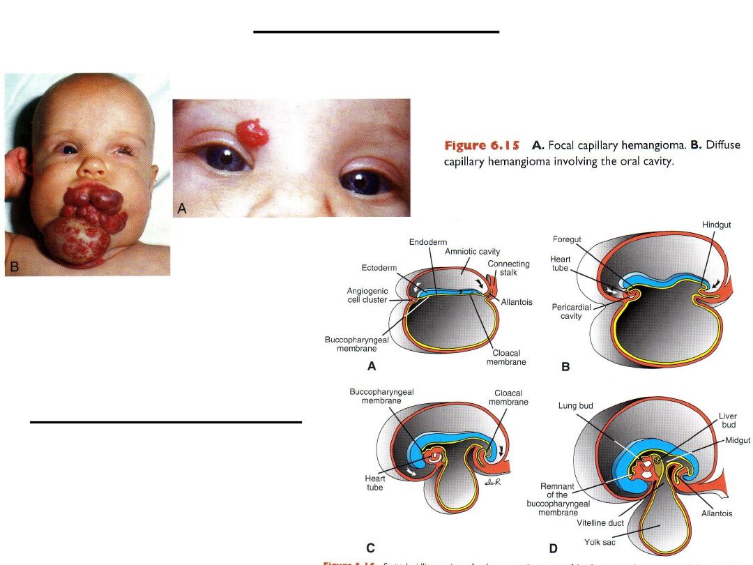
Capillary hemangioma
A .Focal capillary hemangioma .B .Diffuse capillary hemangioma involving the oral cavity.
Endoderm derivatives git
Cephalocaudal folding
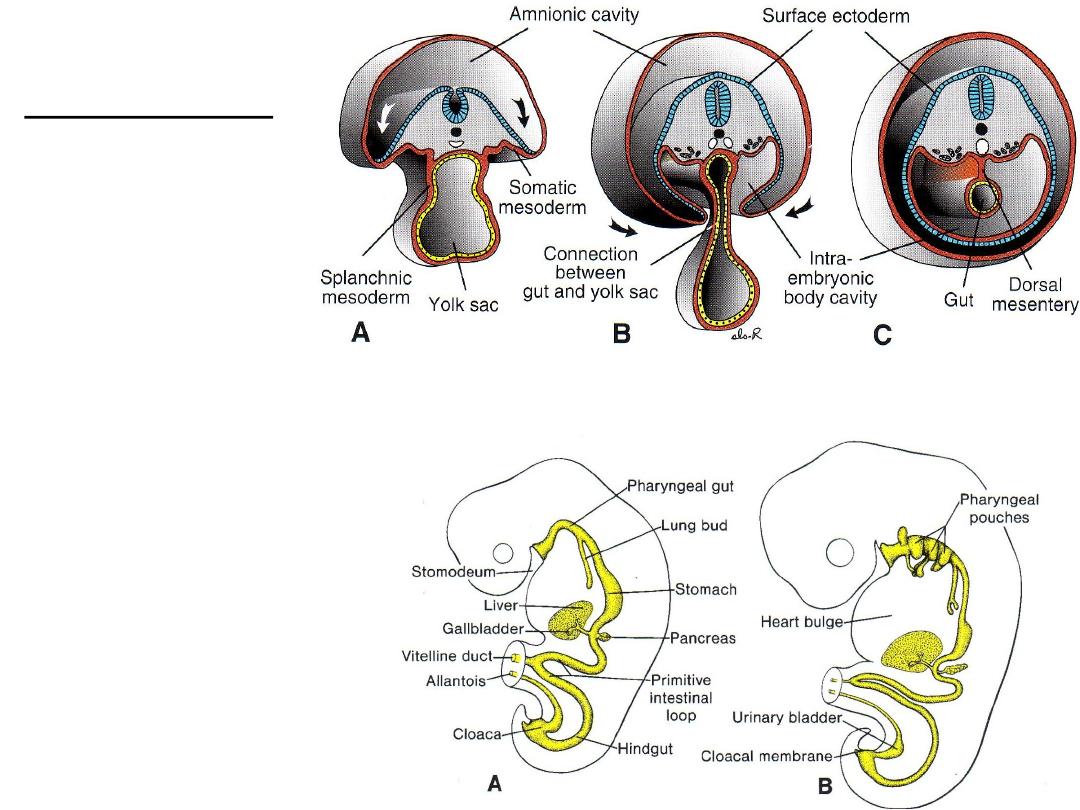
Lateral folding
Derivatives of
endodermal germ
layers

Endodermal derivatives
• Epith lining of GIT
• Epith of respiratory tract
• Parenchyma of: thyroid, parathyroids, liver, pancreas
• Reticular stroma of thymus & tonsils
• Epith lining of urinary bladder & urethra
• Epith lining of tympanic cavity & auditory tube
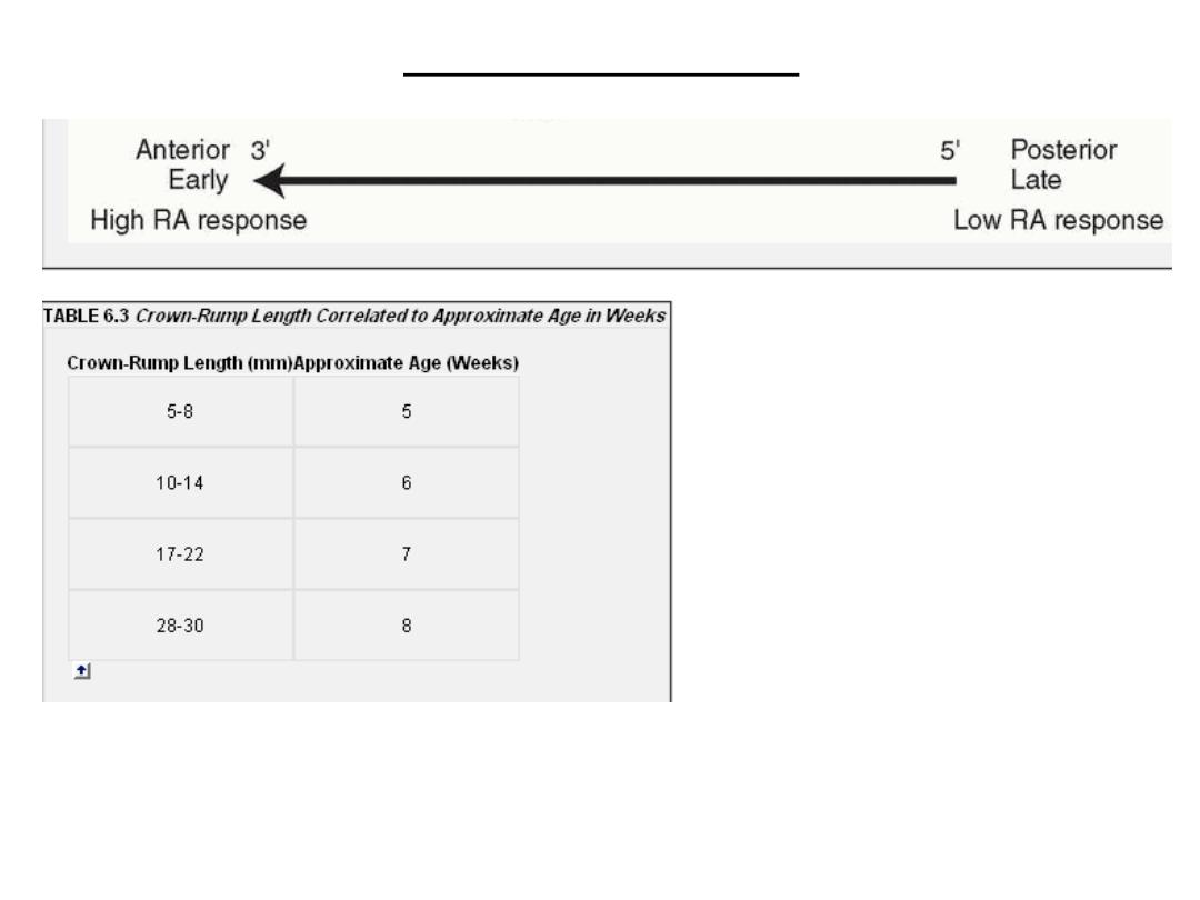
Patterning of the antero-posterior axis: regulations of
homeobox genes
• Craniocaudal patterning of the embryonic axis is controlled by
homeobox
genes.
• These genes are conserved from Drosophila are arranged in
four clusters
,
HOXA
,
HOXB
,
HOXC
, and
HOXD
, on four different chromosomes.
• Genes toward the
3’ end
of the chromosome control development of more
cranial
structures
.
• Those toward the
5’ end
regulate differentiation of more
posterior structures
.
• Together they regulate patterning of the hindbrain and the axis of the embryo.
External appearance during
the second month
• End of 4
th
weeks: 28 somites-external
features: somites+pharyngeal arches
Second month
Age: crown-rump length (CRL) in mm
• Increase in head size
• Formation of: limbs, face, ears, nose, eyes
Crown-Rump Length
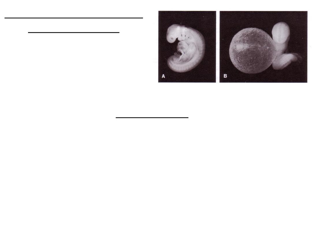
Limbs formation
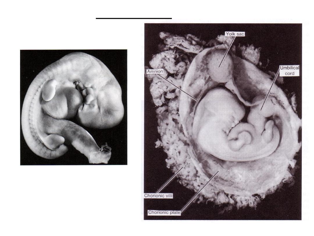
Human embryo (CRL 21 mm, seventh
week) (X4). The chorionic sac is open to
show the embryo in its amniotic sac.
The yolk sac, umbilical cord, and vessels
in the chorionic plate of the placenta are
clearly visible. Note the size of the head
in comparison with the rest of the body .
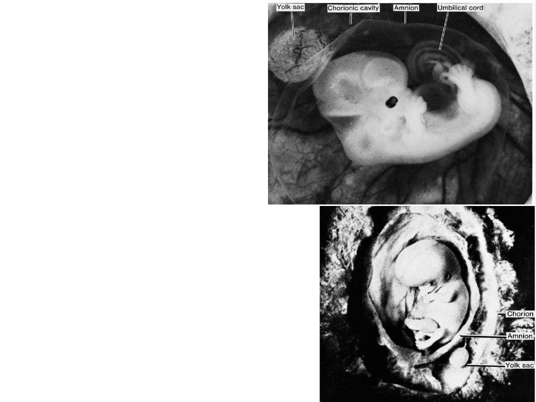
Human embryo (CRL 25 mm, seventh to eighth
weeks). The chorion and the amnion have been
opened. Note the size of the head, the eye, the
auricle of the ear, the well-formed toes, the
swelling in the umbilical cord caused by intestinal
loops, and the yolk sac in the chorionic cavity .