
Usually, there is one cell type for each major hormone formed in the
Anterior Pituitary Gland Contains Several Different Cell Types That Synthesize and Secrete
in the body fluids.
excretion into the urine, thus helping to control the concentration of water
The two hormones secreted by the posterior pituitary play other roles.
their hormonal and reproductive activities.
control growth of the ovaries and testes, as well as
luteinizing hormone,
• Two separate gonadotropic hormones,
control the rates of most intracellular chemical reactions in the body.
thyroxine and triiodothyronine by the thyroid gland, and these hormones
Thyroid-stimulating hormone (thyrotropin)
and fats.
adrenocortical hormones, which affect the metabolism of glucose, proteins,
Adrenocorticotropin (corticotropin)
formation, cell multiplication, and cell differentiation.
Growth hormone
Figure 75–2.
roles in the control of metabolic functions throughout the body, as shown in
pituitary. The hormones of the anterior pituitary play major
pituitary, and two important peptide hormones are secreted
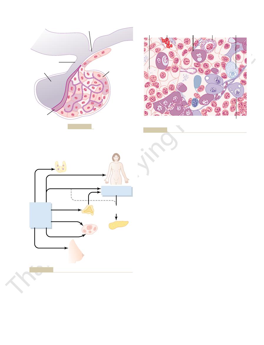
itary from the pharyngeal epithelium explains the epithelioid nature of its cells,
neural tissue outgrowth from the hypothalamus. The origin of the anterior pitu-
invagination of the pharyngeal epithelium, and the posterior pituitary from a
Rathke’s pouch,
Embryologically, the two portions of the pituitary originate from different
is much larger and much more functional in some lower animals.
Between these is a small, relatively avascular
neurohypophysis.
posterior pituitary,
adenohypophysis,
pituitary,
logically, the pituitary gland is divisible into two distinct portions: the
) stalk. Physio-
a bony cavity at the base of the brain, and is
hypophysis,
(Figure 75–1), also
The
Pituitary Gland: Two Distinct Parts–The Anterior and Poste-
Pituitary Gland and
Pituitary Hormones and Their
C
H
A
P
T
E
R
7
5
918
Control by the Hypothalamus
Its Relation to the
Hypothalamus
rior Lobes.
pituitary gland
called the
is a small gland—about 1 cen-
timeter in diameter and 0.5 to 1 gram in weight—
that lies in the sella turcica,
connected to the hypothalamus by the pituitary (or hypophysial
anterior
also known as the
and the
also
known as the
zone called the pars intermedia, which is almost absent in the human being but
sources—the anterior pituitary from
which is an embryonic
and the origin of the posterior pituitary from neural tissue explains the pres-
ence of large numbers of glial-type cells in this gland.
Six important peptide hormones plus several less important ones are secreted
by the anterior
by the posterior
•
promotes growth of the entire body by affecting protein
•
controls the secretion of some of the
•
controls the rate of secretion of
• Prolactin promotes mammary gland development and milk production.
follicle-stimulating hormone and
• Antidiuretic hormone (also called vasopressin) controls the rate of water
• Oxytocin helps express milk from the glands of the breast to the nipples
during suckling and possibly helps in the delivery of the baby at the end of
gestation.
Hormones.
and transplanted to some other part of the body, its
thalamus. Indeed, when the pituitary gland is removed
Pituitary Secretion
Hypothalamus Controls
in the chapter.
terior pituitary gland. This is discussed more fully later
of the hypothalamus. The hormones
nocellular neurons,
pituitary gland itself but are large neurons, called
The bodies of the cells that secrete
Posterior Pituitary Hormones Are Synthesized by Cell Bodies in
acidophilic tumors.
Thus, pituitary tumors that
acidophils.
tions, and milk secretion by the breasts.
hormones for controlling thyroid function, sexual func-
cent of the total; nevertheless, they secrete powerful
about 20 per cent are corticotropes that secrete ACTH.
are somatotropes that secrete growth hormone, and
—gonadotropic hormones, which
—adrenocorticotropin (ACTH)
logical actions. These five cell types are:
cell types, the hormones they produce, and their physio-
(Figure 75–3). Table 75–1 provides a summary of these
hormones, at least five cell types can be differentiated
anterior pituitary gland. With special stains attached to
Pituitary Hormones and Their Control by the Hypothalamus
Chapter 75
919
high-affinity antibodies that bind with the distinctive
1. Somatotropes—human growth hormone (hGH)
2. Corticotropes
3. Thyrotropes—thyroid-stimulating hormone (TSH)
4. Gonadotropes
include both luteinizing hormone (LH) and follicle-
stimulating hormone (FSH)
5. Lactotropes—prolactin (PRL)
About 30 to 40 per cent of the anterior pituitary cells
Each of the other cell types accounts for only 3 to 5 per
Somatotropes stain strongly with acid dyes and are
therefore called
secrete large quantities of human growth hormone are
called
the Hypothalamus.
the posterior pituitary hormones are not located in the
mag-
located in the supraoptic and par-
aventricular nuclei
are then transported in the axoplasm of the neurons’
nerve fibers passing from the hypothalamus to the pos-
Almost all secretion by the pituitary is controlled by
either hormonal or nervous signals from the hypo-
from its normal position beneath the hypothalamus
Hypothalamus
Anterior pituitary
Pars intermedia
Posterior pituitary
Hypophysial stalk
Pituitary gland.
Figure 75–1
Thyroid
gland
Mammary
gland
Increases blood
glucose level
Pancreas
Adrenal cortex
Ovary
ACH
Promotes secretion
of insulin
Anterior
pituitary
gland
Thyrotropin
Growth
Corticotropin
Follicle
stimulating
Luteinizing
Prolactin
adrenal corticosteroid hormones.
Metabolic functions of the anterior pituitary hormones. ACH,
Figure 75–2
Sinusoid
Gamma
(
g
) cell
Alpha
(
a
) cell
Epsilon (
e
)
acidophil cell
Delta (
d
)
basophil cell
Beta (
b
) cell
Guyton AC: Physiology of the Human Body, 6th ed. Philadelphia:
Cellular structure of the anterior pituitary gland. (Redrawn from
Figure 75–3
Saunders College Publishing, 1984.)
hypothalamus. The blood then flows through small
extensive capillary sinuses among the glandular cells.
The anterior pituitary is a highly vascular gland with
Pituitary Gland
Blood Vessels of the Anterior
pituitary hormones.
of the body, and much of this information is used
thalamus. Thus, the hypothalamus is a collecting center
ents, electrolytes, water, and various hormones in the
the hypothalamus. Even the concentrations of nutri-
the hypothalamus. Olfactory stimuli denoting pleasant
ing thought, a portion of the signal is transmitted into
mitted into the hypothalamus. Likewise, when a
exposed to pain, a portion of the pain signal is trans-
sources in the nervous system. Thus, when a person is
The hypothalamus receives signals from many
cussed in the next section of this chapter.
control their secretion. This system of control is dis-
In the anterior pituitary, these releasing
portal vessels.
in Figure 75–4, to the anterior pituitary through
the hypothalamus itself and then conducted, as shown
and terminate in the posterior pituitary. In contrast,
for prolactin) fall to very low levels.
920
Unit XIV
Endocrinology and Reproduction
rates of secretion of the different hormones (except
Secretion from the posterior pituitary is controlled
by nerve signals that originate in the hypothalamus
secretion by the anterior pituitary is controlled by hor-
mones called hypothalamic releasing and hypothala-
mic inhibitory hormones (or factors) secreted within
minute blood vessels called hypothalamic-hypophysial
and inhibitory hormones act on the glandular cells to
person experiences some powerful depressing or excit-
or unpleasant smells transmit strong signal compo-
nents directly and through the amygdaloid nuclei into
blood excite or inhibit various portions of the hypo-
for information concerning the internal well-being
to control secretions of the many globally important
Hypothalamic-Hypophysial Portal
Almost all the blood that enters these sinuses passes
first through another capillary bed in the lower
Table 75–1
IGF, insulin-like
Mammotropes
acids
Lactotropes,
Prolactin (PRL)
Single chain of 198 amino
Stimulates milk secretion and production
by the ovary; stimulates testosterone
(115 amino acids)
production of estrogen and progesterone
(89 amino acids) and
corpus luteum in the ovary; stimulates
Luteinizing hormone (LH)
Glycoprotein of two subunits,
Causes ovulation and formation of the
(112 amino acids)
the testis
(89 amino acids) and
follicles; regulates spermatogenesis in
Gonadotropes
Follicle-stimulating hormone
Glycoprotein of two subunits,
Stimulates development of ovarian
(112 amino acids)
maintains size of follicular cells
(89 amino acids) and
hormones by thyroid follicular cells;
(TSH; thyrotropin)
Thyrotropes
Thyroid-stimulating hormone
Glycoprotein of two subunits,
Stimulates production of thyroid
(ACTH; corticotropin)
and androgens by the adrenal cortex;
Corticotropes
Adrenocorticotropic hormone
Single chain of 39 amino acids
Stimulates production of glucocorticoids
somatotropin)
secretion of IGF-1; stimulates lipolysis;
Somatotropes
Growth hormone (GH;
Single chain of 191 amino acids
Stimulates body growth; stimulates
Cell
Hormone
Chemistry
Physiological Actions
Cells and Hormones of the Anterior Pituitary Gland and Their Physiological Functions
inhibits actions of insulin on
carbohydrate and lipid metabolism
maintains size of zona fasciculata and
zona reticularis of cortex
a
b
(FSH)
a
b
a
b
production by the testis
growth factor
Median eminence
Optic chiasm
Artery
Primary capillary
plexus
Hypothalamic-
hypophysial
portal vessels
Sinuses
Anterior pituitary
gland
Vein
Posterior pituitary
gland
Mamillary body
Hypothalamus
Hypothalamic-hypophysial portal system.
Figure 75–4

along with the target glands. Growth hormone, in
tive target glands that, except for growth hormone,
cortex, ovaries, testicles, and mammary glands. The
ulating target glands, including thyroid gland, adrenal
growth hormone, exert their principal effects by stim-
All the major anterior pituitary hormones, except for
of Growth Hormone
misleading to attempt delineation here.
hormones are still poorly known, so that it would be
The specific loci of the neuronal cell bodies that form
hypothalamic hormones. However, the neuronal cell
and, therefore, causes release of essentially all the
transported to the anterior pituitary gland. Electrical
cific Hypothalamic Releasing and Inhibitory Hormones.
Specific Areas in the Hypothalamus Control Secretion of Spe-
this and subsequent chapters.
itary hormones. Each of the more important hypo-
There are some additional hypothalamic hormones,
(PIH), which causes
causes release of the two gonadotropic hormones,
(GnRH), which
(GHIH), also
growth hormone inhibitory hormone
which causes release of growth hormone, and
Growth hormone–releasing hormone
(CRH), which
(TRH), which
summarized in Table 75–2 and are the following:
hormone probably exerts more control. The major
important, but for prolactin, a hypothalamic inhibitory
itary hormones, it is the releasing hormones that are
rior pituitary hormones. For most of the anterior pitu-
The function of the releasing and
rior Pituitary Secretion.
Hypothalamic Releasing and Inhibitory Hormones Control Ante-
These hormones are immediately absorbed into the
ing and inhibitory hormones into the tissue fluids.
endings in the central nervous system, in that their
The endings of these fibers are different from most
tion of the anterior pituitary hormones. These neurons
Hypothalamic Releasing and Inhibitory Hormones Are Secreted
blood to the anterior pituitary sinuses.
hypothalamic-hypophysial portal blood vessels. These
vessels return to its surface, coalescing to form the
stalk. Small arteries penetrate into the substance
eminence,
ermost portion of the hypothalamus, called the
anterior pituitary sinuses. Figure 75–4 shows the low-
Pituitary Hormones and Their Control by the Hypothalamus
Chapter 75
921
hypothalamic-hypophysial portal blood vessels into the
median
which connects inferiorly with the pituitary
of the median eminence and then additional small
pass downward along the pituitary stalk to supply
into the Median Eminence.
Special neurons in the hypo-
thalamus synthesize and secrete the hypothalamic
releasing and inhibitory hormones that control secre-
originate in various parts of the hypothalamus and
send their nerve fibers to the median eminence and
tuber cinereum, an extension of hypothalamic tissue
into the pituitary stalk.
function is not to transmit signals from one neuron to
another but rather to secrete the hypothalamic releas-
hypothalamic-hypophysial portal system and carried
directly to the sinuses of the anterior pituitary gland.
inhibitory hormones is to control secretion of the ante-
hypothalamic releasing and inhibitory hormones are
1. Thyrotropin-releasing hormone
causes release of thyroid-stimulating hormone
2. Corticotropin-releasing hormone
causes release of adrenocorticotropin
3.
(GHRH),
called somatostatin, which inhibits release of
growth hormone
4. Gonadotropin-releasing hormone
luteinizing hormone and follicle-stimulating
hormone
5. Prolactin inhibitory hormone
inhibition of prolactin secretion
including one that stimulates prolactin secretion and
perhaps others that inhibit release of the anterior pitu-
thalamic hormones is discussed in detail as the specific
hormonal systems controlled by them are presented in
All or
most of the hypothalamic hormones are secreted at
nerve endings in the median eminence before being
stimulation of this region excites these nerve endings
bodies that give rise to these median eminence nerve
endings are located in other discrete areas of the hypo-
thalamus or in closely related areas of the basal brain.
the different hypothalamic releasing or inhibitory
Physiological Functions
functions of each of these pituitary hormones are so
intimately concerned with the functions of the respec-
their functions are discussed in subsequent chapters
Table 75–2
Prolactin-inhibiting hormone (PIH)
Dopamine (a catecholamine)
Inhibits secretion of prolactin by lactotropes
Growth hormone inhibitory hormone
Single chain of 14 amino acids
Inhibits secretion of growth hormone by somatotropes
Growth hormone–releasing hormone
Single chain of 44 amino acids
Stimulates secretion of growth hormone by somatotropes
Corticotropin-releasing hormone (CRH)
Single chain of 41 amino acids
Stimulates secretion of ACTH by corticotropes
Gonadotropin-releasing hormone (GnRH)
Single chain of 10 amino acids
Stimulates secretion of FSH and LH by gonadotropes
Thyrotropin-releasing hormone (TRH)
Peptide of 3 amino acids
Stimulates secretion of TSH by thyrotropes
Hormone
Structure
Primary Action on Anterior Pituitary
Hypothalamic Releasing and Inhibitory Hormones That Control Secretion of the Anterior Pituitary Gland
(GHRH)
(somatostatin)
ACTH, adrenocorticotropic hormone; FSH, follicle-stimulating hormone; LH, luteinizing hormone; TSH, thyroid-stimulating hormone.
for Energy
Growth Hormone Enhances Fat Utilization
proteins.
of amino acid uptake and protein synthesis by cells,
Summary.
sparer.”
for the body’s cells, thus acting as a potent “protein
tissue, and these are used to supply most of the energy
decrease in the breakdown of cell protein. A probable
tion to the increase in protein synthesis, there is a
tion of growth hormone.
In the long run, this may be the most important func-
vitamins, and other requisites for growth are available.
and promotes growth if sufficient energy, amino acids,
tities of RNA. This promotes more protein synthesis
the nucleus, causing the formation of increased quan-
hormone also stimulates the transcription of DNA in
more prolonged periods (24 to 48 hours), growth
Increased Nuclear Transcription of DNA to Form RNA.
cytoplasm.
still increases RNA translation, causing protein to be
tions are not increased in the cells, growth hormone
Enhancement of RNA Translation to Cause Protein Synthesis by
trolling glucose transport through the membrane, as
the increased protein synthesis. This control of amino
the cell membranes to the interior of the cells. This
Enhancement of Amino Acid Transport Through the Cell Mem-
a series of different effects are known, all of which
Growth Hormone Promotes Protein Deposition
serves carbohydrates.
enhances body protein, uses up fat stores, and con-
throughout the body. Thus, in effect, growth hormone
energy; and (3) decreased rate of glucose utilization
acids in the blood, and increased use of fatty acids for
fatty acids from adipose tissue, increased free fatty
most cells of the body; (2) increased mobilization of
hormone has multiple specific metabolic effects,
Aside from its general effect in causing growth, growth
Metabolic Effects
the body can continue to grow throughout life.
bone cannot occur, even though most other tissues of
have united with the shafts, further lengthening of
many of the soft tissues continued to grow. This results
reached, most of the bones stopped lengthening, but
increased proportionately in size; after adulthood was
stages of development, all organs of the treated rat
after the two rats reached adulthood. In the early
did not receive growth hormone. This figure shows
growing littermate rats, one of which received daily
Figure 75–5 shows typical weight charts of two
such as bone growth cells and early muscle cells.
mitosis, with development of greater numbers of cells
all tissues of the body that are capable of growing. It
molecular weight of 22,005. It causes growth of almost
Growth hormone, also called
of Many Body Tissues
almost all tissues of the body.
contrast to other hormones, does not function through
922
Unit XIV
Endocrinology and Reproduction
a target gland but exerts its effects directly on all or
Growth Hormone Promotes Growth
somatotropic hormone
or somatotropin, is a small protein molecule that con-
tains 191 amino acids in a single chain and has a
promotes increased sizes of the cells and increased
and specific differentiation of certain types of cells
injections of growth hormone and the other of which
marked enhancement of growth in the rat given
growth hormone—in the early days of life and even
from the fact that once the epiphyses of the long bones
Growth Hormone Has Several
including (1) increased rate of protein synthesis in
in Tissues
Although the precise mechanisms by which growth
hormone increases protein deposition are not known,
could lead to enhanced protein deposition.
branes.
Growth hormone directly enhances transport
of at least some and perhaps most amino acids through
increases the amino acid concentrations in the cells
and is presumed to be at least partly responsible for
acid transport is similar to the effect of insulin in con-
discussed in Chapters 67 and 78.
the Ribosomes.
Even when the amino acid concentra-
synthesized in greater amounts by the ribosomes in the
Over
Decreased Catabolism of Protein and Amino Acids.
In addi-
reason for this is that growth hormone also mobilizes
large quantities of free fatty acids from the adipose
Growth hormone enhances almost all facets
while at the same time reducing the breakdown of
Growth hormone has a specific effect in causing
the release of fatty acids from adipose tissue and,
0
600
Control
Injected daily with
growth hormone
500
400
300
200
200
0
500
400
300
200
100
Body weight (grams)
Days
hormone with that of a normal littermate.
Comparison of weight gain of a rat injected daily with growth
Figure 75–5

chondrocytes cultured outside the body, proliferation
When growth hormone is supplied directly to cartilage
of Its Effect Through Intermediate
eyes.
lower teeth. Likewise, the bones of the skull can grow
lescence, causing forward protrusion of the chin and
cially true for the membranous bones. For instance, the
under the influence of growth hormone; this is espe-
Therefore, the
hormone strongly stimulates osteoblasts.
Growth
resorption, the thickness of the bone increases.
When the rate of deposition is greater than that of
(discussed in detail in Chapter 79) remove old bone.
of older bone. Simultaneously,
further lengthening of the long bone can occur.
the shaft and the epiphysis at each end, so that no
bone growth. At this time, bony fusion occurs between
used up, so that by late adolescence, no additional epi-
time, the epiphyseal cartilage itself is progressively
the epiphyses farther and farther apart. At the same
into new bone, thus elongating the shaft and pushing
deposition of new cartilage, followed by its conversion
are separated from the shaft. This growth first causes
cartilages, where the epiphyses at the ends of the bone
tion, the long bones grow in length at the epiphyseal
growth. First, in response to growth hormone stimula-
There are two principal mechanisms of bone
bone.
into osteogenic cells, thus causing deposition of new
and (3) a specific effect of converting chondrocytes
(2) increased rate of reproduction of these cells,
cytic and osteogenic cells that cause bone growth,
multiple effects of growth hormone on bone, including
increase growth of the skeletal frame. This results from
all tissues of the body, its most obvious effect is to
enhance the transport of some amino acids into cells,
well. Especially important is insulin’s ability to
olism of growth, but there seem to be other effects as
tive. Part of this requirement for carbohydrates and
excluded from the diet. This shows that adequate
creas; it also fails to cause growth if carbohydrates are
and skeletal muscle to insulin’s effects on carbohy-
tissue glucose utilization. Experimental studies indi-
trations of fatty acids may impair insulin’s actions on
decreased glucose utilization by the cells. However,
We do not know the precise mechanism by which
dependent) diabetes, who are also very resistant to the
diabetogenic,
For these reasons, growth hormone’s effects are called
liver; this leads to increased blood glucose concentra-
ates insulin’s actions to stimulate the uptake and uti-
hormone–induced “insulin resistance,” which attenu-
the liver, and (3) increased insulin secretion.
muscle and fat, (2) increased glucose production by
influence carbohydrate metabolism, including (1)
Growth Hormone Decreases
liver.
This excessive mobilization of fat
ketosis.
formed by the liver and released into the body fluids,
ence of excessive amounts of growth hormone, fat
in minutes under the influence of growth hormone.
fat by growth hormone requires several hours to occur,
increase in lean body mass. However, mobilization of
together with its protein anabolic effect, causes an
Growth hormone’s ability to promote fat utilization,
proteins.
the influence of growth hormone, fat is used for
subsequent utilization for energy. Therefore, under
fatty acids to acetyl coenzyme A (acetyl-CoA) and its
body, growth hormone enhances the conversion of
the body fluids. In addition, in tissues throughout the
therefore, increasing the concentration of fatty acids in
Pituitary Hormones and Their Control by the Hypothalamus
Chapter 75
923
energy in preference to the use of carbohydrates and
whereas enhancement of protein synthesis can begin
“Ketogenic” Effect of Growth Hormone.
Under the influ-
mobilization from adipose tissue sometimes becomes
so great that large quantities of acetoacetic acid are
thus causing
from the adipose tissue also frequently causes a fatty
Carbohydrate Utilization
Growth hormone causes multiple effects that
decreased glucose uptake in tissues such as skeletal
Each of these changes results from growth
lization of glucose in skeletal muscle and fat and to
inhibit gluconeogenesis (glucose production) by the
tion and a compensatory increase in insulin secretion.
and excess secretion of growth hormone
can produce metabolic disturbances very similar to
those found in patients with type II (non-insulin-
metabolic effects of insulin.
growth hormone causes insulin resistance and
growth hormone–induced increases in blood concen-
cate that raising blood levels of fatty acids above
normal rapidly decreases the sensitivity of the liver
drate metabolism.
Necessity of Insulin and Carbohydrate for the Growth-
Promoting Action of Growth Hormone.
Growth hormone
fails to cause growth in an animal that lacks a pan-
insulin activity and adequate availability of carbohy-
drates are necessary for growth hormone to be effec-
insulin is to provide the energy needed for the metab-
in the same way that it stimulates glucose transport.
Growth Hormone Stimulates Cartilage
and Bone Growth
Although growth hormone stimulates increased de-
position of protein and increased growth in almost
(1) increased deposition of protein by the chondro-
physeal cartilage remains to provide for further long
Second, osteoblasts in the bone periosteum and in
some bone cavities deposit new bone on the surfaces
osteoclasts in the bone
bones can continue to become thicker throughout life
jaw bones can be stimulated to grow even after ado-
in thickness and give rise to bony protrusions over the
Growth Hormone Exerts Much
Substances Called “Somatomedins”
(Also Called “Insulin-Like
Growth Factors”)

For instance, the extremely high levels of growth
depletion than with the degree of glucose insufficiency.
chronic conditions, growth hormone secretion seems
an acute decrease in protein intake. Conversely, in
Under acute conditions, hypoglycemia is a far more
child or adolescent, it is about 6 ng/ml. These values
plasma of an adult is between 1.6 and 3 ng/ml; in a
The normal concentration of growth hormone in the
shown in Figure 75–6. Table 75–3 summarizes some of
deep sleep,
exercise;
low concentration of
secretion: (1)
understood, but several factors related to a person’s
increasing and decreasing. The precise mechanisms
per cent of the adolescent level in very old age.
decreases slowly with aging, finally falling to about 25
has proved to be untrue. After adolescence, secretion
then disappeared from the blood at adolescence. This
For many years it was believed that growth hormone
Figure 75–6.
This greatly prolongs the growth-promoting effects of
blood to the tissues, with a half-time of about 20 hours.
result, somatomedin C is released only slowly from the
is produced in response to growth hormone. As a
carrier protein in the blood that, like somatomedin C,
By contrast, somatomedin C attaches strongly to a
having a half-time in the blood of less than 20 minutes.
it is released from the blood into the tissues rapidly,
weakly to the plasma proteins in the blood. Therefore,
one.
local tissue to cause local growth. It is also possible
of the somatomedin hypothesis are still questionable.
hormone required for this is minute. Some aspects
of these cartilage areas, and the amount of growth
bones and other peripheral tissues. Even so, experi-
somatomedin C and other somatomedins, rather
It has been postulated that most, if not all, of the
people. Some other dwarfs (e.g., the Lévi-Lorain
growth hormone is either normal or high, they have
Therefore, even though their plasma concentration of
synthesize significant amounts of somatomedin C.
The pygmies of Africa have a congenital inability to
somatomedin C is about 7500, and its concentration in
(also called IGF-I). The molecular weight of
effects of insulin on growth. Therefore, the somato-
increasing all aspects of bone growth. Many of the
causes the liver (and, to a much less extent,
In brief, it has been found that growth hormone
same cells.
occur. Yet growth hormone injected into the intact
or enlargement of the chondrocytes usually fails to
924
Unit XIV
Endocrinology and Reproduction
animal does cause proliferation and growth of the
other tissues) to form several small proteins
called somatomedins that have the potent effect of
somatomedin effects on growth are similar to the
medins are also called insulin-like growth factors
(IGFs).
At least four somatomedins have been isolated,
but by far the most important of these is somatomedin
C
the plasma closely follows the rate of growth hormone
secretion.
diminished amounts of somatomedin C in the plasma;
this apparently accounts for the small stature of these
dwarf) also have this problem.
growth effects of growth hormone result from
than from direct effects of growth hormone on the
ments have demonstrated that injection of growth
hormone directly into the epiphyseal cartilages of
bones of living animals causes the specific growth
One possibility is that growth hormone can cause
the formation of enough somatomedin C in the
that growth hormone itself is directly responsible
for increased growth in some tissues and that the
somatomedin mechanism is an alternative means of
increasing growth but not always a necessary
Short Duration of Action of Growth Hormone but Prolonged
Action of Somatomedin C.
Growth hormone attaches only
the bursts of growth hormone secretion shown in
Regulation of Growth
Hormone Secretion
was secreted primarily during the period of growth but
Growth hormone is secreted in a pulsatile pattern,
that control secretion of growth hormone are not fully
state of nutrition or stress are known to stimulate
starvation, especially with severe protein
deficiency; (2) hypoglycemia or
fatty acids in the blood; (3)
(4) excitement; and
(5) trauma. Growth hormone also characteristically
increases during the first 2 hours of
as
the factors that are known to influence growth
hormone secretion.
often increase to as high as 50 ng/ml after depletion of
the body stores of proteins or carbohydrates during
prolonged starvation.
potent stimulator of growth hormone secretion than is
to correlate more with the degree of cellular protein
hormone that occur during starvation are closely
related to the amount of protein depletion.
8 am
4 am 8 pm
4 am
30
20
10
0
12
Noon
12
Midnight
8 am
Growth hormone
(ng/ml plasma)
Strenuous
exercise
Sleep
and also the high rate of growth hormone secretion that occurs
demonstrating the especially powerful effect of strenuous exercise
Typical variations in growth hormone secretion throughout the day,
Figure 75–6
during the first few hours of deep sleep.
and release of the hormone into the blood. The long-
cell; within minutes, this causes fusion of the growth
both a short-term and a long-term effect. The short-
cyclic adenosine monophosphate (cAMP). This has
the cell membrane, increasing the intracellular level of
receptors activate the adenylyl cyclase system inside
of the growth hormone cells in the pituitary gland. The
through the inhibitory hormone somatostatin. GHRH
a different neuronal system in the hypothalamus, all
dopamine, and serotonin, each of which is released by
experiments have shown that catecholamines,
mic control of growth hormone secretion. In fact,
emotions, stress, and trauma can all affect hypothala-
In a similar manner, hypothalamic signals depicting
person’s behavioral feeding instincts also alter the rate
areas of the hypothalamus. Therefore, it is reasonable
glycemic states and hunger in hypoglycemic states. The
blood glucose concentration, causing satiety in hyper-
of GHRH is the ventromedial nucleus; this is the
The part of the hypothalamus that causes secretion
amino acids.
amino acids, and somatostatin is composed of 14
these are polypeptides; GHRH is composed of 44
). Both of
growth hormone
growth
hypophysial portal vessels.
They are
growth hormone secretion. It is known that growth
that can affect growth hormone secretion, one can
From the preceding description of the many factors
Hormone Secretion
Somatostatin in the Control of Growth
Hormone–Releasing Hormone, and
Role of the Hypothalamus, Growth
growth hormone. The protein deficiency must also be
tions of protein malnutrition, adequate calories alone
These results demonstrate that under severe condi-
tant decrease in the hormone.
ments for 3 and 25 days, respectively, with a concomi-
hormone concentration. The third and fourth columns
of carbohydrates in their diets, demonstrating that
extreme protein deficiency during the protein malnu-
of adding protein to the diet. The first column shows
ciency on plasma growth hormone and then the effect
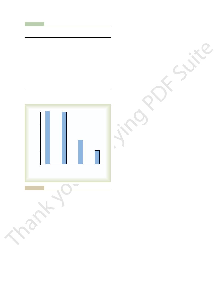
Figure 75–7 demonstrates the effect of protein defi-
Pituitary Hormones and Their Control by the Hypothalamus
Chapter 75
925
very high levels of growth hormone in children with
trition condition called kwashiorkor; the second
column shows the levels in the same children after 3
days of treatment with more than adequate quantities
the carbohydrates did not lower the plasma growth
show the levels after treatment with protein supple-
are not sufficient to correct the excess production of
corrected before the growth hormone concentration
will return to normal.
readily understand the perplexity of physiologists as
they attempt to unravel the mysteries of regulation of
hormone secretion is controlled by two factors
secreted in the hypothalamus and then transported to
the anterior pituitary gland through the hypothalamic-
hormone–releasing hormone and
inhibitory hormone (also called somatostatin
same area of the hypothalamus that is sensitive to
secretion of somatostatin is controlled by other nearby
to believe that some of the same signals that modify a
of growth hormone secretion.
increase the rate of growth hormone secretion.
Most of the control of growth hormone secretion
is probably mediated through GHRH rather than
stimulates growth hormone secretion by attaching to
specific cell membrane receptors on the outer surfaces
term effect is to increase calcium ion transport into the
hormone secretory vesicles with the cell membrane
term effect is to increase transcription in the nucleus
Table 75–3
Factors That Stimulate or Inhibit Secretion of
Growth hormone–releasing
growth factors)
Deep sleep ( stages II and IV)
Somatomedins (insulin-like
Testosterone, estrogen
Growth hormone (exogenous)
Exercise
hormone (somatostatin)
Trauma, stress, excitement
Growth hormone inhibitory
deficiency
Obesity
Starvation or fasting, protein
Aging
acids
acids
Decreased blood free fatty
Increased blood free fatty
Decreased blood glucose
Increased blood glucose
Secretion
Secretion
Stimulate Growth Hormone
Inhibit Growth Hormone
Growth Hormone
hormone
40
30
20
10
0
Protein
deficiency
(kwashiorkor)
Protein
treatment
(25 days)
Protein
treatment
(3 days)
Carbohydrate
treatment
(3 days)
Plasma growth hormone (ng/ml)
on growth hormone secretion in protein-calorie malnutrition. Am J
treatment in lowering growth hormone concentration. (Drawn from
failure of carbohydrate treatment but the effectiveness of protein
of growth hormone in the disease kwashiorkor. Also shown is the
Effect of extreme protein deficiency on the plasma concentration
Figure 75–7
data in Pimstone BL, Barbezat G, Hansen JD, Murray P: Studies
Clin Nutr 21:482, 1968.)

diagnosed, further effects can often be blocked by
death in early adulthood. However, once gigantism is
general deficiency of pituitary hormones usually causes
grows until the gland itself is destroyed. This eventual
ops if they remain untreated, because the gigantism is
In most giants, panhypopituitarism eventually devel-
ally develops.
per cent of giants, full-blown
owing to the hyperglycemia. Consequently, in about 10
The giant ordinarily has
the long bones have become fused with the shafts,
tion occurs before adolescence, before the epiphyses of
tissues grow rapidly, including the bones. If the condi-
quantities of growth hormone are produced. All body
dophilic tumors occur in the gland. As a result, large
become excessively active, and sometimes even aci-
Occasionally,
the acidophilic,
growth
and thyroid hormones.
abnormal sexual functions, the patient can usually be
mones) and has lost all sexual functions. Except for the
adrenocorticotropic, adrenocortical, and thyroid hor-
functions are lost. Thus, the picture is that of a lethargic
corticoids by the adrenal glands, and (3) suppressed
(1) hypothyroidism, (2) depressed production of gluco-
The general effects of adult panhypopituitarism are
birth of her baby.
blood vessels. This abnormality occasionally occurs
destroyed. The third cause is thrombosis of the pituitary
craniopharyngiomas or chromophobe tumors, may
three common abnormalities. Two tumorous conditions,
because of its widespread metabolic functions.
cured if treated early in life. Human growth hormone
pure growth hormone deficiency can be completely
quantities for treatment purposes. Dwarfs who have
Therefore, this hormone is now available in sufficient
ful application of recombinant DNA technology.
Escherichia coli
However, human growth hormone can now be synthe-
hormone deficiency, except on an experimental basis.
pared from human pituitary glands, it was difficult to
In the past, because growth hormone had to be pre-
to distinguish it from the others.
human growth
is not effective in human beings. Therefore, the growth
lower animals (except, to some extent, from primates)
species. For this reason, growth hormone prepared from
only in the one species or, at most, closely related
Treatment with Human Growth Hormone.
of growth by growth hormone.
somatomedin C, which is a key step for the promotion
or high, but there is a hereditary inability to form
dwarf), the rate of growth hormone secretion is normal
of dwarfism (the African pygmy and the Lévi-Lorain
mature sexually and occasionally reproduce. In one type
only growth hormone is deficient; these persons do
sexual functions. In one third of such dwarfs, however,
10 years.
aged 4 to 5 years, and the same person at age 20 years
greatly decreased. A child who has reached the age of
portion to one another, but the rate of development is
popituitarism) during childhood. In general, all the
alized deficiency of anterior pituitary secretion (panhy-
may occur suddenly or slowly at any time during life,
secretion may be congenital (present from birth), or it
of all the anterior pituitary hormones. The decrease in
This term means decreased secretion
Abnormalities of Growth
cells.
growth hormone secretion. Growth hormone, in turn,
need for cellular proteins—for instance, after a severe
nutrition. That is, nutritional deficiency or excess tissue
tissues themselves, especially their level of protein
ing: the major long-term controller of growth hormone
thesis and tissue growth, we can propose the follow-
a composite picture. Yet, because of the extreme secre-
In summary, our knowledge of the regulation of
tion, are uncertain.
somatostatin, which inhibits growth hormone secre-
for essentially all hormones. The nature of this feed-
subject to typical negative feedback control, as is true
This demonstrates that growth hormone secretion is
of endogenous growth hormone secretion decreases.
the blood of an animal over a period of hours, the rate
When growth hormone is administered directly into
hormone.
926
Unit XIV
Endocrinology and Reproduction
by the genes to stimulate the synthesis of new growth
back mechanism and whether it is mediated mainly
through inhibition of GHRH or enhancement of
growth hormone secretion is not sufficient to describe
tion of growth hormone during starvation and its
important long-term effect to promote protein syn-
secretion is the long-term state of nutrition of the
bout of exercise when the muscles’ nutritional status
has been taxed—in some way increases the rate of
promotes synthesis of new proteins while at the same
time conserving the proteins already present in the
Hormone Secretion
Panhypopituitarism.
most often resulting from a pituitary tumor that
destroys the pituitary gland.
Dwarfism.
Most instances of dwarfism result from gener-
physical parts of the body develop in appropriate pro-
10 years may have the bodily development of a child
may have the bodily development of a child aged 7 to
A person with panhypopituitary dwarfism does not
pass through puberty and never secretes sufficient
quantities of gonadotropic hormones to develop adult
Growth hor-
mones from different species of animals are sufficiently
different from one another that they will cause growth
hormone of the human being is called
hormone
obtain sufficient quantities to treat patients with growth
sized by
bacteria as a result of success-
may prove to be beneficial in other metabolic disorders
Panhypopituitarism in the Adult.
Panhypopituitarism first
occurring in adulthood frequently results from one of
compress the pituitary gland until the functioning
anterior pituitary cells are totally or almost totally
when a new mother develops circulatory shock after the
secretion of the gonadotropic hormones so that sexual
person (from lack of thyroid hormones) who is gaining
weight (because of lack of fat mobilization by growth,
treated satisfactorily by administering adrenocortical
Gigantism.
hormone–producing cells of the anterior pituitary gland
height increases so that the person becomes a giant—
up to 8 feet tall.
hyperglycemia, and the beta
cells of the islets of Langerhans in the pancreas are
prone to degenerate because they become overactive
diabetes mellitus eventu-
usually caused by a tumor of the pituitary gland that
microsurgical removal of the tumor or by irradiation of
the pituitary gland.

oxytocin.
(ADH), also
hormones: (1)
capillaries, where they secrete two posterior pituitary
secretory granules. These endings lie on the surfaces of
(hypophysial stalk). The
in Figure 75–9. These tracts pass to the neurohypoph-
of the hypothalamus, as shown
The pituicytes do not secrete hormones; they
pituicytes.
pophysis,
The
Posterior Pituitary Gland
of increased energy.
the muscles; (2) decreased fat deposits; and (3) a feeling
increased protein deposition in the body, especially in
important effects that suggest antiaging actions: (1)
secretion. In fact, multiple tests of growth hormone
Thus, it is highly possible that some of the normal
As one ages, the average plasma concentration of
function of some of the organs, and diminished muscle
increased wrinkling of the skin, diminished rates of
its place. The physical and physiological effects are
ance of a person aged 65. The aged appearance seems
For instance, a 50-year-old person who has been without
hormone, some features of the aging process accelerate.
kidneys, become greatly enlarged.
organs, such as the tongue, the liver, and especially the
Finally, many soft tissue
kyphosis.
vertebrae ordinarily cause a hunched back, which is
normal size. In addition to these effects, changes in the
require size 14 or larger shoes, and the fingers become
nose increases to as much as twice normal size, the feet
excess development of the supraorbital ridges, the
half an inch, the forehead slants forward because of
the lower jaw protrudes forward, sometimes as much as
growth does not cease at adolescence. Consequently,
jawbone, and portions of the vertebrae, because their
nose, bosses on the forehead, supraorbital ridges, lower
membranous bones,
acromegaly.
Figure 75–8, is known as
tissues can continue to grow. This condition, shown in
taller, but the bones can become thicker and the soft
cence—that is, after the epiphyses of the long bones
Acromegaly.
Pituitary Hormones and Their Control by the Hypothalamus
Chapter 75
927
If an acidophilic tumor occurs after adoles-
have fused with the shafts—the person cannot grow
Enlargement is
especially marked in the bones of the hands and feet
and in the
including the cranium,
extremely thickened so that the hands are almost twice
known clinically as
Possible Role of Decreased Growth Hormone Secretion
in Causing Changes Associated with Aging
In people who have lost the ability to secrete growth
growth hormone for many years may have the appear-
to result mainly from decreased protein deposition in
most tissues of the body and increased fat deposition in
mass and strength.
growth hormone in an otherwise normal person
changes approximately as follows:
aging effects result from diminished growth hormone
therapy in older people have demonstrated three
and Its Relation to the
Hypothalamus
posterior pituitary gland, also called the neurohy-
is composed mainly of glial-like cells called
act simply as a supporting structure for large numbers
of terminal nerve fibers and terminal nerve endings
from nerve tracts that originate in the supraoptic and
paraventricular nuclei
ysis through the pituitary stalk
nerve endings are bulbous knobs that contain many
antidiuretic hormone
called vasopressin, and (2)
Acromegalic patient.
Figure 75–8
20 to 40 years
3
5 to 20 years
6
ng/ml
40 to 70 years
1.6

a few minutes.
small amounts to large amounts, or vice versa, in only
centration of ADH in the body fluids can change from
almost total cessation of ADH secretion.Thus, the con-
Conversely, injection of a dilute solution into this
ing the ADH secretion to as high as 20 times normal.
of ADH into the circulating blood, sometimes increas-
hypothalamus, the ADH neurons in the supraoptic and
When a concentrated electrolyte
Regulation of ADH Production
mechanism of the kidneys.
lecting tubules and ducts by osmosis, as explained in
interstitial fluid. Water is then absorbed from the col-
process reverses in another 5 to 10 minutes. Thus, this
minutes. Then, in the absence of ADH, the entire
water permeability. All this occurs within 5 to 10
cell membranes, thus providing many areas of high
phosphorylation of elements in the special vesicles,
of cAMP inside the tubular cell cytoplasm. This causes
that activate adenylyl cyclase and cause the formation
on the cell, it first combines with membrane receptors
When ADH acts
aquaporins.
number of special vesicles that have highly water-
ducts are almost impermeable to water. However,
partially known. Without ADH, the luminal mem-
The precise mechanism by which ADH acts on the
trated urine.
tubular fluid passes through these ducts, thereby con-
the presence of ADH, the permeability of the collect-
causing extreme dilution of the urine. Conversely, in
fore allows extreme loss of water into the urine, also
and ducts become almost impermeable to water, which
Briefly, in the absence of ADH, the collecting tubules
excretion of water by the kidneys (antidiuresis). This
The injection of extremely minute quantities of
Physiological Functions of ADH
functional similarities.
The similarity of the molecules explains their partial
replace isoleucine and leucine of the oxytocin molecule.
except that in vasopressin, phenylalanine and arginine
Oxytocin: Cys-Tyr-Ile-Gln-Asn-Cys-Pro-Leu-GlyNH
Vasopressin: Cys-Tyr-Phe-Gln-Asn-Cys-Pro-Arg-
tides, each containing nine amino acids. Their amino
Both oxytocin and ADH (vasopressin) are polypep-
Chemical Structures of ADH
terminals.
hormone separates almost immediately. The neuro-
because they are only loosely bound to each other, the
physin and the hormone are secreted together, but
absorbed into adjacent capillaries. Both the neuro-
cular nuclei, the hormone is immediately released
When nerve impulses are transmitted downward
its primary hormone.
whereas oxytocin is formed primarily in the paraven-
posterior pituitary gland, requiring several days to
transported in combination with “carrier” proteins
rior pituitary. The reason for this is that the hormones
after a transient decrease for a few days; they are then
pituitary hormones continue to be secreted normally,
but the entire hypothalamus is left intact, the posterior
928
Unit XIV
Endocrinology and Reproduction
If the pituitary stalk is cut above the pituitary gland
secreted by the cut ends of the fibers within the hypo-
thalamus and not by the nerve endings in the poste-
are initially synthesized in the cell bodies of the
supraoptic and paraventricular nuclei and are then
called neurophysins down to the nerve endings in the
reach the gland.
ADH is formed primarily in the supraoptic nuclei,
tricular nuclei. Each of these nuclei can synthesize
about one sixth as much of the second hormone as of
along the fibers from the supraoptic or paraventri-
from the secretory granules in the nerve endings by
the usual secretory mechanism of exocytosis and is
physin has no known function after leaving the nerve
and Oxytocin
acid sequences are the following:
GlyNH
2
2
Note that these two hormones are almost identical
ADH—as small as 2 nanograms—can cause decreased
antidiuretic effect is discussed in detail in Chapter 28.
prevents significant reabsorption of water and there-
ing ducts and tubules to water increases greatly and
allows most of the water to be reabsorbed as the
serving water in the body and producing very concen-
collecting ducts to increase their permeability is only
branes of the tubular epithelial cells of the collecting
immediately inside the cell membrane are a large
permeable pores called
which then causes the vesicles to insert into the apical
process temporarily provides many new pores that
allow free diffusion of water from the tubular fluid
through the tubular epithelial cells and into the renal
Chapter 28 in relation to the urine-concentrating
Osmotic Regulation.
solution is injected into the artery that supplies the
paraventricular nuclei immediately transmit impulses
into the posterior pituitary to release large quantities
artery causes cessation of the impulses and therefore
Optic chiasm
Hypothalamic-
hypophysial
tract
Supraoptic
nucleus
Anterior pituitary
Posterior pituitary
Mamillary body
Paraventricular
nucleus
Hypothalamic control of the posterior pituitary.
Figure 75–9

ture, function, and regulation. Physiol Rev 81:629, 2001.
Gimpl G, Fahrenholz F: The oxytocin receptor system: struc-
Rev 80:1523, 2000.
structure, function, and regulation of secretion. Physiol
Freeman ME, Kanyicska B, Lerant A, Nagy G: Prolactin:
Metab 84:4379, 1999.
Eugster EA, Pescovitz OH: Gigantism. J Clin Endocrinol
insulin-like growth factor systems. Endocr Rev 24:737,
dermal homeostasis: the role of the growth hormone and
Edmondson SR,Thumiger SP, Werther GA,Wraight CJ: Epi-
interactions during stress. Ann N Y Acad Sci 1018:25, 2004.
Dunn AJ, Swiergiel AH, Palamarchouk V: Brain circuits
treatment. Lancet 363:1977, 2004.
related disorders: insights into causation, diagnosis, and
Dattani M, Preece M: Growth hormone deficiency and
adults. Annu Rev Med 54:513, 2003.
Cummings DE, Merriam GR: Growth hormone therapy in
63:141, 2001.
have related and independent roles. Annu Rev Physiol
axis: growth hormone and the insulin-like growth factors
Butler AA, Le Roith D: Control of growth by the somatropic
neurohypophysial system. Physiol Rev 81:1197, 2001.
Burbach JP, Luckman SM, Murphy D, Gainer H: Gene
Science Limited, 2002.
Endocrinology, 3rd ed. Philadelphia: Mosby, Elsevier
Besser GM,
Thorner MO:
Comprehensive Clinical
84:169, 2004.
roendocrine control of body fluid metabolism. Physiol Rev
Antunes-Rodrigues J, de Castro M, Elias LL, et al: Neu-
milk letdown
ning of suckling, milk begins to flow. This mechanism
mammary glands. In less than a minute after the begin-
carried by the blood to the breasts, where it causes
by the posterior pituitary gland. The oxytocin is then
in the hypothalamus, which causes release of oxytocin
This mechanism works as follows: The suckling stim-
can obtain it by suckling.
In lactation, oxytocin causes milk to be expressed from
that is far better understood than its role in delivery.
cause increased secretion of oxytocin. These effects
during labor, especially during the last stage. (3) Stim-
(2) The amount of oxytocin in the plasma increases
indicating a possible effect of oxytocin during delivery.
sectomized animal, the duration of labor is prolonged,
is supported by the following facts: (1) In a hypophy-
partially responsible for causing birth of the baby. This
especially toward the end of gestation. Therefore,
erfully stimulates contraction of the pregnant uterus,
in accordance with its name, pow-
oxytocin,
The
pressure feedback mechanism, refer to Chapter 28.
secretion. For further details about this blood volume-
aortic, and pulmonary regions also stimulates ADH
site occurs, with greatly increased ADH secretion.
tors are unexcited as a result of underfilling, the oppo-
to inhibit ADH secretion. Conversely, when the recep-
overfilling. When excited, they send signals to the brain
The atria have stretch receptors that are excited by
following.
to as high as 50 times normal. The cause of this is the
cent or more; the secretory rate then sometimes rises
tion is decreased blood volume. This occurs especially
One of the stimuli for causing intense ADH secre-
ADH has another name,
fore increasing the arterial pressure. For this reason,
concentrations of ADH have a potent effect of con-
increased water conservation by the kidneys, higher
Whereas minute concentrations of ADH cause
Blood Volume
and Increased ADH Secretion Caused by Low
Vasoconstrictor and Pressor Effects of ADH,
Further details on the role of ADH in controlling
body fluids.
body fluids inhibit them. A feedback control system is
fluids stimulate the supraoptic nuclei, whereas dilute
Regardless of the mechanism, concentrated body
anteroventral wall of the third ventricle.
nuclei), others believe that they are located in the
decreases the signal for ADH secretion. Although
osmosis in the opposite direction, into the cell, and this
extracellular fluid becomes too dilute, water moves by
cause additional ADH secretion. Conversely, when the
ceptor cell,
decreasing its size and initiating
trated, fluid is pulled by osmosis out of the osmore-
When the extracellular fluid becomes too concen-
osmoreceptors.
clear. Yet somewhere in or near the hypothalamus
the extracellular fluid controls ADH secretion is not
The precise way that the osmotic concentration of
Pituitary Hormones and Their Control by the Hypothalamus
Chapter 75
929
are modified neuron receptors called
appropriate nerve signals in the hypothalamus to
some researchers place these osmoreceptors in the
hypothalamus itself (possibly even in the supraoptic
organum vasculosum, a highly vascular structure in the
available to control the total osmotic pressure of the
renal function and body fluid osmolality are presented
in Chapter 28.
stricting the arterioles throughout the body and there-
vasopressin.
strongly when the blood volume decreases 15 to 25 per
Decreased stretch of the baroreceptors of the carotid,
Oxytocic Hormone
Oxytocin Causes Contraction of the Pregnant Uterus.
hormone
many obstetricians believe that this hormone is at least
ulation of the cervix in a pregnant animal elicits
nervous signals that pass to the hypothalamus and
and this possible mechanism for aiding in the birth
process are discussed in much more detail in Chapter
82.
Oxytocin Aids in Milk Ejection by the Breasts.
Oxytocin also
plays an especially important role in lactation—a role
the alveoli into the ducts of the breast so that the baby
ulus on the nipple of the breast causes signals to be
transmitted through sensory nerves to the oxytocin
neurons in the paraventricular and supraoptic nuclei
contraction of myoepithelial cells that lie outside of
and form a latticework surrounding the alveoli of the
is called
or milk ejection. It is discussed
further in Chapter 82 in relation to the physiology of
lactation.
References
regulation in the magnocellular hypothalamo-
involved in corticotropin-releasing factor–norepinephrine
2003.

Behav 77:731, 2002.
thirst: similarities and dissimilarities in signals. Physiol
Stricker EM, Sved AF: Controls of vasopressin secretion and
Curr Opin Pharmacol 3:672, 2003.
Paisley AN, Trainer PJ: Medical treatment in acromegaly.
kidney: from molecules to medicine. Physiol Rev 82:205,
Nielsen S, Frokiaer J, Marples D, et al: Aquaporins in the
understanding. Curr Opin Pharmacol 3:642, 2003.
Murray RD: Adult growth hormone replacement: current
1007:143, 2003.
Steroid regulation of GnRH neurons. Ann N Y Acad Sci
Moenter SM, Defazio RA, Straume M, Nunemaker CS:
Longo VD, Finch CE: Evolutionary medicine: from dwarf
Regul Integr Comp Physiol 285:R715, 2003.
Lohmeier TE: Neurohypophysial hormones. Am J Physiol
phia: WB Saunders, 2003.
Williams Textbook of Endocrinology, 10th ed. Philadel-
Larsen PR, Kronenberg HM, Melmed S, Polonsky KS:
prime time? Ann Intern Med 137:190, 2002.
Isley WL: Growth hormone therapy for adults: not ready for
N Y Acad Sci 1019:294, 2004.
function in mechanosensitive growth factor signaling. Ann
Goldspink G: Age-related muscle loss and progressive dys-
930
Unit XIV
Endocrinology and Reproduction
model systems to healthy centenarians? Science 299:1342,
2003.
2002.