brain activity, the rate of utilization of oxygen by the brain tissue remains within
Oxygen Deficiency as a Regulator of Cerebral Blood Flow.
to maintain a normal, constant level of neuronal activity.
hydrogen ion concentration back toward normal. Thus, this mechanism helps main-
carbonic acid from the tissues; this, along with removal of other acids, reduces the
forming substances away from the brain tissues. Loss of carbon dioxide removes
blood flow, which in turn carries hydrogen ions, carbon dioxide, and other acid-
hydrogen ion concentration greatly depresses neuronal activity. Therefore, it is for-
Such substances include lactic acid, pyruvic acid, and any other acidic material
increases hydrogen ion concentration, will likewise increase cerebral blood flow.
Any other substance that increases the acidity of the brain tissue, and therefore
acid to form hydrogen ions. The hydrogen ions then cause vasodilation of the cere-
water in the body fluids to form carbonic acid, with subsequent dissociation of this
cerebral blood flow.
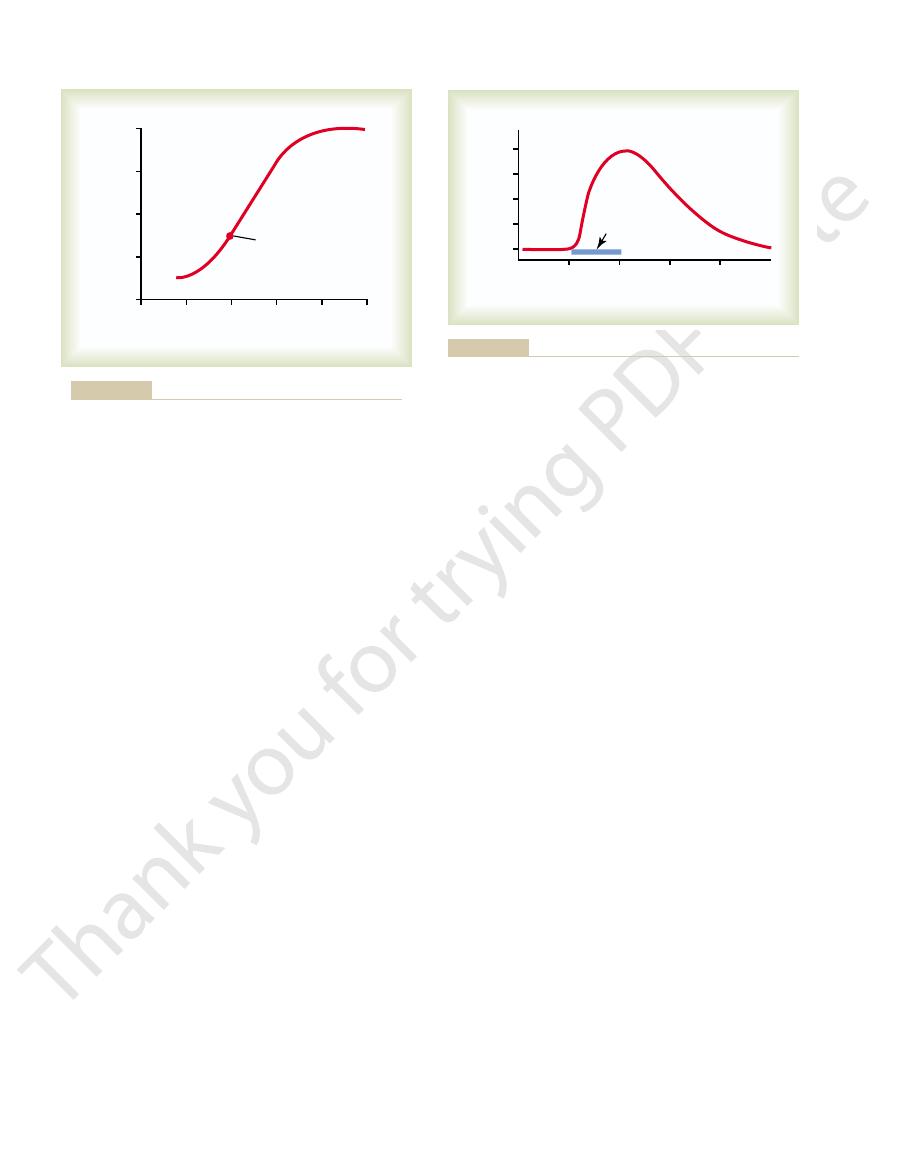
61–1, which shows that a 70 per cent increase in arterial P
fusing the brain greatly increases cerebral blood flow. This is demonstrated in Figure
centration, and (3) oxygen concentration.
trolling cerebral blood flow: (1) carbon dioxide concentration, (2) hydrogen ion con-
metabolism of the tissue. At least three metabolic factors have potent effects in con-
As in most other vascular areas of the body, cerebral blood flow is highly related to
750 to 900 ml/min, or 15 per cent of the resting cardiac output.
liters per 100 grams of brain tissue per minute. For the entire brain, this amounts to
Cerebral Blood Flow
cerebrospinal fluid, either its composition or its fluid pressure, can have equally
cells. Also, on a longer time scale, abnormalities of the
seconds. This occurs because lack of oxygen delivery to
brain function. For instance, total cessation of blood
and its fluids. However, this is far from true because
if it were independent of its blood flow, its metabolism,
Thus far, we have discussed the function of the brain as
Cerebrospinal Fluid, and Brain
C
H
A
P
T
E
R
6
1
761
Cerebral Blood Flow,
Metabolism
abnormalities of any of these can profoundly affect
flow to the brain causes unconsciousness within 5 to 10
the brain cells shuts down most metabolism in these
severe effects on brain function.
Normal Rate of Cerebral Blood Flow
Normal blood flow through the brain of the adult person averages 50 to 65 milli-
Regulation of Cerebral Blood Flow
Increase of Cerebral Blood Flow in Response to Excess Carbon Dioxide or Excess Hydrogen Ion
Concentration.
An increase in carbon dioxide concentration in the arterial blood per-
CO
2
approximately doubles
Carbon dioxide is believed to increase cerebral blood flow by combining first with
bral vessels—the dilation being almost directly proportional to the increase in
hydrogen ion concentration up to a blood flow limit of about twice normal.
formed during the course of tissue metabolism.
Importance of Cerebral Blood Flow Control by Carbon Dioxide and Hydrogen Ions.
Increased
tunate that an increase in hydrogen ion concentration also causes an increase in
tain a constant hydrogen ion concentration in the cerebral fluids and thereby helps
Except during periods of intense
exceptionally high level, such as during strenuous
When mean arterial pressure rises acutely to an
nervous effects.
brain. However, transection of the sympathetic nerves
then into the brain along with the cerebral arteries. This
The cerebral circulatory system has strong
Blood Flow.
Role of the Sympathetic Nervous System in Controlling Cerebral
pressure falls below 60 mm Hg, cerebral blood flow then
180 mm Hg mean arterial pressure. But, if the arterial
and hypotensive patients. Note the extreme constancy
160 to 180 mm Hg. This is demonstrated in Figure 61–3,
tension, autoregulation of cerebral blood flow occurs
cerebral blood flow. And, in people who have hyper-
to as high as 140 mm Hg without significant change in
decreased acutely to as low as 60 mm Hg or increased
and 140 mm Hg. That is, mean arterial pressure can be
Cerebral blood flow is “autoregulated”
intense light is shined into its eyes for one-half minute.
in occipital blood flow recorded in a cat’s brain when
cerebral blood flow, Figure 61–2 shows a typical increase
poral cortex. This measuring procedure can also be used
blood flow, especially in the visual areas of the occipital
opposite side of the brain. Reading a book increases the
to changes in local neuronal activity. For instance,
Using this technique, it has become clear that blood
pressed against the surface of the cortex. The rapidity
purpose, 256 small radioactive scintillation detectors are
tive substance passes through the brain tissue. For this
injected into the carotid artery; then the radioactivity of
radioactive substance, such as radioactive xenon, is
human cerebral cortex simultaneously. To do this, a
ity on the Flow.
Measurement of Cerebral Blood Flow, and Effect of Brain Activ-
ment of mental capability.
bral neuronal activity and, therefore, against derange-
nism for local regulation of cerebral blood flow is a very
can result at these low levels. Thus, the oxygen mecha-
levels below 20 mm Hg. Even coma
blood flow. This is fortuitous because brain function
40 mm Hg) immediately begins to increase cerebral
below about 30 mm Hg (normal value is 35 to
circulatory areas of the body.
nary blood vessels, in skeletal muscle, and in most other
normal. Thus, this local blood flow regulatory mecha-
causes vasodilation, returning the brain blood flow and
ciency mechanism for causing vasodilation immediately
supply this needed amount of oxygen, the oxygen defi-
oxygen per 100 grams of brain tissue per minute. If
The Nervous System: C. Motor and Integrative Neurophysiology
762
Unit XI
narrow limits—almost exactly 3.5 (
± 0.2) milliliters of
blood flow to the brain ever becomes insufficient to
transport of oxygen to the cerebral tissues to near
nism is almost exactly the same in the brain as in coro-
Experiments have shown that a decrease in cerebral
tissue P
O
2
becomes deranged at not much lower values of P
O
2
,
especially so at P
O
2
important protective response against diminished cere-
A method has been developed to record
blood flow in as many as 256 isolated segments of the
each segment of the cortex is recorded as the radioac-
of rise and decay of radioactivity in each tissue segment
is a direct measure of the rate of blood flow through that
segment.
flow in each individual segment of the brain changes as
much as 100 to 150 per cent within seconds in response
simply making a fist of the hand causes an immedi-
ate increase in blood flow in the motor cortex of the
cortex and in the language perception areas of the tem-
for localizing the origin of epileptic attacks because
local brain blood flow increases acutely and markedly
at the focal point of each attack.
Demonstrating the effect of local neuronal activity on
Autoregulation of Cerebral Blood Flow When the Arterial Pressure
Changes.
extremely well between arterial pressure limits of 60
even when the mean arterial pressure rises to as high as
which shows cerebral blood flow measured both in
persons with normal blood pressure and in hypertensive
of cerebral blood flow between the limits of 60 and
does become severely decreased.
sympathetic innervation that passes upward from the
superior cervical sympathetic ganglia in the neck and
innervation supplies both the large brain arteries and
the arteries that penetrate into the substance of the
or mild to moderate stimulation of them usually causes
very little change in cerebral blood flow because the
blood flow autoregulation mechanism can override the
0
20
40
60
80
100
Normal
Cerebral blood flow (times normal)
0.4
2.0
1.6
1.2
0.8
Arterial Pco
2
Arterial Pco
2
and cerebral blood flow.
Relationship between arterial P
Figure 61–1
CO
2
1.0
1.5
0
0.5
110
Blood flow (per cent of normal)
100
120
130
Light shining
in eyes
140
Minutes
Minutes
Increase in blood flow to the occipital regions of a cat’s brain when
Figure 61–2
light is shined into its eyes.

brain to be momentarily contorted by the blow.
neously with the skull, causing no one portion of the
if it is not too intense, moves the entire brain simulta-
simply floats in the fluid. Therefore, a blow to the head,
(only about 4 per cent different), so that the brain
the brain within its solid vault. The brain and the cere-
nected with one another, and the pressure of the fluid
brain and the spinal cord.
subarachnoid space around both the
, and in the
, in the
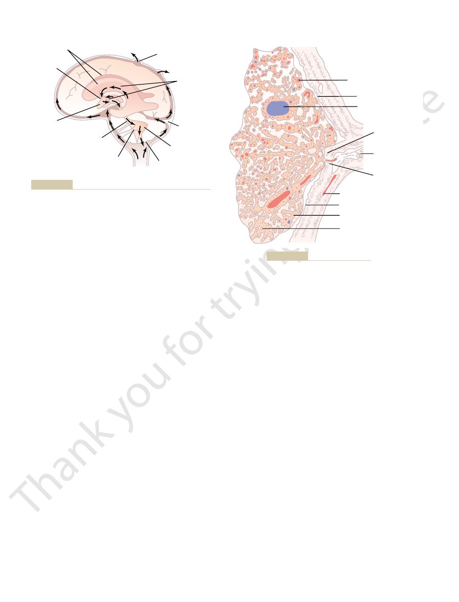
This fluid, as shown in Figure 61–4, is present in the
The entire cerebral cavity enclosing the brain and spinal
Cerebrospinal Fluid System
on the same side as the stroke lesion. Especially devas-
hemisphere on the same side as the blockage, which
In a similar manner, blockage of a
the body.
word formation. In addition, loss of function of neural
speak words because of loss of Broca’s motor area for
bral hemisphere, and he or she also becomes unable to
Wernicke’s speech comprehension area in the left cere-
left side of the brain, the person is likely to become
instance, if the middle cerebral artery is blocked on the
plies the midportion of one brain hemisphere. For
middle cerebral artery
brain area affected. One of the most common types of
brain tissue and further compromising its functions. The
burst; hemorrhage then occurs, compressing the local
In about one quarter of people who develop strokes,
blood flow in the artery, thereby leading to acute loss of
of the blood, causing a blood clot to occur and block
brain. The plaques can activate the clotting mechanism
bance of brain function, a condition called a “stroke.”
arteries in the brain, and as many as 10 per cent even-
Cerebral Blood Vessels Are Blocked
the brain break down, serious brain edema ensues,
transmission of the high pressure to the capillaries. We
develop high blood pressure, and these arterioles
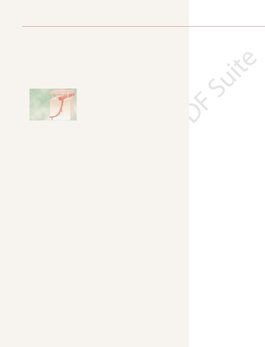
The walls of the small arterioles leading to the brain
capillary blood pressure.
on all sides by “glial feet,” which are small projections
blood capillaries in almost any other tissue of the body.
capillaries is that they are much less “leaky” than the
as great in the gray matter.
of white matter; correspondingly, the number of capil-
where the metabolic needs are greatest. The overall
As is true for almost all other tissues of the body, the
for preventing the occurrence of “cerebral stroke.”
venting vascular hemorrhages into the brain—that is,
smaller brain blood vessels. This is important in pre-
activity, the sympathetic nervous system normally con-
Cerebral Blood Flow, Cerebrospinal Fluid, and Brain Metabolism
Chapter 61
763
exercise or during other states of excessive circulatory
stricts the large- and intermediate-sized brain arteries
enough to prevent the high pressure from reaching the
Cerebral Microcirculation
number of blood capillaries in the brain is greatest
metabolic rate of the brain gray matter where the neu-
ronal cell bodies lie is about four times as great as that
laries and rate of blood flow are also about four times
An important structural characteristic of the brain
One reason for this is that the capillaries are supported
from the surrounding glial cells that abut against all sur-
faces of the capillaries and provide physical support to
prevent overstretching of the capillaries in case of high
capillaries become greatly thickened in people who
remain significantly constricted all the time to prevent
shall see later in the chapter that whenever these
systems for protecting against transudation of fluid into
which can lead rapidly to coma and death.
Cerebral “Stroke” Occurs When
Almost all elderly people have blockage of some small
tually have enough blockage to cause serious distur-
Most strokes are caused by arteriosclerotic plaques
that occur in one or more of the feeder arteries to the
brain function in a localized area.
high blood pressure makes one of the blood vessels
neurological effects of a stroke are determined by the
stroke is blockage of the
that sup-
almost totally demented because of lost function in
motor control areas of the left hemisphere can create
spastic paralysis of most muscles on the opposite side of
posterior cerebral
artery will cause infarction of the occipital pole of the
causes loss of vision in both eyes in the half of the retina
tating are strokes that involve the blood supply to the
midbrain because this can block nerve conduction in
major pathways between the brain and spinal cord,
causing both sensory and motor abnormalities.
cord has a capacity of about 1600 to 1700 milliliters;
about 150 milliliters of this capacity is occupied by cere-
brospinal fluid and the remainder by the brain and cord.
ven-
tricles of the brain
cisterns around the outside of
the brain
All these chambers are con-
is maintained at a surprisingly constant level.
Cushioning Function of the
Cerebrospinal Fluid
A major function of the cerebrospinal fluid is to cushion
brospinal fluid have about the same specific gravity
Hypotension
Hypertension
0
150
Cerebral blood flow (ml/100 g/min)
0
20
40
60
50
100
Mean arterial blood pressure (mm Hg)
Mean arterial blood pressure (mm Hg)
beings. (Modified from Lassen NA: Cerebral blood flow and
to hypertensive level, on cerebral blood flow in different human
Effect of differences in mean arterial pressure, from hypotensive
Figure 61–3
oxygen consumption in man. Physiol Rev 39:183, 1959.)
osmosis of water through the membrane, thus providing
brospinal fluid, which then causes almost immediate
charge. The two of these together increase the quantity
the sodium ion attracts the chloride ion’s negative
plexus. The sodium ions in turn pull along large amounts
fourth ventricle.
portion of the third ventricle, and (4) the roof of the
temporal horn of each lateral ventricle, (3) the posterior
epithelial cells. This plexus projects into (1 and 2) the
section of which is shown in Figure 61–5, is a cauliflower-
choroid plexus
The
other venous sinuses of the cerebrum. Thus, any extra
arachnoidal villi
noid spaces surrounding the cerebrum. From here, the
cord. Almost all the cerebrospinal fluid then flows
subarach-
The cisterna magna is continuous with the
, a fluid space that lies behind the medulla and
, entering the
two lateral foramina of Luschka
small openings,
another minute amount of fluid is added. Finally, the
, where still
from the third ventricle, it flows downward along the
; then, after addition of minute amounts of fluid
third
through the cerebrospinal fluid system. The fluid
choroid plexuses
The arrows in Figure 61–4 show that the main chan-
membranes; and a small amount comes from the brain
. Additional
tricles,
secretion from the choroid plexuses
system. About two thirds or more of this fluid originates
liliters each day, which is three to four times as much as
of Cerebrospinal Fluid
Formation, Flow, and Absorption
; if it occurs on the opposite side,
injury, it is a
blow to the head, such as that experienced by a boxer.
bony protuberances in the base of the skull, are often
temporal lobes, where the brain comes into contact with
The poles and the inferior surfaces of the frontal and
no longer being accelerated by the blow, the vacuum
in the area opposite to the blow. Then, when the skull is
brain momentarily because of the brain’s inertia, creat-
site to the area that is struck, the sudden movement of
same time in unison with the skull. On the side oppo-
that as the skull moves, the fluid pushes the brain at the
struck, the fluid on the struck side is so incompressible
reason for this effect is the following: When the blow is
This phenomenon is known as “contrecoup,” and the
head where the blow is struck but on the opposite side.
severe, it may not damage the brain on the side of the
When a blow to the head is extremely
The Nervous System: C. Motor and Integrative Neurophysiology
764
Unit XI
Contrecoup.
the whole skull causes the skull to pull away from the
ing for a split second a vacuum space in the cranial vault
suddenly collapses and the brain strikes the inner
surface of the skull.
the sites of injury and contusions (bruises) after a severe
If the contusion occurs on the same side as the impact
coup injury
the contusion is a contrecoup injury.
Cerebrospinal fluid is formed at a rate of about 500 mil-
the total volume of fluid in the entire cerebrospinal fluid
as
in the four ven-
mainly in the two lateral ventricles
small amounts of fluid are secreted by the ependymal
surfaces of all the ventricles and by the arachnoidal
itself through the perivascular spaces that surround the
blood vessels passing through the brain.
nels of fluid flow from the
and then
secreted in the lateral ventricles passes first into the
ventricle
aqueduct of Sylvius into the fourth ventricle
fluid passes out of the fourth ventricle through three
and a
midline foramen of Magendie
cisterna
magna
beneath the cerebellum.
noid space that surrounds the entire brain and spinal
upward from the cisterna magna through the subarach-
fluid flows into and through multiple
that project into the large sagittal venous sinus and
fluid empties into the venous blood through pores of
these villi.
Secretion by the Choroid Plexus.
, a
like growth of blood vessels covered by a thin layer of
Secretion of fluid into the ventricles by the choroid
plexus depends mainly on active transport of sodium
ions through the epithelial cells lining the outside of the
of chloride ions as well because the positive charge of
of osmotically active sodium chloride in the cere-
the fluid of the secretion.
Choroid
plexuses
Tentorium
cerebelli
Fourth
ventricle
Foramen of
Magendie
Arachnoidal
villi
Foramen of
Luschka
Aqueduct
of Sylvius
Third
ventricle
Foramen
of Monro
Lateral
ventricles
protruding into the dural sinuses.
choroid plexuses in the lateral ventricles to the arachnoidal villi
The arrows show the pathway of cerebrospinal fluid flow from the
Figure 61–4
Artery
Ependyma
Vein
Taenia
fornicis
Taenia
choroidea
Blood vessel
Ependyma
Villus epithelium
Villus connective tissue
Tela
choroides
Choroid plexus in a lateral ventricle.
Figure 61–5

very simple and is the following: First, the person lies
The usual pro-
hydrocephalus.
villi with abnormal absorptive properties. This is dis-
pressure. This is often caused by abnormally high resist-
villi. This also sometimes elevates the cerebrospinal
brospinal fluid, and they can cause serious blockage of
vault. In both these conditions, large numbers of red
The cerebrospinal fluid pressure also rises consider-
500 millimeters of water (37 mm Hg) or about four
the cerebrospinal fluid back into the blood. As a result,
cause high cerebrospinal fluid pressure, as follows.
cerebrospinal fluid in brain diseases. Such blockage can
become blocked by large particulate matter, by fibrosis,
Conversely, in disease states, the villi sometimes
the cerebral venous sinuses.
more widely, so that under normal conditions, the cere-
brospinal fluid pressure rises still higher, the valves open
sure of the blood in the venous sinuses. Then, if the cere-
fluid pressure is about 1.5 mm Hg greater than the pres-
mally, this valve action of the villi allows cerebrospinal
blood to flow backward in the opposite direction. Nor-
versely, the arachnoidal villi function like “valves” that
formation are seldom a factor in pressure control. Con-
remains very nearly constant, so that changes in fluid
The normal rate of cerebrospinal fluid formation
millimeters of water (10 mm Hg), although this may be
when one is lying in a horizontal position
The normal pressure in the cerebrospinal fluid system
perivascular spaces.
tion occurs in the brain, dead white blood cells and
matter out of the brain. For instance, whenever infec-
In addition to transporting fluid and proteins, the
lar spaces, in effect, are a specialized lymphatic system
into the large cerebral veins. Therefore, perivascu-
arachnoidal
brospinal fluid, to be absorbed through the
arachnoid spaces, the protein then flows with the cere-
into the subarachnoid spaces. On reaching the sub-
brain tissue, excess protein in the brain tissue leaves the
the brain. Because no true lymphatics are present in
elsewhere in the body, a small amount of protein leaks
Lymphatic Function of the Perivascular Spaces.
brain as far as the arterioles and venules go.
exists between it and each vessel. Therefore, perivascu-
perivascular space,
ent to the vessels, so that a space, the
as shown in Figure 61–6. The pia is only loosely adher-
pia mater,
but their ends penetrate inward, carrying with them a
The large arter-
fluid, (2) dissolved protein molecules, and (3) even par-
sinuses. The endothelial cells covering the villi have
arach-
walls and into the venous sinuses. Conglomerates of
arachnoidal villi
The
per cent less; and glucose, about 30 per cent less.
greater than in plasma; potassium ion, approximately 40
equal to that of plasma; chloride ion, about 15 per cent
plasma; sodium ion concentration, also approximately
ing: osmotic pressure, approximately equal to that of
fluid into the capillaries. Therefore, the resulting char-
Cerebral Blood Flow, Cerebrospinal Fluid, and Brain Metabolism
Chapter 61
765
Less important transport processes move small
amounts of glucose into the cerebrospinal fluid and both
potassium and bicarbonate ions out of the cerebrospinal
acteristics of the cerebrospinal fluid become the follow-
Absorption of Cerebrospinal Fluid Through the Arachnoidal Villi.
are microscopic fingerlike inward
projections of the arachnoidal membrane through the
these villi form macroscopic structures called
noidal granulations that can be seen protruding into the
been shown by electron microscopy to have vesicular
passages directly through the bodies of the cells large
enough to allow relatively free flow of (1) cerebrospinal
ticles as large as red and white blood cells into the
venous blood.
Perivascular Spaces and Cerebrospinal Fluid.
ies and veins of the brain lie on the surface of the brain
layer of
the membrane that covers the brain,
lar spaces follow both the arteries and the veins into the
As is true
out of the brain capillaries into the interstitial spaces of
tissue flowing with fluid through the perivascular spaces
villi
for the brain.
perivascular spaces transport extraneous particulate
other infectious debris are carried away through the
Cerebrospinal Fluid Pressure
averages 130
as low as 65 millimeters of water or as high as 195 mil-
limeters of water even in the normal healthy person.
Regulation of Cerebrospinal Fluid Pressure by the Arachnoidal
Villi.
allow cerebrospinal fluid and its contents to flow readily
into the blood of the venous sinuses while not allowing
fluid to begin to flow into the blood when cerebrospinal
brospinal fluid pressure almost never rises more than a
few millimeters of mercury higher than the pressure in
or by excesses of blood cells that have leaked into the
High Cerebrospinal Fluid Pressure in Pathological Conditions of
the Brain.
Often a large brain tumor elevates the cere-
brospinal fluid pressure by decreasing reabsorption of
the cerebrospinal fluid pressure can rise to as much as
times normal.
ably when hemorrhage or infection occurs in the cranial
and/or white blood cells suddenly appear in the cere-
the small absorption channels through the arachnoidal
fluid pressure to 400 to 600 millimeters of water (about
four times normal).
Some babies are born with high cerebrospinal fluid
ance to fluid reabsorption through the arachnoidal villi,
resulting either from too few arachnoidal villi or from
cussed later in connection with
Measurement of Cerebrospinal Fluid Pressure.
cedure for measuring cerebrospinal fluid pressure is
exactly horizontally on his or her side so that the fluid
Arachnoid membrane
Arachnoid trabecula
Subarachnoid space
Pia mater
Perivascular space
Blood vessel
Brain tissue
(Redrawn from Ranson SW, Clark SL: Anatomy of the Nervous
Figure 61–6
Drainage of a perivascular space into the subarachnoid space.
System. Philadelphia: WB Saunders Co, 1959.)

vault, accumulation of extra edema fluid compresses the
most other capillaries of the body.
having large slit-pores between them, as is the case for
That is, the membranes of the
tight junctions.
capillaries are joined to one another. They are joined by
The cause of the low permeability of the blood–
antibodies and non–lipid-soluble drugs, in the cere-
concentrations of therapeutic drugs, such as protein
fore, the blood–cerebrospinal fluid and blood-brain bar-
most non–lipid-soluble large organic molecules. There-
electrolytes such as sodium, chloride, and potassium;
such as alcohol and anesthetics; slightly permeable to
dioxide, oxygen, and most lipid-soluble substances
brain barriers are highly permeable to water, carbon
In general, the blood–cerebrospinal fluid and blood-
activity.
leptin, from the blood into the hypothalamus where
cules that facilitate transport of hormones, such as
The blood-brain barrier also has specific carrier mole-
hormones that regulate thirst, such as angiotensin II.
glucose concentration, as well as receptors for peptide
in the body fluids, such as changes in osmolality and in
diffuse with greater ease into the tissue spaces. The ease
, where substances
, and
respectively.
, exist between the
said that barriers, called the
the usual interstitial fluids of the body. Therefore, it is
in the body. Furthermore, many large molecular sub-
damage the brain at any age. A therapy for many types
skull is still pliable and can be stretched, and it can
to swell tremendously if it occurs in infancy when the
extent inside the ventricles. This will also cause the head
absorbed into the venous sinuses. Fluid therefore col-
The
into a thin shell against the skull. In neonates, the
three ventricles increase greatly. This flattens the brain
lateral and the third ventricles, the volumes of these
babies or from blockage by a brain tumor at any age.
block in the aqueduct of Sylvius
noid space, whereas in noncommunicating hydro-
cephalus.
vault. This condition is frequently divided into
“Hydrocephalus” means excess water in the cranial
of the retina and swells into the cavity of the eye. The
ble than those of the remainder of the retina, so that the
The tissues of the optic disc are much more distensi-
eye, which results in still more retinal edema.
impedes flow of blood in the retinal vein, thereby
the retina; and (3) the pressure in the sheath also
outward fluid flow in the optic nerves, causing accumu-
interior of the eyeball; (2) the high pressure decreases
itself. Therefore, (1) high cerebrospinal fluid pressure
the optic nerve sheath. The retinal artery and vein
rises in the cerebrospinal fluid system, it also rises inside
connects with the sclera of the eye. When the pressure
Anatomically, the dura of the brain
mercury, about 10 mm Hg pressure.
dividing this by 13.6, which is the specific gravity of
sure is said to be 136 millimeters of water pressure or,
136 millimeters above the level of the needle, the pres-
to rise in the tube as high as it will. If it rises to a level
is open to the air at its top. The spinal fluid is allowed
the cranial vault. A spinal needle is then inserted into
The Nervous System: C. Motor and Integrative Neurophysiology
766
Unit XI
pressure in the spinal canal is equal to the pressure in
the lumbar spinal canal below the lower end of the cord,
and the needle is connected to a vertical glass tube that
High Cerebrospinal Fluid Pressure Causes Edema of the Optic
Disc—Papilledema.
extends as a sheath around the optic nerve and then
pierce this sheath a few millimeters behind the eye and
then pass along with the optic nerve fibers into the eye
pushes fluid first into the optic nerve sheath and then
along the spaces between the optic nerve fibers to the
lation of excess fluid in the optic disc at the center of
increasing the retinal capillary pressure throughout the
disc becomes far more edematous than the remainder
swelling of the disc can be observed with an ophthal-
moscope and is called papilledema. Neurologists can
estimate the cerebrospinal fluid pressure by assessing
the extent to which the edematous optic disc protrudes
into the eyeball.
Obstruction to Flow of
Cerebrospinal Fluid Can
Cause Hydrocephalus
commu-
nicating hydrocephalus and noncommunicating hydro-
In communicating hydrocephalus fluid flows
readily from the ventricular system into the subarach-
cephalus fluid flow out of one or more of the ventricles
is blocked.
Usually the noncommunicating type of hydro-
cephalus is caused by a
,
resulting from atresia (closure) before birth in many
As fluid is formed by the choroid plexuses in the two
increased pressure also causes the whole head to swell
because the skull bones have not yet fused.
communicating type of hydrocephalus is usually
caused by blockage of fluid flow in the subarachnoid
spaces around the basal regions of the brain or by block-
age of the arachnoidal villi where the fluid is normally
lects both on the outside of the brain and to a lesser
of hydrocephalus is surgical placement of a silicone tube
shunt all the way from one of the brain ventricles to the
peritoneal cavity where the excess fluid can be absorbed
into the blood.
Blood–Cerebrospinal Fluid and
Blood-Brain Barriers
It has already been pointed out that the concentrations
of several important constituents of cerebrospinal fluid
are not the same as in extracellular fluid elsewhere
stances hardly pass at all from the blood into the cere-
brospinal fluid or into the interstitial fluids of the brain,
even though these same substances pass readily into
blood–cerebrospinal fluid
barrier and the blood-brain barrier
blood and the cerebrospinal fluid and brain fluid,
Barriers exist both at the choroid plexus and at the
tissue capillary membranes in essentially all areas of the
brain parenchyma except in some areas of the hypothal-
amus, pineal gland
area postrema
of diffusion in these areas is important because they
have sensory receptors that respond to specific changes
they bind to specific receptors that control other func-
tions such as appetite and sympathetic nervous system
and almost totally impermeable to plasma proteins and
riers often make it impossible to achieve effective
brospinal fluid or parenchyma of the brain.
cerebrospinal fluid and blood-brain barriers is the
manner in which the endothelial cells of the brain tissue
so-called
adjacent endothelial cells are tightly fused rather than
Brain Edema
One of the most serious complications of abnormal
cerebral fluid dynamics is the development of brain
edema. Because the brain is encased in a solid cranial

human brain. J Clin Invest 112:4, 2003.
Gore JC: Principles and practice of functional MRI of the
Clin Exp Pharmacol Physiol 27:422, 2000.
Ganong WF: Circumventricular organs: definition and role
Rev 78:53, 1998.
tion: role of endothelium and potassium channels. Physiol
Faraci FM, Heistad DD: Regulation of the cerebral circula-
Faraci FM: Vascular protection. Stroke 34:327, 2003.
tionship to local energy demand. News Physiol Sci 16:71,
Duelli R, Kuschinsky W: Brain glucose transporters: rela-
Physiol Rev 83:1183, 2003.
Chesler M: Regulation and modulation of pH in the brain.
of the nervous system. News Physiol Sci 19:110, 2004.
Burmester T, Hankeln T: Neuroglobin: a respiratory protein
84:169, 2004.
roendocrine control of body fluid metabolism. Physiol Rev
Antunes-Rodrigues J, de Castro M, Elias LL, et al: Neu-
of cerebral microcirculation. Trends Neurosci 26:340,
Anderson CM, Nedergaard M: Astrocyte-mediated control
deranged, leading sometimes to coma and even more
erly, and mental function does then become seriously
muscle and liver cells. When this happens, not enough
non-neural cells throughout the body, especially into
overtreated with insulin, the blood glucose concentra-
diabetic patients. Yet, when a diabetic patient is
betes with essentially zero secretion of insulin, glucose
body cells. Therefore, in patients who have serious dia-
membrane is not dependent on insulin, even though
neurons at any given time.
the capillary blood, with a total of only about a 2-minute
from the blood. As is true for oxygen, most of this is
Under normal conditions, almost all the energy
unconsciousness within 5 to 10 seconds.
the blood. Putting these factors together, one can under-
rate of the neurons, so that most neuronal activity
lism. One of the reasons for this is the high metabolic
The brain is not capable of much anaerobic metabo-
alive.
glucose and glycogen. However, it does keep the tissues
bining these with oxygen. This delivers energy only at
olism, which means release of energy by partially
as 30 minutes. During this time, the tissue cells obtain
Therefore, during excessive brain activity, neuronal
centration differences across the neuron membranes.
through the membranes, increasing the need for addi-
a neuron conducts an action potential, these ions move
membrane and potassium ions to the interior. Each time
ions through their membranes, mainly to transport
the neurons, not in the glial supportive tissues. The
lism in non–nervous system tissues.
fore, under resting conditions, brain metabolism per unit
brain is only 2 per cent of the total body mass. There-
metabolism in the body, even though the mass of the
Under resting but awake conditions, the metabolism of
Total Brain Metabolic Rate and Metabolic Rate of Neurons.
nutrients to supply its metabolic needs. However, there
Like other tissues, the brain requires oxygen and food
pressure.
needle puncture, thereby relieving the intracerebral
from the brain tissue and breaks up the vicious circles.
centrated mannitol solution. This pulls fluid by osmosis
a concentrated osmotic substance, such as a very con-
the brain. One such measure is to infuse intravenously
Once these two vicious circles have begun, heroic
pumps of the neuronal tissue cells, thus allowing these
still more fluid leakage. It also turns off the sodium
increases the permeability of the capillaries, allowing
cerebral blood flow also decreases oxygen delivery. This
edema becomes progressively worse. (2) The decreased
illary pressure then causes more edema fluid, so that the
further increase in capillary pressure. The increased cap-
decreases blood flow and causes brain ischemia. The
(1) Edema compresses the vasculature. This in turn
Once brain edema begins, it often initiates two vicious
matized tissues.
, in which the brain tissues and capillaries are
cause is a serious blow to the head, leading to
wall that makes the wall leaky to fluid. A very common
The usual cause of brain edema is either greatly
flow and destruction of brain tissue.
blood vessels, often causing seriously decreased blood
Cerebral Blood Flow, Cerebrospinal Fluid, and Brain Metabolism
Chapter 61
767
increased capillary pressure or damage to the capillary
brain con-
cussion
traumatized so that capillary fluid leaks into the trau-
circles because of the following positive feedbacks:
ischemia in turn causes arteriolar dilation with still
cells to swell in addition.
measures must be used to prevent total destruction of
Another procedure is to remove fluid quickly from the
lateral ventricles of the brain by means of ventricular
Brain Metabolism
are special peculiarities of brain metabolism that
require mention.
the brain accounts for about 15 per cent of the total
mass of tissue is about 7.5 times the average metabo-
Most of this excess metabolism of the brain occurs in
major need for metabolism in the neurons is to pump
sodium and calcium ions to the outside of the neuronal
tional membrane transport to restore proper ionic con-
metabolism can increase as much as 100 to 150 per cent.
Special Requirement of the Brain for Oxygen—Lack of Significant
Anaerobic Metabolism.
Most tissues of the body can live
without oxygen for several minutes and some for as long
their energy through processes of anaerobic metab-
breaking down glucose and glycogen but without com-
the expense of consuming tremendous amounts of
depends on second-by-second delivery of oxygen from
stand why sudden cessation of blood flow to the brain
or sudden total lack of oxygen in the blood can cause
Under Normal Conditions Most Brain Energy Is Supplied by
Glucose.
used by the brain cells is supplied by glucose derived
derived minute by minute and second by second from
supply of glucose normally stored as glycogen in the
A special feature of glucose delivery to the neurons
is that its transport into the neurons through the cell
insulin is required for glucose transport into most other
still diffuses readily into the neurons—which is most
fortunate in preventing loss of mental function in
tion can fall extremely low because the excess insulin
causes almost all the glucose in the blood to be trans-
ported rapidly into the vast numbers of insulin-sensitive
glucose is left in the blood to supply the neurons prop-
often to mental imbalances and psychotic distur-
bances—all caused by overtreatment with insulin.
References
2003.
2001.
in the regulation of endocrine and autonomic function.

organs. Front Biosci 9:290, 2004.
systemic inflammation: role of sensory circumventricular
Roth J, Harre EM, Rummel C, et al: Signaling the brain in
control of cardiovascular function. Physiol Rev 82:131,
Roman RJ: P-450 metabolites of arachidonic acid in the
cerebral blood flow. Eur Neuropsychopharmacol 12:495,
Paulson OB: Blood-brain barrier, brain metabolism and
Cambridge: Cambridge University Press, 1998.
Pardridge WM: An Introduction to the Blood-Brain Barrier.
Clin Exp Pharmacol Physiol 27:443, 2000.
term control of fluid homeostasis and arterial pressure.
osmoreceptor inputs to the area postrema: role in long-
Osborn JW, Collister JP, Carlson SH: Angiotensin and
stroke. N Engl J Med 348:2355, 2003.
Nadareishvili Z, Hallenbeck J: Neuronal regeneration after
Annu Rev Physiol 66:735, 2004.
Logothetis NK, Wandell BA: Interpreting the BOLD signal.
port: a lymphatic perspective. News Physiol Sci 17:227,
Johnston M, Papaiconomou C: Cerebrospinal fluid trans-
Sci 17:27, 2002.
in matching blood flow to metabolic activity. News Physiol
Harder DR, Zhang C, Gebremedhin D: Astrocytes function
The Nervous System: C. Motor and Integrative Neurophysiology
768
Unit XI
2002.
2002.
2002.