and afferent connections with deeper structures of the brain. It is especially
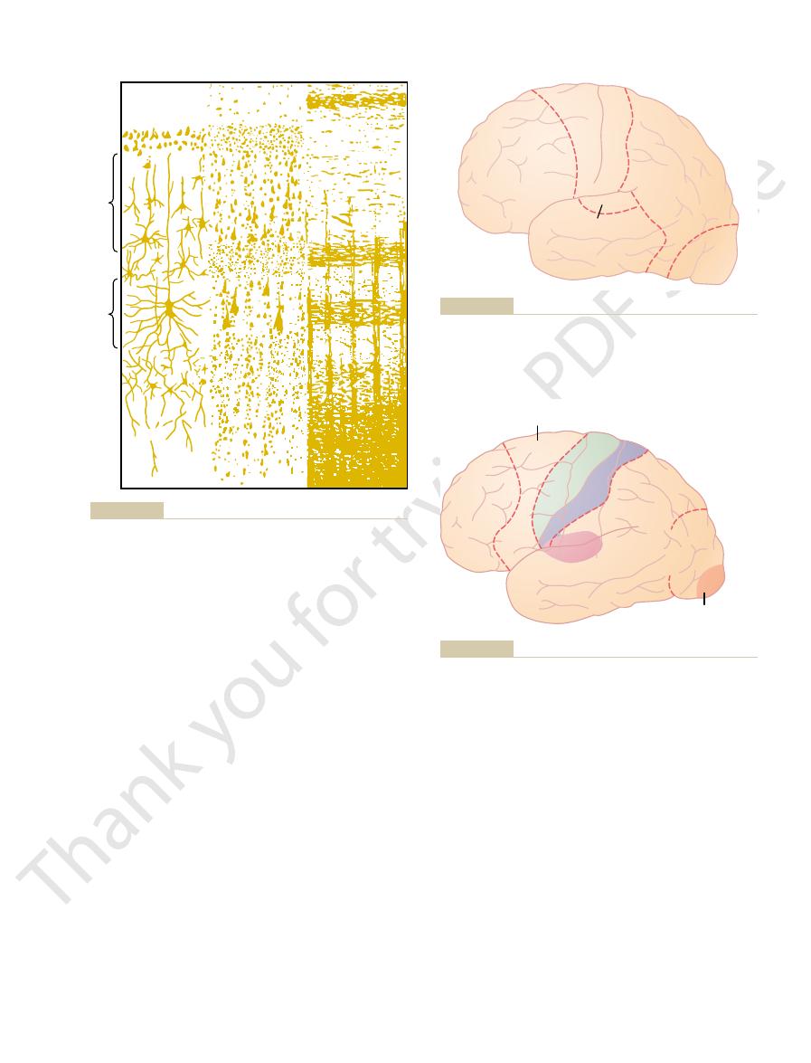
and III making short horizontal connections with adjacent cortical areas.
cortical association functions, with especially large numbers of neurons in layers II
to the thalamus arise in layer VI. Layers I, II, and III perform most of the intra-
brain stem and cord arise generally in layer V; and the tremendous numbers of fibers
the cortex through neurons located in layers V and VI; the very large fibers to the
signals from the body terminate in cortical layer IV. Most of the output signals leave
ters 47 and 51. By way of review, let us recall that most incoming specific sensory
The functions of the specific layers of the cerebral cortex are discussed in Chap-
through long association bundles.
that extend between adjacent areas of the cortex, but note also the
horizontal fibers
the different layers of the cerebral cortex. Note particularly the large number of
To the right in Figure 57–1 is shown the typical organization of nerve fibers within
that pass from one major part of the brain to another.
cord. They also give rise to most of the large subcortical association fiber bundles
They are the source of the long, large nerve fibers that go all the way to the spinal
cortex. The pyramidal cells are larger and more numerous than the fusiform cells.
pyramidal
The
sensory signals within the sensory areas and association areas.
these granule cells, suggesting a high degree of intracortical processing of incoming
The sensory areas of the cortex as well as the
gamma-aminobutyric acid (GABA).
; others are inhibitory and release mainly the inhibitory neurotransmitter
itself. Some are excitatory, releasing mainly the excitatory neurotransmitter
neurons generally have short axons and, therefore, function mainly
The
, the last named for their characteristic pyramidal shape.
pyramidal
, and (3)
), (2)
neurons are of three types: (1)
cerebral cortex, with its successive layers of different types of neurons. Most of the
Figure 57–1
cortex contains about 100 billion neurons.
thick, with a total area of about one quarter of a square meter. The total cerebral
surface of all the convolutions of the cerebrum. This layer is only 2 to 5 millimeters
The functional part of the cerebral cortex is a thin layer of neurons covering the
Physiologic Anatomy of the Cerebral Cortex
forth are presented briefly.
involved in thought processes, memory, analysis of sensory information, and so
of the cortex. In the first part of this chapter, the
nervous system. But we do know the effects of
It is ironic that of all the parts of the brain, we know
Functions of the Brain, Learning
Cerebral Cortex, Intellectual
C
H
A
P
T
E
R
5
7
714
and Memory
least about the functions of the cerebral cortex,
even though it is by far the largest portion of the
damage or specific stimulation in various portions
facts known about cortical function are discussed;
then basic theories of neuronal mechanisms
shows the typical histological structure of the neuronal surface of the
granular (also called stellate
fusiform
granular
as interneurons that transmit neural signals only short distances within the cortex
gluta-
mate
association areas between sensory and motor areas have large concentrations of
and fusiform cells give rise to almost all the output fibers from the
vertical fibers that extend to and from the cortex to lower areas of the brain and
some all the way to the spinal cord or to distant regions of the cerebral cortex
Anatomical and Functional Relations of the Cerebral Cortex to the Thalamus and Other Lower
Centers.
All areas of the cerebral cortex have extensive to-and-fro efferent
sometimes they experienced movements. Occasionally
told their thoughts evoked by the stimulation, and
had been removed. The electrically stimulated patients
Figure
cerebral cortical areas have separate functions.
gists, and neuropathologists have shown that different
Studies in human beings by neurosurgeons, neurolo-
Cortical Areas
through the thalamus, with the principal exception of
. Almost all pathways from the sensory
the thalamus: for this reason, the thalamus and the
entirely lost. Therefore, the cortex operates in close
more, when the thalamic connections are cut, the func-
essentially the same area of the thalamus. Further-
directions, both from the thala-
that connect with specific parts of the thalamus. These
Figure 57–2
necessary for almost all cortical activity.
damaged along with the cortex, the loss of cerebral
bral cortex and the thalamus. When the thalamus is
Cerebral Cortex, Intellectual Functions of the Brain, Learning and Memory
Chapter 57
715
important to emphasize the relation between the cere-
function is far greater than when the cortex alone is
damaged because thalamic excitation of the cortex is
shows the areas of the cerebral cortex
connections act in two
mus to the cortex and then from the cortex back to
tions of the corresponding cortical area become almost
association with the thalamus and can almost be con-
sidered both anatomically and functionally a unit with
cortex together are sometimes called the thalamocor-
tical system
receptors and sensory organs to the cortex pass
some sensory pathways of olfaction.
Functions of Specific
57–3 is a map of some of these functions as determined
by Penfield and Rasmussen from electrical stimulation
of the cortex in awake patients or during neurological
examination of patients after portions of the cortex
they spontaneously emitted a sound or even a word or
gave some other evidence of the stimulation.
I
VIb
VIa
V
IV
III
II
Saunders Co, 1959.)
Brodmann]: Anatomy of the Nervous System. Philadelphia: WB
or polymorphic cells. (Redrawn from Ranson SW, Clark SL [after
granular layer; V, large pyramidal cell layer; and VI, layer of fusiform
external granular layer; III, layer of pyramidal cells; IV, internal
Structure of the cerebral cortex, showing: I, molecular layer; II,
Figure 57–1
N. dorsalis
medialis
Med. geniculate
body indeterminate
Pulvinar
N. lateralis
posterior
Lat.
geniculate
body
N. ventralis
posterolateralis
N. ventralis
lateralis
the thalamus.
Figure 57–2
Areas of the cerebral cortex that connect with specific portions of
Contralateral
vision
Bilateral
vision
Elaboration
of thought
Supplementary
motor synergies
Hand
skills
Eye
turning
Speech
Speech
Sens. II
Hearing
Voluntary motor
Somato
sensor
y
Sp
eech
Memory patterns
York: Hafner Co, 1968.)
Cortex of Man: A Clinical Study of Localization of Function. New
cal regions. (Redrawn from Penfield W, Rasmussen T: The Cerebral
Figure 57–3
Functional areas of the human cerebral cortex as determined by
electrical stimulation of the cortex during neurosurgical operations
and by neurological examinations of patients with destroyed corti-

In Chapter 56, we learned
diately superior to the auditory “names” region and
functions performed in Wernicke
through visual input. In turn, the names are essential
learned mainly through auditory input, whereas the
The names are
poral lobe is an area for naming objects.
4. Area for Naming Objects.
through reading.
In its absence, a person can still have excellent
to make meaning out of the visually perceived words.
sion area. This so-called
book into Wernicke’s area, the language comprehen-
occipital lobe, is a visual association area that feeds
area, lying mainly in the anterolateral region of the
Posterior to the language comprehension
3. Area for Initial Processing of Visual Language
discuss this area much more fully later; it is the most
. We
Wernicke
for language comprehension, called
The major area
2. Area for Language Comprehension.
surroundings.
the coordinates of the visual, auditory, and body
parietal cortex. From all this information, it computes
of the body. This area receives visual sensory informa-
1. Analysis of the Spatial Coordinates of the Body.
Figure 57–5.
functional subareas, which are shown in
all the surrounding sensory areas. However, even the
cortex laterally. As would be expected, it provides a
orly, the visual cortex posteriorly, and the auditory
This association
explanations of the functions of these areas.
. Following are
, (2) the
areas have their specializations. The most important
as from subcortical structures. Yet even the association
areas are called
primary and secondary motor and sensory areas. These
bral cortex that do not fit into the rigid categories of
Figure 57–4 also shows several large areas of the cere-
sequence of tones in the auditory signals.
lines and angles, and other aspects of vision; and (3)
interpretation of color, light intensity, directions of
the shape or texture of an object in one’s hand; (2)
specific sensory signals, such as (1) interpretation of
primary areas, begin to analyze the meanings of the
sensory areas, located within a few centimeters of the
of motor activity. On the sensory side, the secondary
motor cortex and basal ganglia to provide “patterns”
in the primary areas. For instance, the supplementary
The secondary areas make sense out of the signals
sensory organs.
specific sensations—visual, auditory, or somatic—
muscle movements. The primary sensory areas detect
chapters. The primary motor areas have direct con-
vision, and hearing, all of which are discussed in earlier
. This figure shows the major
Figure 57–4
many different sources gives a more general map, as
The Nervous System: C. Motor and Integrative Neurophysiology
716
Unit XI
Putting large amounts of information together from
shown in
primary and secondary premotor and supplementary
motor areas of the cortex as well as the major primary
and secondary sensory areas for somatic sensation,
nections with specific muscles for causing discrete
transmitted directly to the brain from peripheral
and premotor areas function along with the primary
interpretations of the meanings of sound tones and
Association Areas
association areas because they receive
and analyze signals simultaneously from multiple
regions of both the motor and sensory cortices as well
association areas are (1) the parieto-occipitotemporal
association area
prefrontal association area,
and (3) the limbic association area
Parieto-occipitotemporal Association Area.
area lies in the large parietal and occipital cortical
space bounded by the somatosensory cortex anteri-
high level of interpretative meaning for signals from
parieto-occipitotemporal association area has its own
An
area beginning in the posterior parietal cortex and
extending into the superior occipital cortex provides
continuous analysis of the spatial coordinates of all
parts of the body as well as of the surroundings
tion from the posterior occipital cortex and simulta-
neous somatosensory information from the anterior
’s area,
lies behind the primary auditory cortex in the posterior
part of the superior gyrus of the temporal lobe
important region of the entire brain for higher intel-
lectual function because almost all such intellectual
functions are language based.
(Reading).
visual information conveyed by words read from a
angular gyrus area is needed
language comprehension through hearing but not
In the most lateral por-
tions of the anterior occipital lobe and posterior tem-
physical natures of the objects are learned mainly
for both auditory and visual language comprehension
(
’s area located imme-
anterior to the visual word processing area).
Prefrontal Association Area.
that the prefrontal association area functions in close
association with the motor cortex to plan complex
Primary
auditory
Prefrontal
Association
Area
Parieto-
occipito-
temporal
Association
Area
Secondary
auditory
Limbic
Association
Area
Supplemental
and premotor
Primary motor
Primary somatic
Secondary somatic
Secondary
visual
Primary
visual
as primary and secondary motor and sensory areas.
Locations of major association areas of the cerebral cortex, as well
Figure 57–4
nition. Most of our daily tasks involve associations
areas, strangely enough, results in little other abnor-
shown in Figure 57–6. Loss of these face recognition
the medioventral surfaces of the temporal lobes, as
is inability to recognize faces. This
Area for Recognition of Faces
learning itself.
brain. This limbic system provides most of the emo-
system, the
We will learn in Chapter 58
emotions,
behavior,
hemisphere. It is concerned primarily with
poral lobe, in the ventral portion of the frontal lobe,
This area is found in the anterior pole of the tem-
Figures 57–4 and 57–5 show still
are learned simultaneously, they are stored together in
storage area for the first language. If both languages
then learns a new language, the area in the brain where
When a person has already learned one language and
chapter.
association cortex, as we discuss more fully later in the
area also works in close association with Wernicke’s
even short phrases are initiated and executed. This
and partly in the premotor area. It is here that plans
. This area, shown in Figure 57–5, is
word formation
provides the neural circuitry for
Broca’s Area.
store on a short-term basis “working memories” that
, and it is said to
thinking as well as motor types. In fact, the prefrontal
ities. It seems to be capable of processing nonmotor as
. This pre-
The
thalamic feedback circuit for motor planning, which
effective movements. Much of the output from the
information, especially information on the spatial
prefrontal association area. Through this bundle, the
this function, it receives strong input through a
patterns and sequences of motor movements. To aid in
Cerebral Cortex, Intellectual Functions of the Brain, Learning and Memory
Chapter 57
717
massive subcortical bundle of nerve fibers connecting
the parieto-occipitotemporal association area with the
prefrontal cortex receives much preanalyzed sensory
coordinates of the body that is necessary for planning
prefrontal area into the motor control system passes
through the caudate portion of the basal ganglia-
provides many of the sequential and parallel compo-
nents of movement stimulation.
prefrontal association area is also essential to
carrying out “thought” processes in the mind
sumably results from some of the same capabilities of
the prefrontal cortex that allow it to plan motor activ-
well as motor information from widespread areas of
the brain and therefore to achieve nonmotor types of
association area is frequently described simply as
important for elaboration of thoughts
are used to combine new thoughts while they are
entering the brain.
A special region in the frontal cortex,
called Broca’s area,
located partly in the posterior lateral prefrontal cortex
and motor patterns for expressing individual words or
language comprehension center in the temporal
An especially interesting discovery is the following:
the new language is stored is slightly removed from the
the same area of the brain.
Limbic Association Area.
another association area called the limbic association
area.
and in the cingulate gyrus lying deep in the longitu-
dinal fissure on the midsurface of each cerebral
and motivation.
that the limbic cortex is part of a much more extensive
limbic system, that includes a complex set
of neuronal structures in the midbasal regions of the
tional drives for activating other areas of the brain and
even provides motivational drive for the process of
An interesting type of brain abnormality called
prosophenosia
occurs in people who have extensive damage on the
medial undersides of both occipital lobes and along
mality of brain function.
One wonders why so much of the cerebral cortex
should be reserved for the simple task of face recog-
Naming of
objects
Vision
Visual
processing
of words
Spatial
coordinates
of body and
surroundings
Somato-
sensory
Word
formation
Word
formation
Broca‘s
Area
Broca‘s
Area
Limbic
Association
Area
Limbic
Association
Area
Wernicke‘s
Area
Wernicke‘s
Area
Auditory
Behavior,
emotions,
motivation
Language
comprehension
intelligence
Language
comprehension
intelligence
Motor
Planning complex
movements and
elaboration of
thoughts
in the left hemisphere.
and speech production, which in 95
cially Wernicke’s and Broca’s
the cerebral cortex, showing espe-
Figure 57–5
Map of specific functional areas in
areas for language comprehension
per cent of all people are located
demented existence.
functions of the brain. Loss of this area in an
Wernicke’s area for processing most intellectual
word blindness
, or
be able to interpret their meanings. This is the condi-
mainly blocked. Therefore, the person may be able to
passing into Wernicke’s area from the visual cortex is
ences as usual, but the stream of visual experiences
intact, the person can still interpret auditory experi-
while Wernicke’s area in the temporal lobe is still
of the occipital lobe as well. If this region is destroyed
nicke’s area and fusing posteriorly into the visual areas
terior parietal lobe, lying immediately behind Wer-
The
ferent patterns of sensory experiences.
belief is in accord with the importance of Wernicke’s
individual memories may be stored elsewhere. This
believed that activation of Wernicke’s area can call
ment made by a specific person. For this reason, it is
tions such as a specific musical piece, or even a state-
might remember from childhood, auditory hallucina-
thalamus. The types of thoughts that might be experi-
thought. This is particularly true when the stimulation
Electrical stimulation in Wernicke’s area of a con-
a coherent thought. Likewise, the person may be able
After severe damage in Wernicke’s area, a person
special significance in intellectual processes.
Wernicke
, and so forth. It is best known as
area, the
knowing
, the
, the
global importance: the
Therefore, this region has been called by
intelligence.
This area of confluence of the different sensory inter-
poral, parietal, and occipital lobes all come together.
, where the tem-
Figure 57–7
temporal lobe, shown in
The somatic, visual, and auditory association areas all
(a General Interpretative Area)
Wernicke
Temporal Lobe
Function of the Posterior Superior
ment, as we see in Chapter 58.
control of one’s behavioral response to the environ-
that has to do with emotions, brain activation, and
is contiguous with the visual cortex, and the temporal
The occipital portion of this facial recognition area
with other people, and one can see the importance of
The Nervous System: C. Motor and Integrative Neurophysiology
718
Unit XI
this intellectual function.
portion is closely associated with the limbic system
Comprehensive Interpretative
—“
’s Area”
meet one another in the posterior part of the superior
pretative areas is especially highly developed in the
dominant side of the brain—the left side in almost all
right-handed people—and it plays the greatest single
role of any part of the cerebral cortex for the higher
comprehension levels of brain function that we call
different names suggestive of an area that has almost
general interpretative area
gnostic area
tertiary association
area
’s area
in honor of the neurologist who first described its
might hear perfectly well and even recognize different
words but still be unable to arrange these words into
to read words from the printed page but be unable to
recognize the thought that is conveyed.
scious person occasionally causes a highly complex
electrode is passed deep enough into the brain to
approach the corresponding connecting areas of the
enced include complicated visual scenes that one
forth complicated memory patterns that involve more
than one sensory modality even though most of the
area in interpreting the complicated meanings of dif-
Angular Gyrus—Interpretation of Visual Information.
angular gyrus is the most inferior portion of the pos-
see words and even know that they are words but not
tion called dyslexia
.
Let us again emphasize the global importance of
adult usually leads thereafter to a lifetime of almost
Frontal
lobe
Temporal
lobe
Facial
recognition area
Scientific American, Inc. All rights reserved.)
Specializations of the human brain. Sci Am 241:180, 1979.
medial occipital and temporal lobes. (Redrawn from Geschwind N:
Facial recognition areas located on the underside of the brain in the
Figure 57–6
“ 1979 by
Primary
auditory
Prefrontal
area
Broca's
speech
area
Motor
Primary
Somatic
Somatic
Interpretative
areas
Visual
interpretative
areas
Auditory
interpretative
areas
Wernicke‘s area
Primary
visual
in the frontal lobe.
s speech area
superior portion of the temporal lobe. Note also the prefrontal area
, located in the postero-
Wernicke
into a general mechanism for interpretation of sensory experience.
Figure 57–7
Organization of the somatic auditory and visual association areas
All of these feed also into
’s area
and Broca’

prominence of the human prefrontal areas. Yet, efforts
is the locus of “higher intellect” in the human being,
For years, it has been taught that the prefrontal cortex
Prefrontal Association Areas
Higher Intellectual Functions of the
be dominant for some other types of intelligence.
though we speak of the “dominant” hemisphere, this
ences related to use of the limbs and hands. Thus, even
people’s voices, and probably many somatic experi-
significance of “body language” and intonations of
tions between the person and their surroundings, the
experiences (especially visual patterns), spatial rela-
standing and interpreting music, nonverbal visual
regions of the opposite hemisphere, are retained.
Many other types of interpretative capabilities, some
even the ability to think through logical problems.
the ability to perform mathematical operations, and
guage or verbal symbolism, such as the ability to read,
an adult person is destroyed, the person normally loses
When Wernicke’s area in the dominant hemisphere of
occipitotemporal Cortex in the
Functions of the Parieto-
lobe.
tion area, into the already developed Wernicke’s lan-
channeled through the angular gyrus, a visual associa-
the medium of reading develops, the visual informa-
in life, when visual perception of language through
introduction to language is by way of hearing. Later
ondary hearing areas of the temporal lobe. This close
interpretation of language is Wernicke’s area, and this
The sensory area of the dominant hemisphere for
read a book, we do not store the visual images of
for other intellectual purposes. For instance, when we
Wernicke’s Area and in Intellectual Functions
thoughts and motor responses.
spheres. This unitary, cross-feeding organization pre-
purpose, they use mainly fiber pathways in the
trolling motor activities in both hemispheres. For this
hemisphere, these areas receive sensory information
areas, are usually highly developed in only the left
lobe and angular gyrus, as well as many of the motor
people.
10 persons, thus causing right-handedness in most
The motor areas for controlling hands are also
taneously the laryngeal muscles, respiratory muscles,
nant on the left side of the brain. This speech area is
intermediate frontal lobe, is also almost always domi-
speech area (Broca’s area), located far laterally in the
As discussed later in the chapter, the premotor
dominance.
right side alone becomes highly developed, with full
taneously to have dual function, or, more rarely, the
remaining 5 per cent, either both sides develop simul-
lobe and angular gyrus become dominant, and in the
In about 95 per cent of all people, the left temporal
Therefore, the left side normally becomes dominant
opposite, less-used side, learning remains slight.
gains the first start increases rapidly, whereas in the
direct one’s attention to the better developed region,
than the right. Thereafter, because of the tendency to
usually slightly larger than the right, the left side
to one principal thought at a time. Presumably,
The attention of the “mind” seems to be directed
following.
will usually develop dominant characteristics.
in very early childhood, the opposite side of the brain
become dominant over the right side. However, if for
more than one half of neonates. Therefore, it is easy
tually become Wernicke’s area is as much as 50 per
Even at birth, the area of the cortex that will even-
inant one.
per cent of all people, the left hemisphere is the dom-
. In about 95
hemisphere than in the other. Therefore, this hemi-
of the speech and motor control areas, are usually
area and the angular gyrus, as well as the functions
The general interpretative functions of Wernicke’s
Concept of the Dominant Hemisphere
Cerebral Cortex, Intellectual Functions of the Brain, Learning and Memory
Chapter 57
719
much more highly developed in one cerebral
sphere is called the dominant hemisphere
cent larger in the left hemisphere than in the right in
to understand why the left side of the brain might
some reason this left side area is damaged or removed
A theory that can explain the capability of one
hemisphere to dominate the other hemisphere is the
because the left posterior temporal lobe at birth is
normally begins to be used to a greater extent
the rate of learning in the cerebral hemisphere that
over the right.
responsible for formation of words by exciting simul-
and muscles of the mouth.
dominant in the left side of the brain in about 9 of
Although the interpretative areas of the temporal
from both hemispheres and are capable also of con-
corpus
callosum for communication between the two hemi-
vents interference between the two sides of the brain;
such interference could create havoc with both mental
Role of Language in the Function of
A major share of our sensory experience is converted
into its language equivalent before being stored in the
memory areas of the brain and before being processed
the printed words but instead store the words them-
selves or their conveyed thoughts often in language
form.
is closely associated with both the primary and sec-
relation probably results from the fact that the first
tion conveyed by written words is then presumably
guage interpretative area of the dominant temporal
Nondominant Hemisphere
almost all intellectual functions associated with lan-
of which use the temporal lobe and angular gyrus
Psychological studies in patients with damage to the
nondominant hemisphere have suggested that this
hemisphere may be especially important for under-
is primarily for language-based intellectual functions;
the so-called nondominant hemisphere might actually
principally because the main difference between the
brains of monkeys and of human beings is the great

thermore, because neurological tests can easily assess
human beings can communicate with one another. Fur-
with moral laws.
rare diseases; and (7) control our activities in accord
cated mathematical, legal, or philosophical problems;
actions before they are performed; (5) solve compli-
is decided; (4) consider the consequences of motor
plan for the future; (3) delay action in response to
memory, we have the abilities to (1) prognosticate; (2)
storing different types of temporary memory, such as
intelligence. In fact, studies have shown that the pre-
“working memory.” This could well explain the many
needed for subsequent thoughts is called the brain’s
This ability of the prefrontal areas to keep track of
to take place.
track of these bits even in temporary memory, proba-
information. Psychological tests have shown that pre-
. This means
of a “Working Memory.”
distractions.
, whereas people with func-
so at most. One of the results is that people without
still think, they show little concerted thinking in logical
goals.
include motor action, so be it. If they do not, then
thought patterns for attaining goals. If these goals
We learned earlier in this chapter that the
Inability to Progress Toward Goals or to Carry Through Sequen-
behavior, which is discussed in detail in Chapter 58.
association cortex. This limbic area helps to control
limbic association cortex, rather than of the prefrontal
shown in Figures 57–4 and 57–5, this area is part of the
the underside of the brain. As explained earlier and
These two characteristics probably result
frontal association areas.
From this information, let us try to piece together a
purpose.
performed throughout life, but often without
7. The patients could also still perform most of the
exhilaration to madness.
any long trains of thought, and their moods
language, but they were unable to carry through
6. The patients could still talk and comprehend
for the occasion, often including loss of morals
5. Their social responses were often inappropriate
sometimes markedly, and, in general, they lost
4. Their level of aggressiveness was decreased,
parallel tasks at the same time.
3. They became unable to learn to do several
tasks to reach complex goals.
2. They became unable to string together sequential
problems.
1. The patients lost their ability to solve complex
lobes from top to bottom. Subsequent studies in these
a blunt, thin-bladed knife through a small opening in
. This was done by inserting
and the remainder of the brain, that is, by a procedure
drugs for treating psychiatric conditions, it was found
Several decades ago, before the advent of modern
become nonfunctional, as follows.
important intellectual functions of their own. These
areas do, however, have less definable but nevertheless
does destruction of the prefrontal areas.The prefrontal
superior temporal lobe (Wernicke’s area) and the
the brain have not been successful. Indeed, destruction
The Nervous System: C. Motor and Integrative Neurophysiology
720
Unit XI
to show that the prefrontal cortex is more important
in higher intellectual functions than other portions of
of the language comprehension area in the posterior
adjacent angular gyrus region in the dominant hemi-
sphere causes much more harm to the intellect than
functions can best be explained by describing what
happens to patients in whom the prefrontal areas have
that some patients could receive significant relief from
severe psychotic depression by severing the neuronal
connections between the prefrontal areas of the brain
called prefrontal lobotomy
the lateral frontal skull on each side of the head and
slicing the brain at the back edge of the prefrontal
patients showed the following mental changes:
ambition.
and little reticence in relation to sex and
excretion.
changed rapidly from sweetness to rage to
usual patterns of motor function that they had
coherent understanding of the function of the pre-
Decreased Aggressiveness and Inappropriate Social Re-
sponses.
from loss of the ventral parts of the frontal lobes on
tial Thoughts.
prefrontal association areas have the capability of
calling forth information from widespread areas of the
brain and using this information to achieve deeper
the thought processes attain intellectual analytical
Although people without prefrontal cortices can
sequence for longer than a few seconds or a minute or
prefrontal cortices are easily distracted from their
central theme of thought
tioning prefrontal cortices can drive themselves to
completion of their thought goals irrespective of
Elaboration of Thought, Prognostication, and Performance of
Higher Intellectual Functions by the Prefrontal Areas—Concept
Another function that has been
ascribed to the prefrontal areas by psychologists and
neurologists is elaboration of thought
simply an increase in depth and abstractness of the dif-
ferent thoughts put together from multiple sources of
frontal lobectomized lower animals presented with
successive bits of sensory information fail to keep
bly because they are distracted so easily that they
cannot hold thoughts long enough for memory storage
many bits of information simultaneously and to cause
recall of this information instantaneously as it is
functions of the brain that we associate with higher
frontal areas are divided into separate segments for
one area for storing shape and form of an object or a
part of the body and another for storing movement.
By combining all these temporary bits of working
incoming sensory signals so that the sensory informa-
tion can be weighed until the best course of response
(6) correlate all avenues of information in diagnosing
Function of the Brain in
Communication—Language
Input and Language Output
One of the most important differences between human
beings and lower animals is the facility with which
sequence is the following: (1) reception in the primary
pathway involved in hearing and speaking. This
communication. The upper half of the figure shows the
Figure 57–8 shows two principal pathways for
Summary.
either total or partial inability to speak distinctly.
55 and 56. Destruction of any of these regions can cause
contractions, making liberal use of basal ganglial and
, and
activate these muscles,
intensities of the sequential sounds.The
sible for the intonations, timing, and rapid changes in
tongue, larynx, vocal cords, and so forth that are respon-
Finally, we have the act of articulation,
lips, mouth, respiratory system, and other accessory
fore, the
hemisphere, as shown in Figures 57–5 and 57–8. There-
, which lies
s speech area
instead of noises. This effect, called
Loss of Broca’s Area Causes Motor Aphasia.
ate sequences of words to express the thought. The
lesion is less severe, the person may be able to formu-
the thoughts that are to be communicated. Or, if the
nicke’s aphasia or global aphasia is unable to formulate
tant for this ability. Therefore, a person with either Wer-
the brain. Again, it is Wernicke’s area in the posterior
The formation of thoughts and even most choices of
itself.
involves two principal stages of mentation: (1) forma-
The process of speech
fissure, the person is likely to be almost totally
inferiorly into the lower areas of the temporal lobe, and
extends (1) backward into the angular gyrus region, (2)
When the lesion in Wernicke’s area is widespread and
Wernicke
Therefore, this type of aphasia is called
Wernicke
that is expressed. This results most frequently when
Wernicke’s Aphasia and Global Aphasia.
word blindness
word deafness
or, more commonly,
These effects are called, respectively,
We noted earlier in the
Sensory Aspects of Communication.
eyes, and, second, the
(language input), involving the ears and
There are two aspects to communication: first, the
nication. From this, one will see immediately how the
, function of the cortex in commu-
Figure 57–8
review, with the help of anatomical maps of neural path-
segment of brain cortex function. Therefore, we will
the ability of a person to communicate with others, we
Cerebral Cortex, Intellectual Functions of the Brain, Learning and Memory
Chapter 57
721
know more about the sensory and motor systems
related to communication than about any other
ways in
principles of sensory analysis and motor control apply
to this art.
sensory aspect
motor aspect (language output),
involving vocalization and its control.
chapter that destruction of portions of the auditory or
visual association areas of the cortex can result in inabil-
ity to understand the spoken word or the written word.
auditory receptive
aphasia and visual receptive aphasia
and
(also called dyslexia).
Some people
are capable of understanding either the spoken word or
the written word but are unable to interpret the thought
’s area in the posterior superior temporal gyrus
in the dominant hemisphere is damaged or destroyed.
’s
aphasia.
(3) superiorly into the superior border of the sylvian
demented for language understanding or communica-
tion and therefore is said to have global aphasia.
Motor Aspects of Communication.
tion in the mind of thoughts to be expressed as well
as choice of words to be used and then (2) motor
control of vocalization and the actual act of vocalization
words are the function of sensory association areas of
part of the superior temporal gyrus that is most impor-
late the thoughts but unable to put together appropri-
person sometimes is even fluent with words but the
words are jumbled.
Sometimes
a person is capable of deciding what he or she wants to
say but cannot make the vocal system emit words
motor aphasia,
results from damage to Broca’
in the prefrontal and premotor facial region of the cere-
bral cortex—about 95 per cent of the time in the left
skilled motor patterns for control of the larynx,
muscles of speech are all initiated from this area.
Articulation.
which means the muscular movements of the mouth,
facial and laryn-
geal regions of the motor cortex
and the cerebellum, basal ganglia
sensory cortex all
help to control the sequences and intensities of muscle
cerebellar feedback mechanisms described in Chapters
s area
Angular gyrus
Motor cortex
Arcuate fasciculus
Broca’s area
Wernicke’s area
Primary auditory area
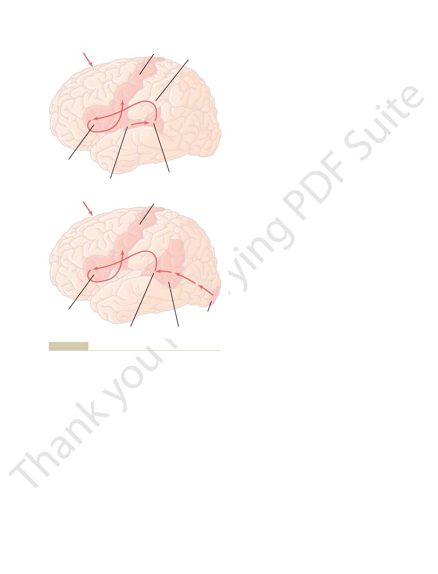
SPEAKING A HEARD WORD
Motor cortex
Broca’s area
Wernicke’
Primary
visual area
SPEAKING A WRITTEN WORD
American, Inc. All rights reserved.)
tions of the human brain. Sci Am 241:180, 1979.
speaking the same word. (Redrawn from Geschwind N: Specializa-
ing the same word, and
Figure 57–8
Brain pathways for (top) perceiving a heard word and then speak-
(bottom) perceiving a written word and then
“ 1979 by Scientific

subconscious level, and the anterior commissure
activities. The corpus callosum is required for the
storage, communication, and control of motor
independent capabilities for consciousness, memory
explain. Thus, the two halves of the brain have
questioned why he said this, the boy could not
necessary to speak the words “No way!” But when
This response required function of Wernicke’s
immediately and with full emotion said, “No way!”
for the right half of his brain to see, the boy
instance, when the command “kiss” was written
anterior commissure that was not sectioned. For
temporal cortices and adjacent areas, were still
two sides of the brain for emotions, the anterior
occurred in the left side of the brain as well. This
In this case, a subconscious emotional response
The effect was quite different when an emotional
spoken word. Furthermore, the right cortex could
hemisphere. Conversely, the right side of the brain
conscious portions of the brain. For example, in a
3. Finally, people whose corpus callosum is
decision making.
dominant hemisphere. Therefore, somatic and
hemisphere into Wernicke’s area in the left
2. Cutting the corpus callosum prevents transfer of
functions of the left hand and arm, even though
in the left hemisphere, lose control over the right
intellectual functions of Wernicke’s area, located
the opposite side of the brain. Therefore, the
of information from Wernicke’s area of the
1. Cutting the corpus callosum blocks transfer
between the two hemispheres are the following.
sphere. Important examples of such cooperation
Thus, one of the functions of the corpus callosum
brain creates recognition in the opposite hemisphere.
chiasm split but the corpus callosum intact, it is found
recognize the objects. However, on repeating
same objects. The answer to this is that the left eye
with its right eye while its left eye is covered. Next,
to the cerebral hemisphere on the side of the eye. Then
gitudinally, so that signals from each eye can go only
the experiments: A monkey is first prepared by cutting
the corpus callosum and anterior commissure. These
mystery.
long time, the function of the corpus callosum was a
to discern deficits in brain function. Therefore, for a
destroyed in laboratory animals, it was at first difficult
hemispheres. However, when the corpus callosum was
corpus callosum, it was assumed from the beginning
, are interconnected by fibers that pass
ral lobes; these temporal areas, including especially
Fibers in the
Hemispheres
Between the Two Cerebral
and Other Information
Thoughts, Memories, Training,
Commissure to Transfer
of recognition in Wernicke’s area. From here, the
rather than in the primary auditory area. Then the infor-
reading and then speaking in response. The initial recep-
The lower figure illustrates the comparable steps in
the motor cortex to control the speech muscles.
mation; and (6) transmission of appropriate signals into
motor programs in Broca’s area for control of word for-
; (5) activation of the skilled
of signals from Wernicke’s area to Broca’s area by way
thoughts and the words to be spoken; (4) transmission
area; (3) determination, also in Wernicke’s area, of the
words; (2) interpretation of the words in Wernicke’s
The Nervous System: C. Motor and Integrative Neurophysiology
722
Unit XI
auditory area of the sound signals that encode the
of the arcuate fasciculus
tive area for the words is in the primary visual area
mation passes through early stages of interpretation in
the angular gyrus region and finally reaches its full level
sequence is the same as for speaking in response to the
spoken word.
Function of the Corpus
Callosum and Anterior
corpus callosum provide abundant bi-
directional neural connections between most of the
respective cortical areas of the two cerebral hemi-
spheres except for the anterior portions of the tempo-
the amygdala
through the anterior commissure.
Because of the tremendous number of fibers in the
that this massive structure must have some important
function to correlate activities of the two cerebral
Properly designed psychological experiments have
now demonstrated extremely important functions for
functions can best be explained by describing one of
the corpus callosum and splitting the optic chiasm lon-
the monkey is taught to recognize different objects
the right eye is covered and the monkey is tested
to determine whether its left eye can recognize the
cannot
the same experiment in another monkey with the optic
invariably that recognition in one hemisphere of the
and the anterior commissure is to make information
stored in the cortex of one hemisphere available to
corresponding cortical areas of the opposite hemi-
dominant hemisphere to the motor cortex on
motor cortex that initiates voluntary motor
the usual subconscious movements of the left
hand and arm are normal.
somatic and visual information from the right
visual information from the left side of the body
frequently fails to reach this general interpretative
area of the brain and therefore cannot be used for
completely sectioned have two entirely separate
teenage boy with a sectioned corpus callosum,
only the left half of his brain could understand
both the written word and the spoken word
because the left side was the dominant
could understand the written word but not the
elicit a motor action response to the written word
without the left cortex ever knowing why the
response was performed.
response was evoked in the right side of the brain:
undoubtedly occurred because the areas of the
communicating with each other through the
area and the motor areas for speech in the left
hemisphere because these left-side areas were
two sides to operate cooperatively at the superficial
plays an important additional role in unifying the
emotional responses of the two sides of the brain.

the various details of an integrated thought, such
, as follows:
type of information that is stored. One of these classi-
“working memory,” that includes mainly short-term
prefrontal lobes) another type of memory, called
ries, we also discussed earlier (in connection with the
can be recalled up to years or even a lifetime later.
, which, once stored,
long-term memory
away; and (3)
, which last for days to weeks but then fade
into longer-term memories; (2)
, which includes memories that last for
cussing these, let us use a common classification of
hours, days, months, or years. For the purpose of dis-
ries last for only a few seconds, whereas others last for
We know that some memo-
sensitized
. We will learn later that special areas in the basal
pathways, and the process is called
memory. It results from
ing and storing the memory traces. This is
important consequences such as pain or pleasure, the
Conversely, for incoming information that causes
memory.
This is a type of
information; the resulting effect is called
information that is of no consequence.This results from
nately, the brain has the capability to learn to ignore
of the brain would be exceeded within minutes. Fortu-
to remember all this information, the memory capacity
tion from all our senses. If our minds attempted
That is, our brain is inundated with sensory informa-
memories, not positive.
thoughts or experiences, probably the greater share
tion” of Synaptic Transmission.
Positive and Negative Memory—“Sensitization” or “Habitua-
However, most memory that we associate with intel-
changed synaptic conduction in lower brain centers.
process. Also, long-term memories result from
tion, and these reflex changes are part of the memory
nervous system. Even spinal cord reflexes can change
thinking mind to reproduce the memories.
established, they can be selectively activated by the
. They are important because once the traces are
ity. The new or facilitated pathways are called
Physiologically, memories are stored in the brain by
Facilitation and Synaptic Inhibition
roundings or our sequential thoughts.
enter into one’s overall awareness of a particular
block wall, and (4) other individual characteristics that
of vision, (2) the feeling of the texture of silk, (3) visual
thought, such as (1) specific localization of sensations
However, specific stimulated areas of the cerebral
areas of the body, and other general characteristics.
crude modalities of sensation, localization to gross
it such qualities as pleasure, displeasure, pain, comfort,
to determine the general nature of the thought, giving
system, thalamus, and reticular formation are believed
of thoughts. The stimulated areas of the limbic
formation of the brain stem. This is called the
cortex, thalamus, limbic system, and upper reticular
many parts of the nervous system at the same time,
thought results from a “pattern” of stimulation of
thought in terms of neural activity as follows: A
We might formulate a provisional definition of a
inability to perceive visual form or color.
cephalon can cause excruciating pain. Conversely, a
areas of the hypothalamus, amygdala, and mesen-
more than mild pain, whereas stimulation of certain
almost entirely on lower centers; the thought of pain
brain stem. Some crude thoughts probably depend
mus, limbic system, and reticular formation of the
signals in many portions of the cerebral cortex, thala-
of awareness of the surroundings.
thoughts, but it does reduce the
know little about the mechanisms of memory. We
ness, thoughts, memory, and learning is that we do not
and Memory
Cerebral Cortex, Intellectual Functions of the Brain, Learning and Memory
Chapter 57
723
Thoughts, Consciousness,
Our most difficult problem in discussing conscious-
know the neural mechanisms of a thought and we
know that destruction of large portions of the cerebral
cortex does not prevent a person from having
depth of the thoughts
and also the degree
Each thought certainly involves simultaneous
is probably a good example because electrical stimu-
lation of the human cortex seldom elicits anything
type of thought pattern that does require large
involvement of the cerebral cortex is that of vision,
because loss of the visual cortex causes complete
probably involving most importantly the cerebral
holistic
theory
cortex determine discrete characteristics of the
on the surface of the body and of objects in the fields
recognition of the rectangular pattern of a concrete
instant. Consciousness can perhaps be described as our
continuing stream of awareness of either our sur-
Memory—Roles of Synaptic
changing the basic sensitivity of synaptic transmission
between neurons as a result of previous neural activ-
memory
traces
Experiments in lower animals have demonstrated
that memory traces can occur at all levels of the
at least slightly in response to repetitive cord activa-
lectual processes is based on memory traces in the
cerebral cortex.
Although we often think
of memories as being positive recollections of previous
of our memories are negative
inhibition of the synaptic pathways for this type of
habituation.
negative
brain has a different automatic capability of enhanc-
positive
facilitation of the synaptic
memory sensitiza-
tion
limbic regions of the brain determine whether infor-
mation is important or unimportant and make the sub-
conscious decision whether to store the thought as a
memory trace or to suppress it.
Classification of Memories.
memories that divides memories into (1) short-term
memory
seconds or at most minutes unless they are converted
intermediate long-term
memories
In addition to this general classification of memo-
memory that is used during the course of intellectual
reasoning but is terminated as each stage of the
problem is resolved.
Memories are frequently classified according to the
fications divides memory into declarative memory and
skill memory
1. Declarative memory basically means memory of
as memory of an important experience that

ions can diffuse into the habituated terminal, and
less, much smaller than normal amounts of calcium
calcium channel closure is not fully known. Neverthe-
the terminal membrane, though the cause of this
Molecular Mechanism of Intermediate Memory
habituation has occurred, this pathway can be con-
thereafter. It is especially interesting that even after
the facilitator terminal. Thus, the noxious stimulus
minutes, hours, days, or, with more intense training, up
stronger and stronger; and it will remain strong for
gressively weaker, the ease of transmission becomes
minal is stimulated, then instead of the transmitted
Conversely, if a noxious stimulus excites the facili-
explained previously. It is a type of
, as was
ceases. This phenomenon is
sion at first is great, but it becomes less and less intense
stimulation of the facilitator terminal, signal transmis-
. When the
The other terminal is a
the neuron that is to be stimulated; this is called the
synaptic terminals. One terminal is from a sensory
. In this figure, there are two
Figure 57–9
Neuronal Membrane
the Presynaptic Terminal or Postsynaptic
Memory Based on Chemical Changes in
several weeks. These mechanisms are so important
brane, changes that can persist for a few minutes up to
physical changes, or both, in either the synapse presy-
long-term memories. Experiments in primitive animals
become more permanent; then they are classified as
minutes or even weeks. They will eventually be lost
Intermediate Long-Term Memory
Circuits of this type could lead to short-term memory.
or inhibition lasting for seconds up to several minutes.
subsequent neuron. The neurotransmitter chemicals
This occurs at synapses that lie on terminal nerve
this theory. Another possible explanation of short-
berating neurons.
to think about the numbers or facts.
Short-term memory is typified by one’s memory of 7
Short-Term Memory
stroke.
the body, the arms, and the racquet required to hit
the racquet, and (3) deduce rapidly the motions of
automatic memories to (1) sight the ball, (2)
developed for hitting a tennis ball, including
activities of the person’s body, such as all the skills
one’s deductions that were left in the person’s
meaning of the experience, and (5) memory of
causes of the experience, (4) memory of the
memory of time relationships, (3) memory of
includes (1) memory of the surroundings, (2)
The Nervous System: C. Motor and Integrative Neurophysiology
724
Unit XI
mind.
2. Skill memory is frequently associated with motor
calculate the relationship and speed of the ball to
the ball as desired—all of these activated instantly
based on previous learning of the game of
tennis—then moving on to the next stroke of the
game while forgetting the details of the previous
to 10 numerals in a telephone number (or 7 to 10 other
discrete facts) for a few seconds to a few minutes at a
time but lasting only as long as the person continues
Many physiologists have suggested that this short-
term memory is caused by continual neural activity
resulting from nerve signals that travel around and
around a temporary memory trace in a circuit of rever-
It has not yet been possible to prove
term memory is presynaptic facilitation or inhibition.
fibrils immediately before these fibrils synapse with a
secreted at such terminals frequently cause facilitation
Intermediate long-term memories may last for many
unless the memory traces are activated enough to
have demonstrated that memories of the intermediate
long-term type can result from temporary chemical or
naptic terminals or the synapse postsynaptic mem-
that they deserve special description.
shows a mechanism of memory studied
especially by Kandel and his colleagues that can cause
memories lasting from a few minutes up to 3 weeks in
the large snail Aplysia
input neuron and terminates directly on the surface of
sensory terminal.
presynaptic
ending that lies on the surface of the sensory terminal,
and it is called the facilitator terminal
sensory terminal is stimulated repeatedly but without
with repeated stimulation until transmission almost
habituation
negative memory
that causes the neuronal circuit to lose its response to
repeated events that are insignificant.
tator terminal at the same time that the sensory ter-
signal into the postsynaptic neuron becoming pro-
to about 3 weeks even without further stimulation of
causes the memory pathway through the sensory
terminal to become facilitated for days or weeks
verted back to a facilitated pathway with only a few
noxious stimuli.
Mechanism for Habituation.
At the molecular level,
the habituation effect in the sensory terminal results
from progressive closure of calcium channels through
much less sensory terminal transmitter is therefore
released because calcium entry is the principal stim-
ulus for transmitter release (as was discussed in
Chapter 45).
Facilitator
terminal
Facilitator
terminal
Serotonin
cAMP
cAMP
Noxious
stimulus
Sensory
stimulus
Sensory
terminal
Sensory
terminal
Calcium
channels
Calcium
ions
Figure 57–9
Memory system that has been discovered in the snail Aplysia.

memory that can be recalled weeks or years later, it
For short-term memory to be converted into long-term
Consolidation of Memory
neurons in the memory circuits; however, recent
that very little “learning” is achieved in adult human
for the remainder of life. Until recently, it was believed
nected to the covered eye—will degenerate, and the
for many weeks after birth, neurons in alternate stripes
For example, if one eye of a newborn animal is covered
the human nervous system. This is a type of learning.
“use it or lose it” that governs the final number of
Therefore, soon after birth, there is a principle of
eventually disappear.
when insufficient connectivity occurs, the entire
retrogradely from the stimulated cells. Furthermore,
nerve growth factors
few weeks. Thus, the number of neuronal connections
cells, the new axons themselves will dissolute within a
appropriate subsequent neurons, muscle cells, or gland
other neurons. If the new axons fail to connect with
great excess of neurons, and the neurons send out
year or so of life, many parts of the brain produce a
During the first few weeks, months, and perhaps even
Often Change Significantly During Learning
Number of Neurons and Their Connectivities
memory traces.
Thus, in several different ways, the structural capa-
permit transmission of stronger signals.
4. Changes in structures of the dendritic spines that
3. Increase in number of presynaptic terminals.
2. Increase in number of transmitter vesicles
transmitter substance.
1. Increase in vesicle release sites for secretion of
The most important of the physical structural
tivity for transmitting nervous signals.
fore, it appears that development of true long-term
nor will the permanent memory trace develop. There-
not occur if a drug is given that blocks DNA stimula-
long-term memory traces. The structural changes will
the Development of Long-Term Memory
mechanisms of long-term memory.
these enhance or suppress signal conduction. Again,
instead of only chemical changes, at the synapses, and
structural changes
degree. However, long-term memory is generally
and true long-term memory. The distinction is one of
There is no obvious demarcation between the more
Long-Term Memory
tially the same memory effects.
presynaptic neuronal membrane, but leading to essen-
conditions, can cause long-term changes in
sources acting on a single neuron, under appropriate
Their studies have shown that stimuli from separate
suggested still another mechanism of synaptic memory.
, have
by Byrne and colleagues, also in the snail
terminal, and this establishes the memory trace. Studies
Thus, in a very indirect way, the associative effect of
synapse, thereby markedly facilitating synaptic
sensory synaptic terminal. These calcium ions
activation of the calcium channels, allowing
5. The prolonged action potential causes prolonged
4. Lack of potassium conductance causes a greatly
weeks.
the channels for potassium conductance. The
synaptic terminal membrane; this in turn blocks
3. The cyclic AMP activates a
membrane. And, finally, the adenyl cyclase causes
sensory terminal membrane, and these receptors
2. The serotonin acts on
1. Stimulation of the facilitator presynaptic terminal
Cerebral Cortex, Intellectual Functions of the Brain, Learning and Memory
Chapter 57
725
Mechanism for Facilitation.
In the case of facilitation,
at least part of the molecular mechanism is believed
to be the following:
at the same time that the sensory terminal is
stimulated causes serotonin release at the
facilitator synapse on the surface of the sensory
terminal.
serotonin receptors in the
activate the enzyme adenyl cyclase inside the
formation of cyclic adenosine monophosphate
(cAMP) also inside the sensory presynaptic
terminal.
protein kinase that
causes phosphorylation of a protein that itself is
part of the potassium channels in the sensory
blockage can last for minutes up to several
prolonged action potential in the synaptic
terminal because flow of potassium ions out of the
terminal is necessary for rapid recovery from the
action potential.
tremendous quantities of calcium ions to enter the
cause greatly increased transmitter release by the
transmission to the subsequent neuron.
stimulating the facilitator terminal at the same time
that the sensory terminal is stimulated causes pro-
longed increase in excitatory sensitivity of the sensory
Aplysia
membrane
properties of the post-synaptic neuron instead of in the
prolonged types of intermediate long-term memory
believed to result from actual
,
let us recall experiments in primitive animals (where
the nervous systems are much easier to study) that
have aided immensely in understanding possible
Structural Changes Occur in Synapses During
Electron microscopic pictures taken from invertebrate
animals have demonstrated multiple physical struc-
tural changes in many synapses during development of
tion of protein replication in the presynaptic neuron;
memory depends on physically restructuring the
synapses themselves in a way that changes their sensi-
changes that occur are the following:
released.
bility of synapses to transmit signals appears to
increase during establishment of true long-term
numerous axon branches to make connections with
is determined by specific
released
neuron that is sending out the axon branches might
neurons and their connectivities in respective parts of
of the cerebral visual cortex—neurons normally con-
covered eye will remain either partially or totally blind
beings and animals by modification of numbers of
research suggests that even adults use this mechanism
to at least some extent.

physical skills required in many types of sports. This
ization or symbolic types of intelligence. For instance,
search and find the memory at a later date. The possi-
the memories. That is, the memory process not only
“search” the memory storehouses and thus “read out”
anterograde amnesia. A possible explanation of this is
that hippocampal lesions can cause both. However,
anterograde amnesia, which suggests that these two
In some people who have hippocampal lesions,
are deeply engrained, and elements of these memories
than for events of the distant past. The reason for this
When retrograde amnesia occurs, the degree of
worthy of memory.
ture, have proved especially important in making the
medial nuclei of the thalamus, another limbic struc-
thoughts that are either pleasant or unpleasant. The
tions of the person. Among these motivations is the
. All these
reward centers
and stimuli that cause pleasure, happiness, or sense of
in Chapter 58. Sensory stimuli or thoughts that cause
“punishment” areas of the limbic system, as explained
important output pathways from the “reward” and
the brain to store new memories? The probable
anterograde amnesia
This is called
types of information that are the basis of intelligence.
longer than a few minutes. Therefore, these people are
memory, or even in intermediate memory lasting
removal, these people have virtually no capability
before removal of the hippocampi. However, after
person’s memory for information stored in the brain
patients. This procedure does not seriously affect the
lateral ventricle. The two hippocampi have been
then upward into the lower, inside surface of the
The hippocampus is
Memory Process
Role of Specific Parts of the Brain in the
one is to be able to “search” the memory store at a
other memories of the same type. This is necessary if
solidation, the new memories are not stored randomly
the new information unprocessed. Thus, during con-
these similarities and differences, rather than to store
are compared for similarities and differences, and part
to help process the new information. The new and old
mation. During this process, similar types of informa-
fatigue.
mation studied only superficially. It also explains why
more fixed in the memory stores. This explains why a
fore, over a period of time, the important features of
information that catches the mind’s attention. There-
rehearse newfound information, especially newfound
consolidation. The brain has a natural tendency to
into Long-Term Memory.
Rehearsal Enhances the Transference of Short-Term Memory
rehearsal of the short-term memory as follows.
anesthesia, or any other effect that temporarily
concussion, sudden application of deep general
experience will not be remembered. Likewise, brain
an electrically induced brain convulsion, the sensory
For instance, if a strong sensory impression is made on
are responsible for the long-term type of memory. This
physical, and anatomical changes in the synapses that
must become “consolidated.” That is, the short-term
The Nervous System: C. Motor and Integrative Neurophysiology
726
Unit XI
memory if activated repeatedly will initiate chemical,
process requires 5 to 10 minutes for minimal consoli-
dation and 1 hour or more for strong consolidation.
the brain but is then followed within a minute or so by
blocks the dynamic function of the brain can prevent
consolidation.
Consolidation and the time required for it to occur
can probably be explained by the phenomenon of
Psychological studies have
shown that rehearsal of the same information again
and again in the mind accelerates and potentiates the
degree of transfer of short-term memory into long-
term memory and therefore accelerates and enhances
sensory experiences become progressively more and
person can remember small amounts of information
studied in depth far better than large amounts of infor-
a person who is wide awake can consolidate memories
far better than a person who is in a state of mental
New Memories Are Codified During Consolidation.
One of the
most important features of consolidation is that new
memories are codified into different classes of infor-
tion are pulled from the memory storage bins and used
of the storage process is to store the information about
in the brain but are stored in direct association with
later date to find the required information.
Hippocampus Promotes Storage of Memories—Anterograde
Amnesia After Hippocampal Lesions.
the most medial portion of the temporal lobe cortex,
where it folds first medially underneath the brain and
removed for the treatment of epilepsy in a few
thereafter for storing verbal and symbolic types of
memories (declarative types of memory) in long-term
unable to establish new long-term memories of those
.
But why are the hippocampi so important in helping
answer is that the hippocampi are among the most
pain or aversion excite the limbic punishment centers,
reward excite the limbic
together provide the background mood and motiva-
drive in the brain to remember those experiences and
hippocampi especially and to a lesser degree the dorsal
decision about which of our thoughts are important
enough on a basis of reward or punishment to be
Retrograde Amnesia—Inability to Recall Memories from the
Past.
amnesia for recent events is likely to be much greater
difference is probably that the distant memories have
been rehearsed so many times that the memory traces
are stored in widespread areas of the brain.
some degree of retrograde amnesia occurs along with
types of amnesia are at least partially related and
damage in some thalamic areas may lead specifically
to retrograde amnesia without causing significant
that the thalamus may play a role in helping the person
requires the storing of memories but also an ability to
ble function of the thalamus in this process is discussed
further in Chapter 58.
Hippocampi Are Not Important in Reflexive Learning.
People
with hippocampal lesions usually do not have difficulty
in learning physical skills that do not involve verbal-
these people can still learn the rapid hand and

Annu Rev Psychol 55:235, 2004.
Wixted JT: The psychology and neuroscience of forgetting.
synaptogenesis and awareness. Trends Neurosci 27:250,
Shors TJ: Memory traces of trace memories: neurogenesis,
83:803, 2003.
daloid complex: anatomy and physiology. Physiol Rev
Sah P, Faber ES, Lopez De Armentia M, Power J: The amyg-
biol 14:198, 2004.
amygdala and hippocampal complex. Curr Opin Neuro-
Phelps EA: Human emotion and memory: interactions of the
Nat Rev Neurosci 5:361, 2004.
receptors, place cells and hippocampal spatial memory.
Nakazawa K, McHugh TJ, Wilson MA, Tonegawa S: NMDA
York: Guilford Press, 1998.
Miller BL, Cummings JL: The Human Frontal Lobes. New
Rev 84:87, 2004.
Lynch MA: Long-term potentiation and memory. Physiol
biol 29:117, 2004.
Leuner B, Shors TJ: New spines, new memories. Mol Neuro-
traced. Sci Am June: 50, 1994.
itive emotional experiences, such as fear, have been
LeDoux JE: Emotion, memory and the brain: the neural
Science, 4th ed. New York: McGraw-Hill, 2000.
Kandel ER, Schwartz JH, Jessell TM: Principles of Neural
dialogue between genes and synapses. Science 294:1030,
Kandel ER: The molecular biology of memory storage: a
cessing. Curr Opin Neurobiol 14:233, 2004.
Hamann S, Canli T: Individual differences in emotion pro-
Churchill Livingstone, 1997.
Haines DE: Fundamental Neuroscience. New York:
perception. J Neurophysiol 90:539, 2003.
Guillery RW: Branching thalamic afferents link action and
ronal Plasticity. New York: Plenum Press, 1998.
Ehrlich YM: Molecular and Cellular Mechanisms of Neu-
is the engram? Annu Rev Psychol 55:51, 2004.
Dudai Y: The neurobiology of consolidations, or, how stable
robiol 14:163, 2004.
memory: physiology and brain imaging. Curr Opin Neu-
Dick P, Katsuyuki S: The prefrontal cortex and working
Brain Res Rev 45:30, 2004.
systems and cellular memory consolidation. Brain Res
Dash PK, Hebert AE, Runyan JD: A unified theory for
New York: John Wiley, 1999.
Conlon R, Hobson JA: Understanding the Human Mind.
semantic processing. Annu Rev Neurosci 25:151, 2002.
Bookheimer S:
Functional MRI of language:
new
synaptic plasticity and learning. Mol Neurobiol 29:131,
Blank T, Nijholt I, Spiess J: Molecular determinants mediat-
forward. Nat Rev Neurosci 4:829, 2003.
Baddeley A: Working memory: looking back and looking
required tasks over and over again, rather than on
; it depends on physically repeating the
Cerebral Cortex, Intellectual Functions of the Brain, Learning and Memory
Chapter 57
727
type of learning is called skill learning or reflexive
learning
symbolical rehearsing in the mind.
References
ing effects of acute stress on hippocampus-dependent
2004.
approaches to understanding the cortical organization of
2001.
routes underlying the formation of memories about prim-
2004.