signals from the medial half of the retina of the opposite eye. The respective
from the lateral half of the ipsilateral retina, whereas layers I, IV, and VI receive
of six nuclear layers. Layers II, III, and V (from ventral to dorsal) receive signals
are kept apart in the dorsal lateral geniculate nucleus. This nucleus is composed
responding points on the two retinas. However, the signals from the two eyes
chiasm are derived from one eye and half from the other eye, representing cor-
). This relay function is so accurate that
geniculate nucleus serves two principal functions: First, it relays visual infor-
as shown in Figure 51–1. The dorsal lateral
lateral geniculate body,
geniculate nucleus,
The optic nerve fibers of the new visual system terminate in the
of the Thalamus
Function of the Dorsal Lateral Geniculate Nucleus
mammals.
primitive animals, even visual form is detected by the older system, using the
aspects of visual form, colors, and other conscious vision. Conversely, in many
human beings, the new system is responsible for perception of virtually all
mission of visual signals into the visual cortex located in the occipital lobes. In
Thus, the visual pathways can be divided roughly into an
to help control some of the body’s behavioral functions.
of the thalamus and surrounding basal regions of the brain, presumably
tional movements of the two eyes; and (4) into the
superior colliculus,
the pupillary light reflex; (3) into the
to elicit
body with night and day; (2) into the
suprachiasmatic nucleus of the hypothalamus,
Visual fibers also pass to several older areas of the brain: (1) from the optic
of the medial occipital lobe.
there,
The fibers of each optic tract
optic tracts.
opposite sides, where they join the fibers from the
optic chiasm,
nerves.
The visual
Figure 51–1 shows the principal visual pathways
Visual Pathways
Neurophysiology of Vision
The Eye: III. Central
C
H
A
P
T
E
R
5
1
640
from the two retinas to the visual cortex.
nerve signals leave the retinas through the optic
At the
the optic nerve fibers
from the nasal halves of the retinas cross to the
opposite temporal retinas to form the
then synapse in the dorsal lateral geniculate nucleus of the thalamus, and from
geniculocalcarine fibers pass by way of the optic radiation (also called the
geniculocalcarine tract) to the primary visual cortex in the calcarine fissure area
tracts to the
presumably to
control circadian rhythms that synchronize various physiologic changes of the
pretectal nuclei in the midbrain,
reflex movements of the eyes to focus on objects of importance and to activate
to control rapid direc-
ventral lateral geniculate
nucleus
old system
to the midbrain and base of the forebrain and a new system for direct trans-
superior colliculus in the same manner that the visual cortex is used in
dorsal lateral
located at the dorsal end of the thalamus and also called
simply the
mation from the optic tract to the visual cortex by way of the optic radiation
(also called the geniculocalcarine tract
there is exact point-to-point transmission with a high degree of spatial fidelity
all the way from the retina to the visual cortex.
It will be recalled that half the fibers in each optic tract after passing the optic
retinal areas of the two eyes connect with neurons that are superimposed over
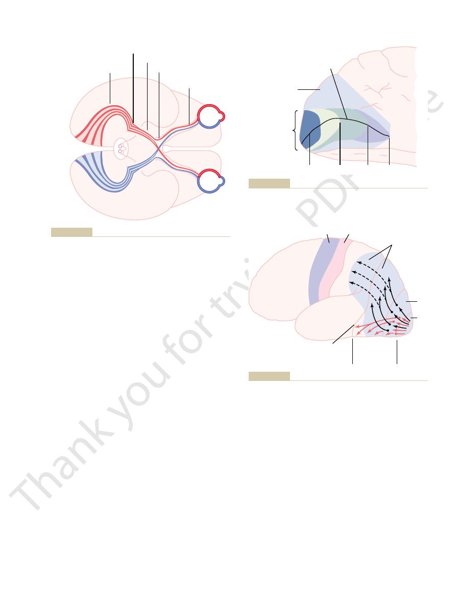
Figure 51–2) lies in the
The primary visual cortex (see
Primary Visual Cortex.
secondary visual areas.
systems, the visual cortex is divided into a
primarily on the medial aspect of the occipital lobes.
Figures 51–2 and 51–3 show the
of the Visual Cortex
Organization and Function
rather than at high velocity.
tion, but at only a moderate velocity of conduction
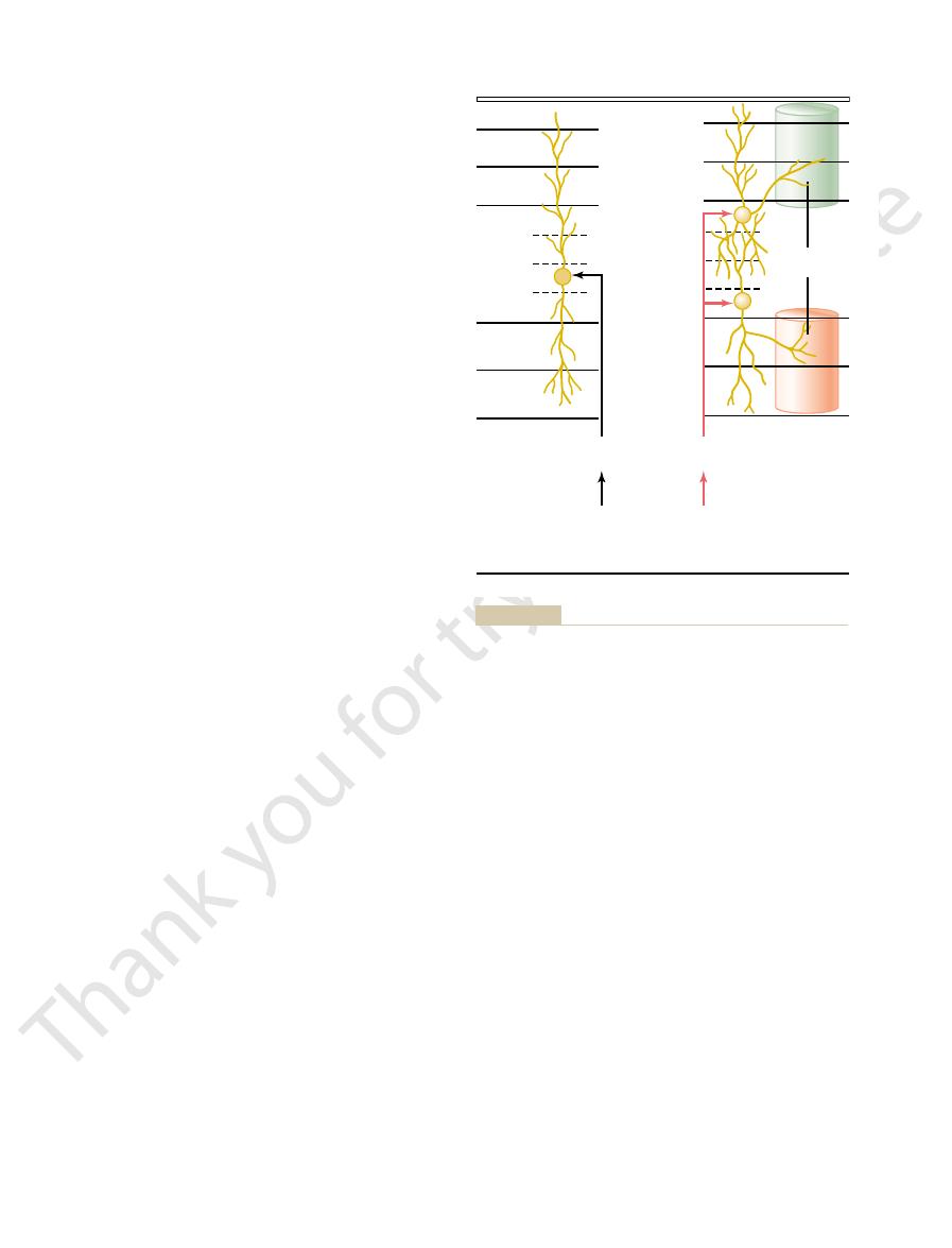
These neurons receive their input almost entirely from
large numbers of small to medium-sized neurons.
parvocellular layers
spread widely in the retina. (2) Layers III through VI
are not many Y ganglion cells, and their dendrites
transmitting only black-and-white information. Also,
the visual cortex. However, this system is color blind,
the large type Y retinal ganglion cells. This magnocel-
neurons. These receive their input almost entirely from
magnocellular layers
divided in another way: (1) Layers I and II are called
Finally, the dorsal lateral geniculate nucleus is
light the visual information that is allowed to pass.
portions of the dorsal lateral geniculate nucleus. It is
stimulated, can turn off transmission through selected
Both of these are inhibitory and, when
geniculate nucleus, and (2)
sources: (1)
much of the signal is allowed to pass to the cortex. The
signals to the visual cortex—that is, to control how
geniculate nucleus is to “gate” the transmission of
The second major function of the dorsal lateral
one another in the paired layers, and similar parallel
The Eye: III. Central Neurophysiology of Vision
Chapter 51
641
transmission is preserved all the way to the visual
cortex.
nucleus receives gating control signals from two major
corticofugal fibers returning in a backward
direction from the primary visual cortex to the lateral
reticular areas of the mes-
encephalon.
assumed that both of these gating circuits help high-
because they contain large
lular system provides a rapidly conducting pathway to
its point-to-point transmission is poor because there
are called
because they contain
the type X retinal ganglion cells that transmit color
and convey accurate point-to-point spatial informa-
visual cortex located
Like the cortical representations of the other sensory
primary
visual cortex and
calcarine fissure area, extend-
ing forward from the occipital pole on the medial
Optic radiation
Optic chiasm
Optic tract
Optic nerve
Left eye
Right eye
Visual cortex
Lateral geniculate body
Superior
colliculus
ified from Polyak SL: The Retina. Chicago: University of Chicago,
Principal visual pathways from the eyes to the visual cortex. (Mod-
Figure 51–1
1941.)
Macula
Secondary
visual areas
Calcarine fissure
Primary
visual cortex
20
∞
60
∞
90
∞
cortex.
calcarine fissure area
Visual cortex in the
Figure 51–2
of the medial occipital
Motor cortex
Somatosensory area I
Form,
3-D position,
motion
18
17
Primary
visual
cortex
Secondary
visual
cortex
Visual
detail,
color
ital lobe and the ventral portion of the posterior temporal lobe.
are transmitted mainly into the anteroventral portion of the occip-
superior portions of the occipital lobe and posterior portions of the
dimensional position, and motion are transmitted mainly into the
parietal cortices. Note that the signals representing form, third-
secondary visual areas on the lateral surfaces of the occipital and
Transmission of visual signals from the primary visual cortex into
Figure 51–3
parietal lobe. By contrast, the signals for visual detail and color
much greater distances.
layers V and VI excite neurons that transmit signals
the cortex. Conversely, the signals that pass inward to
signals that pass outward to layers I, II, and III even-
mation at successive stations along the pathway. The
inward along each vertical column unit. This process-
After the optic signals terminate in layer IV, they are
1000 or more neurons.
represents a functional unit. One can roughly calculate
motor and analytical cortical regions). Each column
a diameter of 30 to 50 micrometers. The same vertical
vertical columns of neuronal cells, each column having
The visual
Vertical Neuronal Columns in the Visual Cortex.
surface of the cortex and to deeper layers. It is these
IV, shown to the right in Figure 51–4. From there, these
, the shallowest and deepest portions of layer
ent from the Y signals. They terminate in layers IVa
retina, also terminate in layer IV, but at points differ-
nerve fibers, derived from the X ganglion cells in the
The visual signals from the medium-sized optic
levels.
, and
subdivisions. The rapidly conducted signals from the Y
mainly in layer IV. But this layer, too, is organized into
sensory systems, the geniculocalcarine fibers terminate
shown in Figure 51–4. Also, as is true for the other
the primary visual cortex has six distinct layers, as
Visual Cortex
and so forth—up to more than a dozen areas. The
ary visual areas have specific designations—V-3, V-4,
or simply V-2. The other, more distant second-
next. Therefore, Brodmann’s area 18 is called
(see Figure 51–3), which is where
Brodmann’s area 18
instance, on all sides of the primary visual cortex is
ted to these areas for analysis of visual meanings. For
shown in Figure 51–3. Secondary signals are transmit-
lateral surfaces of the occipital and parietal cortex, as
cortex. Most of these areas also fold outward over the
anterior, superior, and inferior to the primary visual
visual association areas,
areas, also called
The secondary visual
Secondary Visual Areas of the Cortex.
area has a grossly striated appearance.
. Still another name is the
The primary visual cortex is also called
area, the fovea has several hundred times as much rep-
the highest degree of visual acuity. Based on retinal
fovea transmits its signals. The fovea is responsible for
resents the macula. It is to this region that the retinal
and the lower portion inferiorly.
the calcarine fissure on the medial occipital lobe. The
occipital pole, as shown in Figure 51–2, while signals
nus of direct visual signals from the eyes. Signals from
aspect of each occipital cortex. This area is the termi-
The Nervous System: B. The Special Senses
642
Unit X
the macular area of the retina terminate near the
from the more peripheral retina terminate at or in con-
centric half circles anterior to the pole but still along
upper portion of the retina is represented superiorly
Note in the figure the especially large area that rep-
resentation in the primary visual cortex as do the most
peripheral portions of the retina.
visual area
I
striate cortex because this
lie lateral,
virtually all signals from the primary visual cortex pass
visual
area II,
importance of all these areas is that various aspects
of the visual image are progressively dissected and
analyzed.
Layered Structure of the Primary
Like almost all other portions of the cerebral cortex,
retinal ganglion cells terminate in layer IVc
a
from there they are relayed vertically both outward
toward the cortical surface and inward toward deeper
and IVc
b
signals are transmitted vertically both toward the
X ganglion pathways that transmit the accurate point-
to-point type of vision as well as color vision.
cortex is organized structurally into several million
columnar organization is found throughout the cere-
bral cortex for the other senses as well (and also in the
that each of the visual vertical columns has perhaps
further processed as they spread both outward and
ing is believed to decipher separate bits of visual infor-
tually transmit signals for short distances laterally in
“Color Blobs” in the Visual Cortex.
Interspersed among the
primary visual columns as well as among the columns
(c
b
)
I
II
III
IV
Color
“blobs”
LGN
(parvocellular)
Retinal
"X"
ganglion
Very Accurate, Color
LGN
(magnocellular)
Retinal
"Y"
ganglion
Fast, Black and White
(a)
(b)
(c
a
)
V
VI
blobs,” which are necessary for detection of color.
color. Note especially the areas of the visual cortex called “color
inate in the parvocellular layers (layers III through VI) of the LGN;
the left side of the figure originate in the magnocellular layers of
Six layers of the primary visual cortex. The connections shown on
Figure 51–4
the lateral geniculate nucleus (LGN) and transmit rapidly chang-
ing black-and-white visual signals. The pathways to the right orig-
they transmit signals that depict accurate spatial detail as well as

The visual cortex detects not only the
areas, the greater the degree of stimulation.
that is, the greater the sharpness of contrast and the
does not occur, and the intensity of stimulation of most
from dark to light or light to dark, mutual inhibition
any border in the visual scene where there is a change
retinal receptors mutually inhibit one another. But at
glion cells as well, because equally stimulated adjacent
rather than with noncontrasting areas. We saw in
in the visual scene,
Thus, the visual signal in the primary visual cortex is
occur along the sharp borders of the visual pattern.
left in Figure 51–5. To the right is shown the spatial
place on the wall a large solid cross, as shown to the
primary visual cortex detect? To answer this, let us now
fore, the question must be asked, What does the
the illumination of the wall is bright or weak. There-
visual cortex will be stimulated, regardless of whether
at a blank wall, only a few neurons in the primary
of the Visual Image
Neuronal Patterns of
it means.
mining detailed colors of objects, and deciphering
reading, determining the texture of surfaces, deter-
concerned with such visual feats as recognizing letters,
dissect out color as well. Therefore, this pathway is
detail. Separate portions of this pathway specifically
inferior, ventral,
Figure 51–3, passing from the primary visual cortex
The red arrows in
2. Analysis of Visual Detail and Color.
color.
of the retinal Y ganglion cells, transmitting rapid
pathway are mainly from the large Y optic nerve fibers
dimensional aspects of somatosensory signals. The
cortex, the signals overlap with signals from the pos-
cortex, the signals flow generally into the
whether it is moving. After leaving the primary visual
as motion in the scene. In other words, this pathway
in the space around the body. This pathway also ana-
demonstrated in Figure 51–3 by the black arrows, ana-
One of the analytical pathways,
pathways in the secondary visual areas.
cortex, the visual information is analyzed in two major
Figure 51–3 shows that after leaving the primary visual
(2) The Accurate Color Pathway
Visual Information—(1) The Fast
Two Major Pathways for Analysis of
stereopsis.
fuse with each other (be brought into “register”). The
turn, the deciphered information is used to adjust the
ding points from the two retinas fit with each other. In
register” with each other—that is, whether correspon-
two visual images from the two separate eyes are “in
nating with signals from the second eye. This cortical
eye enter the columns of every other stripe, alter-
stripe about 0.5 millimeter wide; the signals from one
is interlaced with stripes of neuronal columns, each
layer IV of the primary visual cortex. In fact, layer IV
the lateral geniculate nucleus. These signals still
eyes are relayed through separate neuronal layers in
Interaction of Visual Signals from the Two Separate Eyes.
deciphering color.
activated specifically by color signals. Therefore, it is
They receive
color blobs.
The Eye: III. Central Neurophysiology of Vision
Chapter 51
643
of some of the secondary visual areas are special
column-like areas called
lateral signals from adjacent visual columns and are
presumed that these blobs are the primary areas for
Recall that the visual signals from the two separate
remain separated from each other when they arrive in
area deciphers whether the respective areas of the
directional gaze of the separate eyes so that they will
information observed about degree of register of
images from the two eyes also allows a person to dis-
tinguish the distance of objects by the mechanism of
“Position” and “Motion” Pathway;
1. Analysis of Third-Dimensional Position, Gross Form, and
Motion of Objects.
lyzes the third-dimensional positions of visual objects
lyzes the gross physical form of the visual scene as well
tells where every object is during each instant and
posterior
midtemporal area and upward into the broad occipi-
toparietal cortex. At the anterior border of the parietal
terior somatic association areas that analyze three-
signals transmitted in this position-form-motion
signals but depicting only black and white with no
into secondary visual areas of the
and
medial regions of the occipital and temporal cortex,
show the principal pathway for analysis of visual
from all this information what the object is and what
Stimulation During Analysis
Analysis of Contrasts in the Visual Image.
If a person looks
pattern of the most excited neurons in the visual
cortex. Note that the areas of maximum excitation
concerned mainly with contrasts
Chapter 50 that this is true of most of the retinal gan-
neurons is proportional to the gradient of contrast—
greater the intensity difference between light and dark
Visual Cortex Also Detects Orientation of Lines and Borders—
“Simple” Cells.
existence of lines and borders in the different areas of
the retinal image but also the direction of orientation
Retinal image
Cortical stimulation
to a retinal image of a dark cross.
Pattern of excitation that occurs in the visual cortex in response
Figure 51–5

or excessive use of tobacco.
retina, or from toxic conditions such as lead poisoning
pressure in the eyeball), from allergic reactions in the
than the optic disc area. Such blind spots are called
Occasionally, blind
in the figure.
degrees lateral to the central point of vision, as shown
try charts, a
left eye is plotted as shown in Figure 51–6. In all perime-
seen and when it cannot. Thus, the field of vision for the
and forth in all areas of the field of vision, and the
Then a small dot of light or a small object is moved back
looking toward a central spot directly in front of the eye.
This is done by having the
perimetry.
retina, one charts the field of vision for each eye by a
To diagnose blindness in specific portions of the
given instant. The area seen to the nasal side is called
is the visual area seen by an eye at a
The
Fields of Vision; Perimetry
head, and avoidance. This vision is believed to be sub-
These reactions include turning the eyes, turning the
scene, or, rarely, even to some gross patterns of vision.
to changes in light intensity, to movement in the visual
“blind” people can still, at times, react subconsciously
However, psychological studies demonstrate that such
being causes loss of conscious vision, that is, blindness.
Visual Cortex
Effect of Removing the Primary
detected by complex and hypercomplex cells.
simple cells, whereas more complex contrasts are
colors,” excite specific neuronal cells. It is presumed
on the fact that contrasting colors, called “opponent
The mechanism of color contrast analysis depends
has changed the color entering the eyes.
the color of the “white” changes with the light, and
that is, when the color of an illuminating light changes,
sible for the phenomenon called “color constancy”;
white area within the visual scene. In fact, this con-
area. All these colors can also be contrasted against a
area against a red area, or a green area against a yellow
red area is often contrasted against a green area, a blue
detected: by means of color contrast. For instance, a
Detection of Color
ical pathway of the visual cortex, progressively more
visual scene. Thus, as one goes farther into the analyt-
that have other characteristics. That is, these neurons
cific lengths, by specific angulated shapes, or by images
areas, are stimulated only by lines or borders of spe-
columns, as well as neurons in some secondary visual
complex cells.
be stimulated if the line has the same direction. These
or vertically in the field, the same few neurons will still
same direction but are not position-specific. That is,
the visual signal progresses farther away from layer IV,
ally or Vertically in the Visual Field—“Complex” Cells.
They are found mainly in layer IV of the primary
simple cells.
set of cells. These neuronal cells are called
tion of a line, specific neuronal cells are stimulated. A
there is a contrast edge. Thus, for each such orienta-
horizontal or lies at some degree of inclination. This is
of each line or border—that is, whether it is vertical or
The Nervous System: B. The Special Senses
644
Unit X
believed to result from linear organizations of mutu-
ally inhibiting cells that excite second-order neurons
when inhibition occurs all along a line of cells where
line oriented in a different direction excites a different
visual cortex.
Detection of Line Orientation When a Line Is Displaced Later-
As
some neurons respond to lines that are oriented in the
even if a line is displaced moderate distances laterally
cells are called
Detection of Lines of Specific Lengths, Angles, or Other Shapes.
Some neurons in the outer layers of the primary visual
detect still higher orders of information from the
characteristics of each visual scene are deciphered.
Color is detected in much the same way that lines are
trasting against white is believed to be mainly respon-
appropriate computation in the brain allows red to be
interpreted as red even though the illuminating light
that the initial details of color contrast are detected by
Removal of the primary visual cortex in the human
served by neuronal pathways that pass from the optic
tracts mainly into the superior colliculi and other por-
tions of the older visual system.
field of vision
the nasal field of vision, and the area seen to the lateral
side is called the temporal field of vision.
process called
subject look with one eye closed and the other eye
subject indicates when the spot of light or object can be
blind spot caused by lack of rods and cones
in the retina over the optic disc is found about 15
Abnormalities in the Fields of Vision.
spots are found in portions of the field of vision other
sco-
tomata; they frequently are caused by damage to the
optic nerve resulting from glaucoma (too much fluid
60
70
50
60
40
50
30
40
20
10
30
90
80
70
60
50
80
70
60
50
40
40
30
30
20
20
10
10
20
80
70
80
75
45
15
345
330
315
300
285
255
270
240
225
210
195
180
165
150
135
Left
Optic
disc
Right
120
105
60
30
0
Perimetry chart, showing the field of vision for the left eye.
Figure 51–6

tical eye fields automatically “lock” the eyes on a given
To summarize, posterior “involuntary” occipital cor-
or may become totally unable to do so.
bilaterally in an animal, the animal has difficulty
visual cortex. When this fixation area is destroyed
eyes to “lock” on the object of attention once it is
Conversely, the fixation mechanism that causes the
a short time, which then allows the eyes to be moved.
and move them to another point. It is usually neces-
person to “unlock” the eyes from one point of fixation
51–8. Bilateral dysfunction or destruction of these
tical regions of the frontal lobes, as shown in Figure
The voluntary fixation movements are controlled by
involuntary fixation mechanism.
eyes firmly on the object once it has been found; this
The second is an involuntary mechanism that holds the
voluntary fixation mechanism.
vision; this is called the
find the object on which he or she wants to fix the
controlled by two neuronal mechanisms. The first of
portion of the field of vision. Fixation movements are
are those that cause the eyes to “fix” on a discrete
Fixation Movements of the Eyes
mitted from the body’s equilibrium control centers in
of the oculomotor nerves. Strong signals are also trans-
the pretectal and the superior colliculus areas, the ocu-
superior colliculus areas of the brain stem. From both
oculomotor apparatus, showing spread of signals from
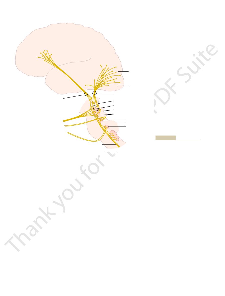
Figure 51–8 demonstrates cortical control of the
pair relaxes while the other contracts.
fasciculus.
peripheral nerves to the ocular muscles. Shown, too,
also shows brain stem nuclei for the third, fourth, and
Figure 51–7
downward. The oblique muscles function mainly to
to move the eyes from side to side. The superior and
The medial and lateral recti contract
inferior obliques.
Figure 51–7: (1) the
are controlled by three pairs of muscles, shown in
The eye movements
To make full use of the visual abilities of the eyes,
Their Control
opposite side of the head. This condition is known as
lesion; as a result, neither eye can see objects to the
the optical system of the eye; this condition is called
retina is blinded, which means that the person is blind
opposite optic tract. Therefore, the nasal half of each
optic chiasm
blindness of the affected eye.
Vision.
Effect of Lesions in the Optic Pathway on the Fields of
central areas.
pigment deposits in the degenerated areas. Retinitis pig-
tions of the retina degenerate, and excessive melanin
In this disease, por-
The Eye: III. Central Neurophysiology of Vision
Chapter 51
645
Still another condition that can be diagnosed by
perimetry is retinitis pigmentosa.
mentosa usually causes blindness in the peripheral field
of vision first and then gradually encroaches on the
Destruction of an entire optic nerve causes
Destruction of the
prevents the crossing
of impulses from the nasal half of each retina to the
in the temporal field of vision for each eye because the
image of the field of vision is inverted on the retina by
bitemporal hemianopsia. Such lesions frequently result
from tumors of the pituitary gland pressing upward
from the sella turcica on the bottom of the optic chiasm.
Interruption of an optic tract denervates the corre-
sponding half of each retina on the same side as the
homonymous hemianopsia.
Eye Movements and
almost equally as important as interpretation of the
visual signals from the eyes is the cerebral control
system for directing the eyes toward the object to be
viewed.
Muscular Control of Eye Movements.
medial and lateral recti, (2) the
superior and inferior recti, and (3) the superior and
inferior recti contract to move the eyes upward or
rotate the eyeballs to keep the visual fields in the
upright position.
Neural Pathways for Control of Eye Movements.
sixth cranial nerves and their connections with the
are interconnections among the brain stem nuclei by
way of the nerve tract called the medial longitudinal
Each of the three sets of muscles to each
eye is reciprocally innervated so that one muscle of the
visual areas in the occipital cortex through occipito-
tectal and occipitocollicular tracts to the pretectal and
lomotor control signals pass to the brain stem nuclei
the brain stem into the oculomotor system (from the
vestibular nuclei by way of the medial longitudinal
fasciculus).
Perhaps the most important movements of the eyes
these allows a person to move the eyes voluntarily to
is called the
a cortical field located bilaterally in the premotor cor-
areas makes it difficult or almost impossible for a
sary to blink the eyes or put a hand over the eyes for
found is controlled by secondary visual areas in the
occipital cortex, located mainly anterior to the primary
keeping its eyes directed toward a given fixation point
Lateral
rectus
Inferior oblique
Medial
longitudinal
fasciculus
Superior oblique
Nuclei
N.III
N.IV
N.VI
Superior rectus
Inferior rectus
Medial rectus
Figure 51–7
Extraocular muscles of the eye and their innervation.
important information. Similar saccades occur when a
the visual scene is not moving past the eyes, but the
cadic movements of the eyes for each line. In this case,
process of reading, a person usually makes several sac-
presses the visual image during saccades, so that the
allocated to the fixation sites. Also, the brain sup-
moving the eyes, with 90 per cent of the time being
The saccades occur so rapidly that no
movements.
saccades,
of two to three jumps per second. The jumps are called
the visual field, jumping from one to the next at a rate
in a car, the eyes fix on one highlight after another in
ually before the eyes, such as when a person is riding
When a visual scene is moving contin-
Saccadic Movement of the Eyes—A Mechanism of Successive
region. This involuntary fixation capability is mostly
strated in Figure 51–9, which shows by the dashed lines
These drifting and flicking motions are demon-
center of the fovea.Thus, an automatic response moves
reaction occurs, producing a flicking movement that
drifts as far as the edge of the fovea, a sudden reflex
to drift slowly across the cones. Each time the spot
the cones, and the drifting movements cause the spot
region of the retina, the tremulous movements cause
When a spot of light has become fixed on the foveal
flicking move-
direction or another, and (3) sudden
slow drift
ocular muscles, (2) a
at a rate of 30 to 80 cycles per second caused
almost imperceptible movements: (1) a
The eyes normally have three types of continuous but
The involuntary locking type of fixation
Mechanism of Involuntary Locking Fixation—Role of the Supe-
cortices.
cortical “voluntary” eye fields located in the frontal
fixation, voluntary signals must be transmitted from
of the image across the retinas. To unlock this visual
The Nervous System: B. The Special Senses
646
Unit X
spot of the visual field and thereby prevent movement
rior Colliculi.
discussed in the previous section results from a nega-
tive feedback mechanism that prevents the object of
attention from leaving the foveal portion of the retina.
continuous
tremor
by successive contractions of the motor units in the
of the eyeballs in one
ments that are controlled by the involuntary fixation
mechanism.
the spot to move back and forth at a rapid rate across
moves the spot away from this edge back toward the
the image back toward the central point of vision.
the slow drifting across the fovea and by the solid lines
the flicks that keep the image from leaving the foveal
lost when the superior colliculi are destroyed.
Fixation Points.
and the movements are called opticokinetic
more than 10 per cent of the total time is spent in
person is not conscious of the movements from point
to point.
Saccadic Movements During Reading.
During the
eyes are trained to move by means of several succes-
sive saccades across the visual scene to extract the
Visual
association
areas
Primary
visual cortex
Occipitotectal and
occipitocollicular tracts
Pretectal nuclei
Visceral nucleus III nerve
Superior colliculus
Oculomotor nucleus
Frontotectal tract
Voluntary fixation
area
Involuntary
fixation
area
III nerve
IV nerve
VI nerve
Medial longitudinal
Trochlear nucleus
Abducens nucleus
Vestibular nuclei
Neural pathways for control of
Figure 51–8
conjugate movement of the eyes.

ter at the same time. Furthermore, the nearer the
each other, it is still impossible for all corresponding
ity. Therefore, even when the two eyes are fused with
side, and the closer the object, the greater the dispar-
object, and the left eye a little more of the left-hand
two retinas are not exactly the same. That is, the right
eyes are more than 2 inches apart, the images on the
In Chapter 49, it is pointed out that because the two
Distances of Visual Objects
Neural Mechanism of Stereopsis for Judging
visual cortex disappears.
excitation of the specific “interference” neurons in the
responding points of the two retinas are in register,
eyes so that fusion can be re-established. Once the cor-
“fused.” This excitation presumably provides the
images are not “in register”—that is, are not precisely
neurons in the visual cortex. Interactions occur
body, and these signals in turn are relayed to parallel
The visual cortex plays an important role in fusion. It
other on “corresponding points” of the two retinas.
To make the visual perceptions more meaningful, the
the Two Eyes
of the Visual Images from
visual, auditory, or somatic.
respect to external disturbances, whether they are
global role in orienting the eyes, head, and body with
liculi are intact. Therefore, the superior colliculi play a
of the eyes, head, and body, but only if the superior col-
stroking of the side of the body, cause similar turning
visual disturbances, such as strong sounds or even
the direction of the disturbance. Other types of non-
visual disturbance, signals are relayed from the supe-
visual pathway, but their function is unclear.)
type W optic nerve fibers. These represent the oldest
the other going to the superior colliculi. (The superior
fibers, with one branch going to the visual cortex and
ments are branches from the rapidly conducting Y
The optic nerve fibers from the eyes to the colliculi
from the body and acoustic signals from the ears.
directional movement of the eyes, the superior colli-
motor nuclei to turn the eyes. To help in this
racy. Even so, the principal direction of a flash of light
in the primary visual cortex, although with less accu-
have also been destroyed. To support this function, the
direction. This does not occur if the superior colliculi
Even after the visual cortex has been destroyed, a
Visual Disturbance
for Turning the Eyes and Head Toward a
Superior Colliculi Are Mainly Responsible
pursuit system for controlling eye movements.
almost exactly. This represents a high degree of auto-
seconds, the eyes develop progressively smoother
ment as that of the object. Then, after another few
so, the eyes begin to jump by means of saccades in
be unable to fixate on it. However, after a second or
rate of several times per second, the eyes at first may
course of movement for the eyes. For instance, if an
pursuit movement.
can also remain fixed on a moving object, which is
The eyes
painting to another, and so forth.
occur in upward, sideways, downward, and angulated
person observes a painting, except that the saccades
The Eye: III. Central Neurophysiology of Vision
Chapter 51
647
directions one after another from one highlight of the
Fixation on Moving Objects—“Pursuit Movement.”
called
A highly developed cortical
mechanism automatically detects the course of move-
ment of an object and then rapidly develops a similar
object is moving up and down in a wavelike form at a
approximately the same wavelike pattern of move-
movements and finally follow the wave movement
matic subconscious computational ability by the
sudden visual disturbance in a lateral area of the visual
field often causes immediate turning of the eyes in that
various points of the retina are represented topo-
graphically in the superior colliculi in the same way as
in a peripheral retinal field is mapped by the colliculi,
and secondary signals are transmitted to the oculo-
culi also have topological maps of somatic sensations
that are responsible for these rapid turning move-
colliculi and other regions of the brain stem are also
strongly supplied with visual signals transmitted in
In addition to causing the eyes to turn toward a
rior colliculi through the medial longitudinal fascicu-
lus to other levels of the brain stem to cause turning
of the whole head and even of the whole body toward
“Fusion”
visual images in the two eyes normally fuse with each
was pointed out earlier in the chapter that correspon-
ding points of the two retinas transmit visual signals to
different neuronal layers of the lateral geniculate
between these cortical neurons to cause interference
excitation in specific neurons when the two visual
signal that is transmitted to the oculomotor apparatus
to cause convergence or divergence or rotation of the
eye sees a little more of the right-hand side of the
points in the two visual images to be exactly in regis-
Voluntary
movement to
fixation site
American Physiological Society, 1960.)
VE (eds): Handbook of Physiology. vol. 2, sec. 1. Washington, DC,
Central control of the eye movements. In Field J, Magoun HW, Hall
resent sudden flicking movements.) (Modified from Whitteridge D:
lines represent slow drifting movements, and the solid lines rep-
ing” eye movements that move the spot back toward the center
Figure 51–9
Movements of a spot of light on the fovea, showing sudden “flick-
of the fovea whenever it drifts to the foveal edge. (The dashed
until they reach the eye. There, the sympathetic fibers
with postganglionic neurons. Postganglionic sympa-
segment of the spinal cord. From there, sympathetic
The sympathetic innervation of the eye originates in
into the eyeball. These nerves excite (1)
pathetic neurons, which in turn send fibers through
which lies immediately behind the eye. There, the pre-
third nerve
Edinger-Westphal nucleus
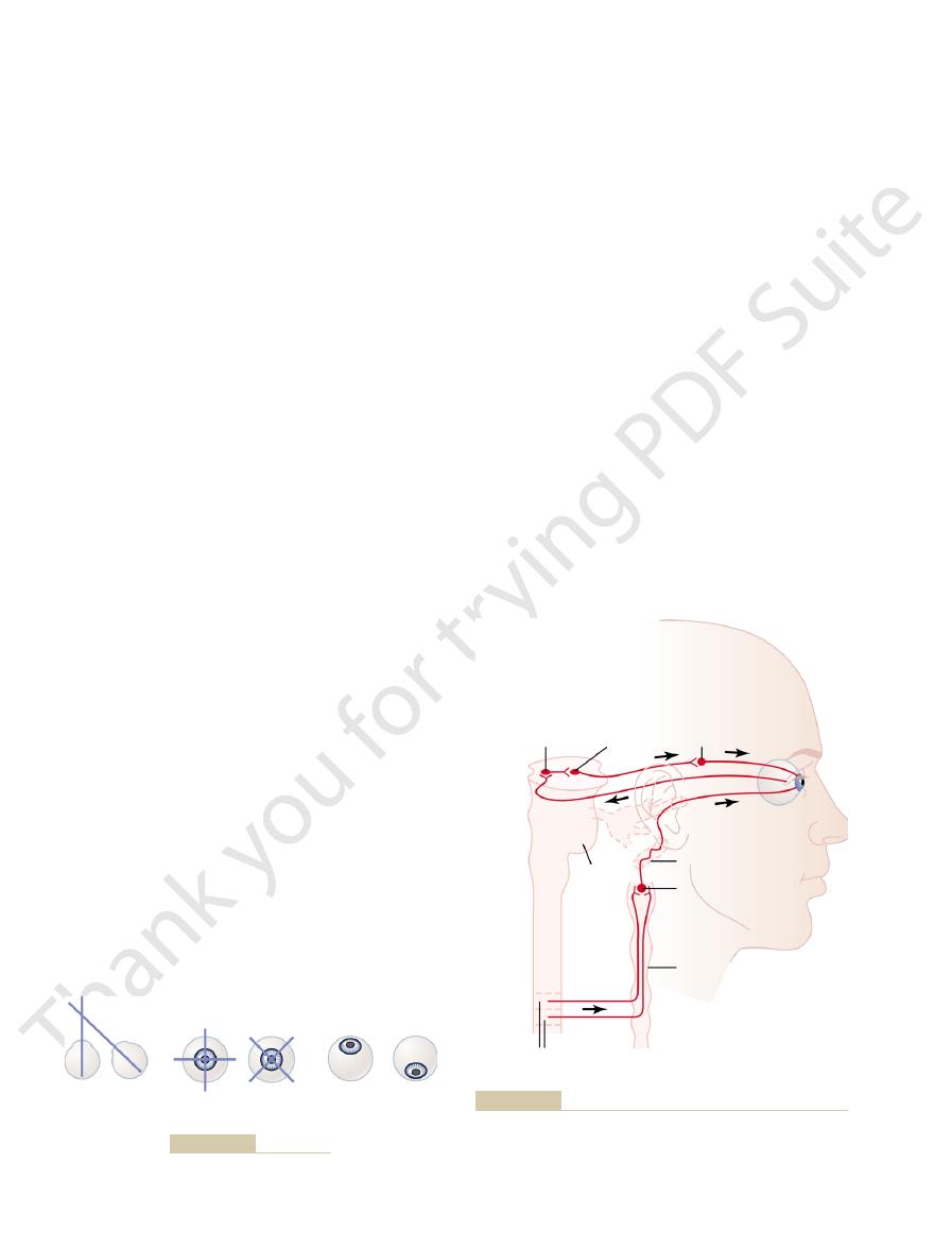
shown in Figure 51–11. The parasympathetic pregan-
both parasympathetic and sympathetic nerve fibers, as
The eye is innervated by
Autonomic Nerves to the Eyes.
Pupillary Aperture
Autonomic Control of
normally receive signals from the repressed eye.
fact, even anatomically, the numbers of neuronal con-
nervous system synaptic connections from the eyes. In
young children. This demonstrates that visual acuity is
becomes blinded, vision in the repressed eye can
remaining 20/400 or less. If the dominant eye then
the repressed eye develops only slightly, sometimes
and is never used for precise vision. The visual acuity of
is used all the time, and the other eye becomes repressed
the object of attention. In other patients, one eye alone
patients with strabismus, the eyes alternate in fixing on
Suppression of the Visual Image from a Repressed Eye.
never fuse.
neuronal control pathways themselves, so that the eyes
movements of the eyes become abnormally “set” in the
never simultaneously. Soon the patterns of conjugate
the other fails do so, or they both fixate satisfactorily but
same object, one of the eyes fixates satisfactorily while
young child’s early efforts to fixate the two eyes on the
fusion mechanism of the visual system. That is, in a
Strabismus is often caused by abnormal “set” of the
of the different types of strabismus often occur.
tical strabismus.
torsional strabismus,
izontal strabismus,
types of strabismus are shown in Figure 51–10: (1)
dinates: horizontal, vertical, or rotational. The basic
cross-eye,
Strabismus, also called
for stereopsis.
are excited by nonregister or register. This phenome-
in register for objects 25 meters away. Thus, the dis-
objects 2 meters away; still another set of pathways is
each side of the central pathway. Therefore, some optic
The neuronal cellular mechanism for stereopsis is
stereopsis,
object is to the eyes, the less the degree of register. This
The Nervous System: B. The Special Senses
648
Unit X
degree of nonregister provides the neural mechanism
for
an important mechanism for judging
the distances of visual objects up to about 200 feet
(60 meters).
based on the fact that some of the fiber pathways from
the retinas to the visual cortex stray 1 to 2 degrees on
pathways from the two eyes are exactly in register for
tance is determined by which set or sets of pathways
non is called depth perception, which is another name
Strabismus
squint or
means lack
of fusion of the eyes in one or more of the visual coor-
hor-
(2)
and (3) ver-
Combinations of two or even all three
In a few
develop only to a slight extent in adults but far more in
highly dependent on proper development of central
nections diminish in the visual cortex areas that would
Accommodation and
glionic fibers arise in the
(the visceral nucleus portion of the third cranial nerve)
and then pass in the
to the ciliary ganglion,
ganglionic fibers synapse with postganglionic parasym-
ciliary nerves
the ciliary muscle that controls focusing of the eye lens
and (2) the sphincter of the iris that constricts the
pupil.
the intermediolateral horn cells of the first thoracic
fibers enter the sympathetic chain and pass upward to
the superior cervical ganglion, where they synapse
thetic fibers from these then spread along the surfaces
of the carotid artery and successively smaller arteries
Horizontal
strabismus
Torsional
strabismus
Vertical
strabismus
Figure 51–10
Basic types of strabismus.
Pretectal
region
Edinger-
Westphal
nucleus
Upper thoracic segments
of spinal cord
Ciliary
ganglion
N.III
N.II
Pons
Carotid plexus
Superior cervical
sympathetic
ganglion
Cervical
sympathetic
trunk
the light reflex. (Modified from Ranson SW, Clark SL: Anatomy of
Autonomic innervation of the eye, showing also the reflex arc of
Figure 51–11
the Nervous System: Its Development and Function, 10th ed.
Philadelphia: WB Saunders, 1959.)

the same time. This is called the
near object, the signals that cause accommodation of
other pathway. For instance, when the eyes fixate on a
Edinger-Westphal nucleus is stimulated through some
Yet the pupils can constrict a little more if the
pupils to remain mostly constricted, in addition to their
lost, the nucleus becomes chronically active, causing the
of the inhibitory type. When their inhibitory effect is
tectal area to the Edinger-Westphal nucleus are mostly
The final nerve fibers in the pathway through the pre-
nerves.
pretectal region of the brain stem, although it can result
and so forth.The block usually occurs in the
encephalitis,
central nervous system syphilis, alcoholism,
the pupillary reflexes. Such blocks frequently occur as a
the Edinger-Westphal nucleus, thus sometimes blocking
Pupillary Reflexes or Reactions in Central Nervous System
change in the amount of light entering the eye.
is about 30 to 1—that is, up to as much as 30 times
of pupillary diameter, the range of light and dark adap-
and 8 millimeters on the large side. Therefore, because
as explained in Chapter 50. The limits of pupillary
adapt extremely rapidly to changing light conditions,
The function of the light reflex is to help the eye
ited, which results in dilation of the pupil.
iris. Conversely, in darkness, the reflex becomes inhib-
and, finally, back through
Edinger-Westphal nucleus
tal nuclei. From here, secondary impulses pass to the
When light impinges on the retina, a few of the result-
strated by the upper two black traces in Figure 51–11.
The neuronal pathway for this reflex is demon-
the pupils constrict, a reaction called the
When light is shone into the eyes,
Pupillary Light Reflex.
mydriasis.
Conversely,
miosis.
pupillary aperture; this is called
the pupillary sphincter muscle, thereby decreasing the
Control of Pupillary Diameter
parasympathetic nerve fibers to the eyes.
Edinger-Westphal nucleus, and finally by way of
pretectal area in the brain stem, then through the
Brodmann’s cortical areas 18 and 19 and transmission
ments of the eyes, with analysis of the visual signals in
The brain cortical areas that control accommoda-
provide appropriate focus.
direction. This could give a rapid clue as to which
The visual image becomes clearer when the
the time at a frequency up to twice per second.
4. It has been found that
is different from the clarity of focus on the edges.
retina, the clarity of focus in the depth of the fovea
Because the fovea lies in a hollowed-out depression
strengthen the lens of the eye.
must converge. The neural mechanisms for
2. When the eyes fixate on a near object, the eyes
lens stronger or weaker.
focus, and this clue relays information to the
more than red rays. The eyes appear to be able to
That is, red light rays focus slightly posteriorly to
a fraction of a second. Second, different types of
fixation point, the lens changes its strength in the
First, when the eyes suddenly change distance of the
some of the known features are the following.
this rapid and accurate focusing of the eye is unclear,
focus on a near object, the lens usually accommodates
degree of visual acuity. When the eyes have been
causes decreased power. How does a person adjust
of the lens, as explained in Chapter 49, and relaxation
muscle. Contraction causes increased refractive power
tial for a high degree of visual acuity. Accommodation
The accommodation mechanism—that is, the mecha-
Control of Accommodation
Horner’s syndrome.
pupil) as well as several extraocular muscles of the eye,
The Eye: III. Central Neurophysiology of Vision
Chapter 51
649
innervate the radial fibers of the iris (which open the
which are discussed subsequently in relation to
(Focusing the Eyes)
nism that focuses the lens system of the eye—is essen-
results from contraction or relaxation of the eye ciliary
accommodation to keep the eyes in focus all the time?
Accommodation of the lens is regulated by a nega-
tive feedback mechanism that automatically adjusts
the refractive power of the lens to achieve the highest
focused on some far object and must then suddenly
for best acuity of vision within less than 1 second.
Although the precise control mechanism that causes
proper direction to achieve a new state of focus within
clues help to change the lens strength in the proper
direction:
1. Chromatic aberration appears to be important.
blue light rays because the lens bends blue rays
detect which of these two types of rays is in better
accommodation mechanism whether to make the
convergence cause a simultaneous signal to
3.
that is slightly deeper than the remainder of the
It
has been suggested that this also gives clues about
which way the strength of the lens needs to be
changed.
the degree of
accommodation of the lens oscillates slightly all
oscillation of the lens strength is changing in
the appropriate direction and becomes poorer
when the lens strength is changing in the wrong
way the strength of the lens needs to change to
tion closely parallel those that control fixation move-
of motor signals to the ciliary muscle through the
Stimulation of the parasympathetic nerves also excites
stimulation of the sympathetic nerves excites the
radial fibers of the iris and causes pupillary dilation,
called
pupillary light
reflex.
ing impulses pass from the optic nerves to the pretec-
parasympathetic nerves to constrict the sphincter of the
diameter are about 1.5 millimeters on the small side
light brightness on the retina increases with the square
tation that can be brought about by the pupillary reflex
Disease.
A few central nervous system diseases damage
nerve transmission of visual signals from the retinas to
result of
from destruction of some small fibers in the optic
failure to respond to light.
the lens and those that cause convergence of the two
eyes cause a mild degree of pupillary constriction at
pupillary reaction to

low can you go? Curr Biol 13:R840, 2003.
van Wezel RJ, van der Smagt MJ: Motion processing: how
saliency. Curr Opin Neurobiol 13:428, 2003.
Treue S: Visual attention: the where, what, how and why of
control by the cerebral cortex. Curr Opin Neurol 17:17,
Pierrot-Deseilligny C, Milea D, Muri RM: Eye movement
5:218, 2004.
the voluntary control of eye movement. Nat Rev Neurosci
Munoz DP, Everling S: Look away: the anti-saccade task and
Neurosci 5:229, 2004.
fixational eye movements in visual perception. Nat Rev
Martinez-Conde S, Macknik SL, Hubel DH: The role of
Biol 13:R906, 2003.
Mante V, Carandini M: Visual cortex: seeing motion. Curr
York: Oxford University Press, 1999.
Leigh RJ, Lee DS: The Neurology of Eye Movements. New
system. J Neurophysiol 91:591, 2004.
Krauzlis RJ: Recasting the smooth pursuit eye movement
in the human cortex. Annu Rev Neurosci 23:315, 2000.
Kastner S, Ungerleider LG: Mechanisms of visual attention
ments. Physiol Rev 80:953, 2000.
Hikosaka O, Takikawa Y, Kawagoe R: Role of the basal
tion. New York: Springer, 1997.
Hendee WA, Wells PNT: The Perception of Visual Informa-
selectivity in the visual cortex. Annu Rev Neurosci 23:441,
Ferster D, Miller KD: Neural mechanisms of orientation
wired up to the cortex? Curr Biol 14:R14, 2004.
Derrington AM, Webb BS: Visual system: how is the retina
cepts of orbital anatomy. Ann N Y Acad Sci 956:17, 2002.
Demer JL: The orbital pulley system: a revolution in con-
retina. Annu Rev Neurosci 23:743, 2000.
Dacey DM: Parallel pathways for spectral coding in primate
Neurobiol 13:655, 2003.
of three-dimensional eye and head movements. Curr Opin
Crawford JD, Martinez-Trujillo JC, Klier EM: Neural control
Eye Res 23:31, 2004.
pathways and retinogeniculate projections. Prog Retin
Chalupa LM, Gunhan E: Development of on and off retinal
Acad Sci 1004:40, 2003.
sensory innervation of extraocular eye muscles. Ann N Y
Buttner-Ennever JA, Eberhorn A, Horn AK: Motor and
14:R195, 2004.
Burr D: Eye movements: keeping vision stable. Curr Biol
Research. New York: Plenum Press, 1999.
Becker W, Reubel H, Mergner T: Current Oculomotor
Walter de Gruyter, 1998.
Backharis W, Kliegl R, Werner JS: Color Vision. Berlin:
affected by Horner’s syndrome.
Fourth, sweating (which requires sympathetic nerve
normally. Third, the blood vessels on the corresponding
Therefore, destruction of the sympathetic nerves makes
superior eyelid and innervated by the sympathetics.
diameter than the pupil of the opposite eye. Second, the
sympathetic nerve fibers to the pupillary dilator muscle,
of the following effects: First, because of interruption of
Horner’s syndrome,
in the cervical sympathetic chain. This causes the clini-
occasionally interrupted. Interruption frequently occurs
The sympathetic nerves to the eye are
Horner’s Syndrome.
syphilis.
The Nervous System: B. The Special Senses
650
Unit X
accommodation. A pupil that fails to respond to light
but does respond to accommodation and is also very
small (an Argyll Robertson pupil) is an important diag-
nostic sign of central nervous system disease—often
cal condition called
which consists
the pupil remains persistently constricted to a smaller
superior eyelid droops because it is normally main-
tained in an open position during waking hours partly
by contraction of smooth muscle fibers embedded in the
it impossible to open the superior eyelid as widely as
side of the face and head become persistently dilated.
signals) cannot occur on the side of the face and head
References
2000.
ganglia in the control of purposive saccadic eye move-
2004.