leaving the lens.) Finally, if the lens has exactly the proper curvature, parallel light
the rays enter the lens, and half as they exit from the opposite side. (At this time,
of the rays. Half the bending occurs when
center, which is called
sively more angulated interface. The outer rays bend more and more toward the
refracted. Toward either edge of the lens, however, the light rays strike a progres-
pendicular to the lens surface and, therefore, pass through the lens without being
lens. The light rays passing through the center of the lens strike the lens exactly per-
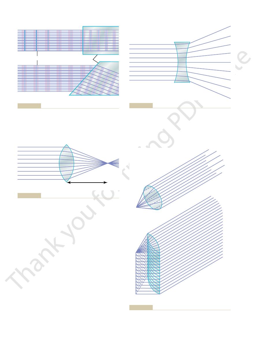
Figure 49–2 shows parallel light rays entering a convex
Application of Refractive Principles to Lenses
This bending of light rays at an angulated interface is known as
the direction in which light travels is always
tical but angulated to the right. Because
front to move ahead of the lower portion, so that the wave front is no longer ver-
travels at a velocity of 200,000 km/sec. This causes the upper portion of the wave
tinues to travel at a velocity of 300,000 km/sec, while that which entered the glass
glass ahead of the upper edge. The wave front in the upper portion of the beam con-
beam first strikes the angulated interface, the lower edge of the beam enters the
of 1.00, and are entering a block of glass having a refractive index of 1.50. When the
In this particular figure, the light rays are leaving air, which has a refractive index
the rays bend if the refractive indices of the two media are different from each other.
If the light rays pass through an angulated interface as shown in Figure 49–1
distances between wave fronts.
ity of transmission and shorter wavelength, as shown in the figure by the shorter
without deviating from their course. The only effect that occurs is decreased veloc-
to the beam, the rays enter the second medium
When light rays traveling forward in a beam (as shown in Figure 49–1
Refraction of Light Rays at an Interface Between Two Media with Different Refractive Indices.
200,000 km/sec, the refractive index of this glass is 300,000 divided by 200,000, or
itself is 1.00. Thus, if light travels through a particular type of glass at a velocity of
velocity of light in air to the velocity in the substance. The refractive index of air
and liquids. The refractive index of a transparent substance is the
about 300,000 km/sec, but they travel much slower through transparent solids
Refractive Index of a Transparent Substance.
physical principles is presented; then the optics of the eye is discussed.
depth of focus, and so forth. A brief review of these
including the physics of light refraction, focusing,
oughly familiar with the basic principles of optics,
system of the eye, the student must first be thor-
of Optics
The Eye: I. Optics of Vision
C
H
A
P
T
E
R
4
9
613
Physical Principles
Before it is possible to understand the optical
Refraction of Light
Light rays travel through air at a velocity of
ratio of the
1.50.
A) strike an
interface that is perpendicular
B,
perpendicular to the plane of the wave front, the direction of travel of the light beam
bends downward.
refraction. Note
particularly that the degree of refraction increases as a function of (1) the ratio of
the two refractive indices of the two transparent media and (2) the degree of angu-
lation between the interface and the entering wave front.
Convex Lens Focuses Light Rays.
convergence
the student should pause and analyze why the rays bend toward the center on
Conversely, light
focal line.
bending occurs in one plane but not the other. Thus, par-
of the lens but not from the top or the bottom. That is,
the cylindrical lens bends light rays from the two sides
lens. Note that
lens and a convex
Figure 49–4 shows both a convex
light rays.
light rays, but the convex lens
the center of the lens. Thus, the concave lens
effect in the convex lens, and it causes the peripheral
ahead of the rays in the center. This is opposite to the
refract. The rays at the edge of the lens enter the lens
is perpendicular to the beam and, therefore, do not
effect of a concave lens on parallel light rays. The rays
Figure 49–3 shows the
single point, which is called the
The Nervous System: B. The Special Senses
614
Unit X
rays passing through each part of the lens will be bent
exactly enough so that all the rays will pass through a
focal point.
Concave Lens Diverges Light Rays.
that enter the center of the lens strike an interface that
light rays to diverge from the light rays that pass through
diverges
converges
Cylindrical Lens Bends Light Rays in Only One Plane—Comparison
with Spherical Lenses.
spherical
cylindrical
allel light rays are bent to a
rays that pass through the spherical lens are refracted
Wave fronts
A
B
Glass
that light rays striking an angulated glass surface are bent.
the glass is shortened to about two thirds that in air. It also shows
. This figure
and a glass surface angulated to the light rays
Light rays entering a glass surface perpendicular to the light rays
Figure 49–1
(A)
(B)
demonstrates that the distance between waves after they enter
Light from
distant source
Focal length
showing that parallel light rays are focused to a
Bending of light rays at each surface of a convex spherical lens,
Figure 49–2
focal point.
Light from
distant source
showing that parallel light rays are
Bending of light rays at each surface of a concave spherical lens,
Figure 49–3
diverged.
A
B
B, Line focus
A, Point focus
Figure 49–4
of parallel light rays by a spherical convex lens.
of parallel light rays by a cylindrical convex lens.
the lens.
lens, and
is the focal length of the lens for parallel rays,
point source of light, and distance of focus is expressed
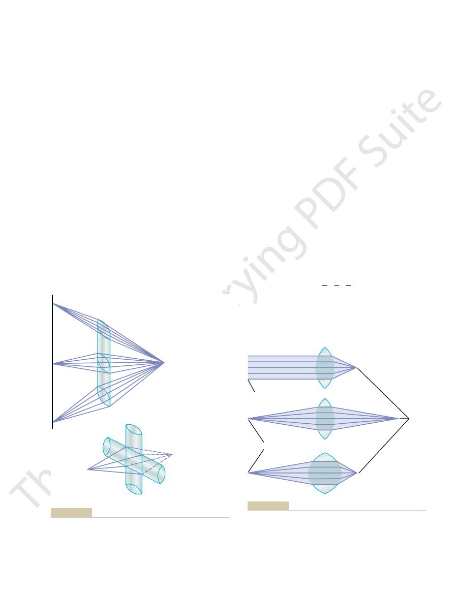
The relation of focal length of the lens, distance of the
provided the lens changes its convexity.
rays can be focused at the same distance beyond a lens,
This demonstrates that both parallel rays and diverging
as that from the lens in the first diagram, in which the
the figure. In this diagram, the distance from the lens at
The bottom diagram of Figure 49–6 shows light rays
for parallel rays.
of light that are already diverging enter a convex lens,
the lens as do parallel rays. In other words, when rays
from the point source, it can be seen from the diagram
the lens itself. Because these rays are diverging outward
In the middle diagram, the light rays that enter the
demonstrates this focusing of parallel light rays.
of the lens. The diagram at the top of Figure 49–6
The distance beyond a convex lens at which
Focal Length of a Lens
same refractive power.
two cylindrical lenses crossed at right angles to each other
light rays come to a single-point focus. In other words,
tal lens converges the top and bottom rays. Thus, all the
pass through the two sides of the lens, and the horizon-
The vertical cylindrical lens converges the light rays that
convex cylindrical lenses at right angles to each other.
Figure 49–5
Combination of Two Cylindrical Lenses at Right Angles
light rays in one plane.
cylindrical
cylindrical lenses
focal point at an appropriate distance.
to the lens, the light rays will impinge on a common
nifying glass. If such a lens is placed in a beam of sun-
The spherical lens is demonstrated by an ordinary mag-
focal line.
closer to the opposite side of the tube, a certain distance
tube full of water. If the test tube is placed in a beam of
The cylindrical lens is well demonstrated by a test
central ray, and all the rays come to a
The Eye: I. Optics of Vision
Chapter 49
615
at all edges of the lens (in both planes) toward the
focal point.
sunlight and a piece of paper is brought progressively
will be found at which the light rays come to a
light and a piece of paper is brought progressively closer
Concave
diverge light rays in only
one plane in the same manner that convex
lenses converge
Equals a Spherical Lens.
B shows two
perform the same function as one spherical lens of the
parallel rays
converge to a common focal point is called the focal
length
convex lens are not parallel but are diverging because
the origin of the light is a point source not far away from
that they do not focus at the same distance away from
the distance of focus on the other side of the lens is
farther from the lens than is the focal length of the lens
that are diverging toward a convex lens that has far
greater curvature than that of the other two lenses in
which the light rays come to focus is exactly the same
lens is less convex but the rays entering it are parallel.
by the following formula:
in which f
a is the distance of the point source of light from the
b is the distance of focus on the other side of
f
a
b
1
=
+
1
1
A
B
Point source of light
Point source of light
Point focus
Line focus
each other, demonstrating that one lens converges light rays in
Two cylindrical convex lenses at right angles to
Focusing of light from a point source to a line focus by a cylin-
Figure 49–5
A,
drical lens. B,
one plane and the other lens converges light rays in the plane at
a right angle. The two lenses combined give the same point focus
as that obtained with a single spherical convex lens.
Focal
points
Light from distant source
Point source
stronger the lens is, the nearer to the lens the point focus is.
(i.e., has a much shorter focal length), demonstrating that the
has far more refractive power than either of the other two lenses
entering the middle lens are diverging; the effect of parallel versus
but the light rays entering the top lens are parallel, whereas those
The two upper lenses of this figure have the same focal length,
Figure 49–6
diverging rays on the focal distance is shown. The bottom lens
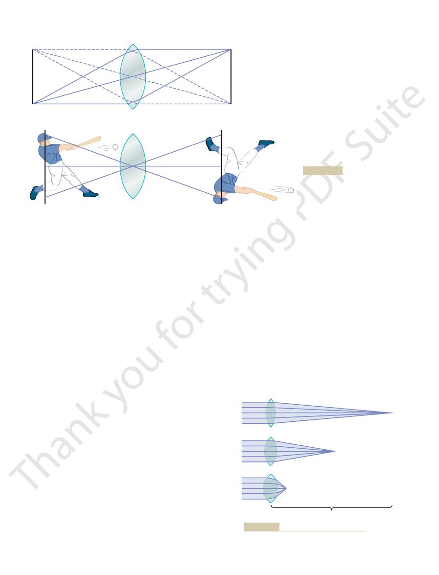
refractive power of +10 diopters.
come to a focal point 0.5 meter beyond the lens. A lens
said to have a strength of +2 diopters, and the light rays
twice as much as a lens with a power of +1 diopter, it is
8. If the lens is capable of bending parallel light rays
a refractive power of +1 diopter, as shown in Figure
length. Thus, a spherical lens that converges parallel
The refractive power in diopters of a
diopters.
This refractive power is measured in
refractive power.
The more a lens bends light rays, the greater is its
Measurement of the Refractive
reversed. This is the method by which the lens of a
object, and the two lateral sides of the image are
However,
the object, as demonstrated in Figure 49
focus distance from the lens, one can see an image of
the lens center. If a white sheet of paper is placed at the
bright, some are very weak, and they vary in color. Each
of point sources of light. Some of these points are very
Any object in front of the lens is, in reality, a mosaic
point source and the center of the lens.
refracted in either direction, the light rays from each
sources of light to the left. Because light rays pass
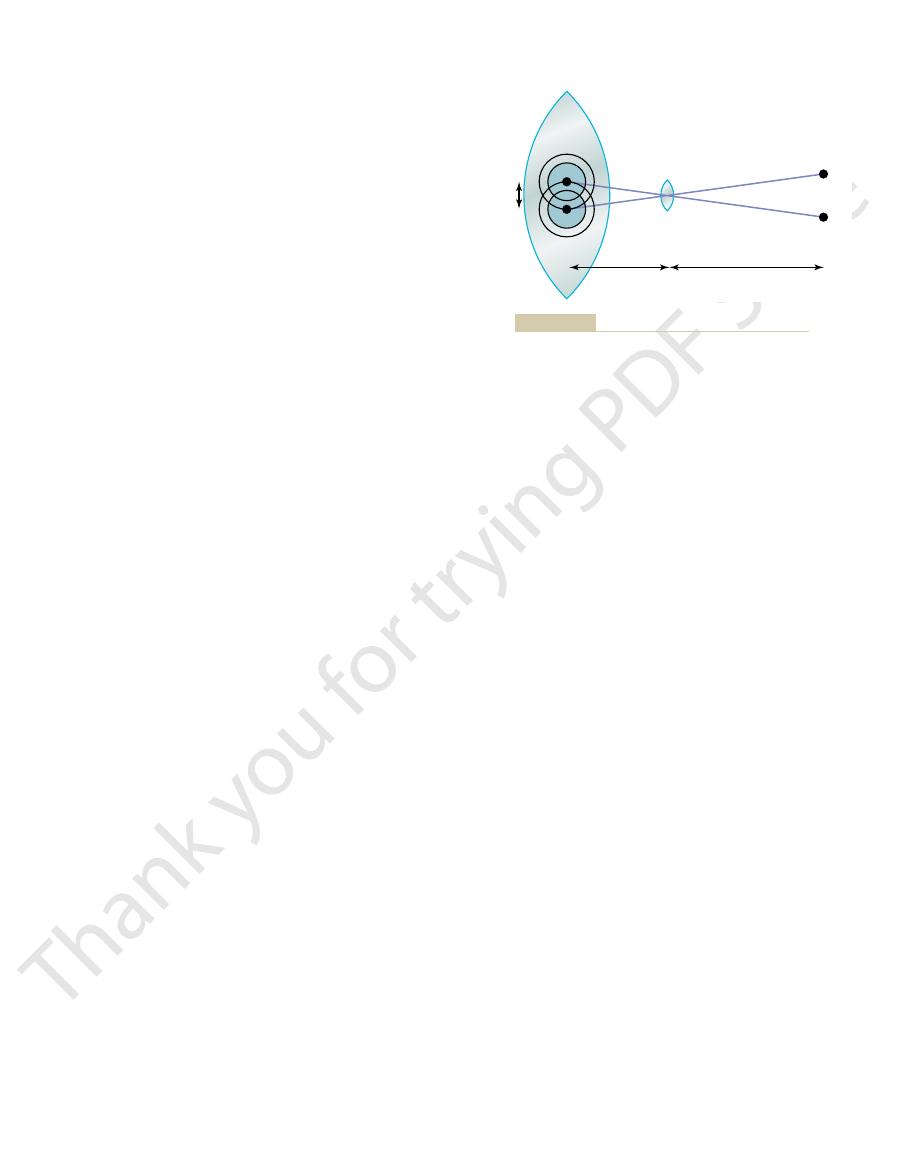
Figure 49
Formation of an Image by a
The Nervous System: B. The Special Senses
616
Unit X
Convex Lens
–7A shows a convex lens with two point
through the center of a convex lens without being
point source of light are shown to come to a point focus
on the opposite side of the lens directly in line with the
point source of light on the object comes to a separate
point focus on the opposite side of the lens in line with
–7B.
this image is upside down with respect to the original
camera focuses images on film.
Power of a Lens—“Diopter”
“
”
terms of
convex lens is equal to 1 meter divided by its focal
light rays to a focal point 1 meter beyond the lens has
49–
capable of converging parallel light rays to a focal point
only 10 centimeters (0.10 meter) beyond the lens has a
Point sources
Focal points
A
B
Two point sources of light
Figure 49–7
A,
focused at two separate points on
opposite sides of the lens. B, For-
mation of an image by a convex
spherical lens.
1
diopter
2
diopters
10
diopters
1 meter
in addition to its strength. If a cylindrical lens focuses
of the cylindrical lens must be stated
the same manner as the strengths of spherical lenses,
The strengths of cylindrical lenses are computed in
in a lens system with zero refractive power.
convex lenses. Thus, placing a 1-diopter concave lens
10 diopters.
much as a +10-diopter lens converges them, this lens is
1. Likewise, if the concave lens diverges light rays as
them, the concave lens is said to have a dioptric strength
point. However, if a concave lens diverges light rays at
because the light rays diverge, rather than focus to a
The refractive power of concave lenses cannot be
Effect of lens strength on the focal distance.
Figure 49–8
stated in terms of the focal distance beyond the lens
the same rate that a 1-diopter convex lens converges
of –
said to have a strength of –
Concave lenses “neutralize” the refractive power of
immediately in front of a 1-diopter convex lens results
except that the axis
around the lens, pulling the lens edges toward the
10, about 70
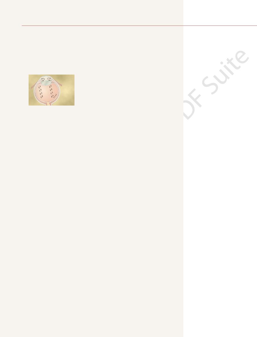
of the lens capsule. However, as shown in Figure
spherical shape, owing mainly to the elastic retraction
with no tension on its capsule, it assumes an almost
uid. When the lens is in a relaxed state
lled with viscous, proteinaceous, but
In a young person, the lens is composed of a strong
lens. The mechanism is as follows.
To do this, the shape of the lens is changed from that
of 14 diopters.
34 diopters; this in an
In children, the refractive power of the lens of the eye
Mechanism of “Accommodation”
to the object. However, the mind perceives objects in
retina. The image is inverted and reversed with respect
that a glass lens can focus an image on a sheet of paper,
cussed later in the chapter.
of the internal lens is that, in response to nervous
total refractive power of the eye. But the importance
on each side, is only 20 diopters, about one third the
eye, as it normally lies in the eye surrounded by
The total refractive power of the internal lens of the
humor.
markedly different from that of air, while the refrac-
by the eye lens). The principal reason
to exist, with its central point 17 millimeters in front of
reduced eye, a single refractive surface is considered
This is useful in simple calculations. In the
eye.
be one single lens, the optics of the normal eye may be
average), 1.40; and the vitreous humor, 1.34.
the aqueous humor, 1.33; the crystalline lens (on
humor. The internal index of air is 1; the cornea, 1.38;
of the lens of the eye, and (4) the interface between
of the cornea and the aqueous humor, (3) the interface
cornea, (2) the interface between the posterior surface
is composed of four refractive interfaces: (1) the inter-
lm. The lens system of the eye
a variable aperture system (the pupil), and a retina
to the usual photographic camera. It has a lens system,
9, is optically equivalent
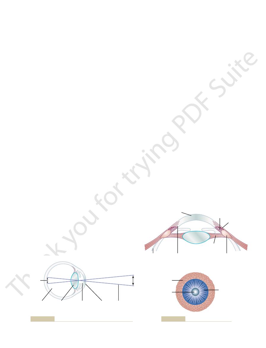
The eye, shown in Figure 49
90 degrees.
its axis is said to be 0 degrees. If it is vertical, its axis is
1 diopter. If the focused line is horizontal,
them, it has a
as a +1-diopter cylindrical lens
lens, it has a strength of +1 diopter. Conversely, if a cylin-
The Eye: I. Optics of Vision
Chapter 49
617
parallel light rays to a line focus 1 meter beyond the
drical lens of a concave type diverges light rays as much
converges
strength of –
Optics of the Eye
The Eye as a Camera
–
that corresponds to the fi
face between air and the anterior surface of the
between the aqueous humor and the anterior surface
the posterior surface of the lens and the vitreous
Reduced Eye.
If all the refractive surfaces of the eye are
algebraically added together and then considered to
simplified and represented schematically as a “reduced
”
the retina and a total refractive power of 59 diopters
when the lens is accommodated for distant vision.
About two thirds of the 59 diopters of refractive
power of the eye is provided by the anterior surface of
the cornea (not
for this is that the refractive index of the cornea is
tive index of the eye lens is not greatly different
from the indices of the aqueous humor and vitreous
fluid
signals from the brain, its curvature can be increased
markedly to provide “accommodation,” which is dis-
Formation of an Image on the Retina.
In the same manner
the lens system of the eye can focus an image on the
the upright position despite the upside-down orienta-
tion on the retina because the brain is trained to con-
sider an inverted image as the normal.
can be increased voluntarily from 20 diopters to about
“accommodation”
of a moderately convex lens to that of a very convex
elastic capsule fi
transparent fl
49–
suspensory ligaments attach radially
Image
Total refractive power = 59 diopters
Vitreous
humor
1.34
Lens
1.40
Aqueous
humor
1.33
Cornea
1.38
Air
1.00
Object
The eye as a camera. The numbers are the refractive indices.
Figure 49–9
Choroid
Circular
fibers
Sclerocorneal
junction
Meridional
fibers
Cornea
Ciliary muscle
Suspensory
ligaments
Lens
Suspensory
ligaments
Sclera
Figure 49–10
Mechanism of accommodation (focusing).
focus, the retina can be displaced considerably from
lens system. When a lens system has great depth of
In other words, the upper lens
blur circle.
eye the size of each spot will increase greatly, becom-
will not change much in the upper eye, but in the lower
ward to an out-of-focus position, the size of each spot
however, that if the retina is moved forward or back-
in perfect focus. It is evident from the diagrams,
quently, in both eyes, the retina sees two spots of light
pupillary aperture and focuses on the retina. Conse-
sources of light; light from each passes through the
is small, and in the lower eye, the aperture is large. In
lary apertures. In the upper eye, the pupillary aperture
Figure 49
ing Pupillary Diameter.
fold as a result of changes in pupillary aperture.
ters and as large as 8 millimeters in diameter. The
of the pupil. The pupil of the
The amount of light that enters the eye through the
eye in Chapter 51.
light. The re
The major function of the iris is to increase the amount
focused for near-seeing (e.g., for reading).
segment focused for far-seeing and the lower segment
To see clearly both in the distance and nearby, an
s eyes. The eyes can
constant distance; this distance depends on the physi-
modating, a condition known as
Thereafter, the lens remains almost totally nonaccom-
decreases to essentially 0 diopters at age 70 years.
by the time a person reaches 45 to 50 years; it then
with age. The power of accommodation decreases
teins. The ability of the lens to change shape decreases
larger and thicker and becomes far less elastic, partly
As a person grows older, the lens grows
mechanism; the neurology of this is discussed in
ing the ciliary muscle, but this effect is so weak that it
the eye to keep the object constantly in focus. (Sym-
quently, as a distant object moves toward the eye, the
than when the eye has less refractive power. Conse-
refractive power, the eye focuses on objects nearer
and increase its refractive power. With this increased
ligaments, thus allowing the lens to become thicker
bers, which relaxes the lens
nucleus in the brain stem, as explained in Chapter 51.
The ciliary muscle is controlled almost entirely by
Accommodation Is Controlled by Parasympathetic Nerves.
elasticity of the lens capsule.
shape, like that of a balloon, because of the natural
lens capsule, and the lens assumes a more spherical
Thus, contraction of either set of smooth muscle
to pull less on the lens capsule.
of ligament attachments; this also allows the ligaments
like action occurs, decreasing the diameter of the circle
attachments so that when they contract, a sphincter-
tension on the lens. The circular
toward the edges of the cornea, thereby releasing the
bers contract, the
When these muscle
The
circular fibers.
ciliary muscle,
However, also located at the lateral attachments of
under normal conditions of the eye.
border of the choroid and retina. The tension on the
outer circle of the eyeball. These ligaments are con-
The Nervous System: B. The Special Senses
618
Unit X
stantly tensed by their attachments at the anterior
ligaments causes the lens to remain relatively flat
the lens ligaments to the eyeball is the
which itself has two separate sets of smooth muscle
fibers—meridional fibers and
meridional fibers extend from the peripheral ends of
the suspensory ligaments to the corneoscleral junction.
fi
peripheral
insertions of the lens ligaments are pulled medially
ligaments’
fibers are
arranged circularly all the way around the ligament
fibers in the ciliary muscle relaxes the ligaments to the
parasympathetic nerve signals transmitted to the eye
through the third cranial nerve from the third nerve
Stimulation of the parasympathetic nerves contracts
both sets of ciliary muscle fi
number of parasympathetic impulses impinging on
the ciliary muscle must be progressively increased for
pathetic stimulation has an additional effect in relax-
plays almost no role in the normal accommodation
Chapter 51.)
Presbyopia.
because of progressive denaturation of the lens pro-
from about 14 diopters in a child to less than 2 diopters
“presbyopia.”
Once a person has reached the state of presbyopia,
each eye remains focused permanently at an almost
cal characteristics of each person’
no longer accommodate for both near and far vision.
older person must wear bifocal glasses with the upper
Pupillary Diameter
of light that enters the eye during darkness and to
decrease the amount of light that enters the eye in day-
flexes for controlling this mechanism are
considered in the discussion of the neurology of the
pupil is proportional to the area of the pupil or to the
square of the diameter
human eye can become as small as about 1.5 millime-
quantity of light entering the eye can change about 30-
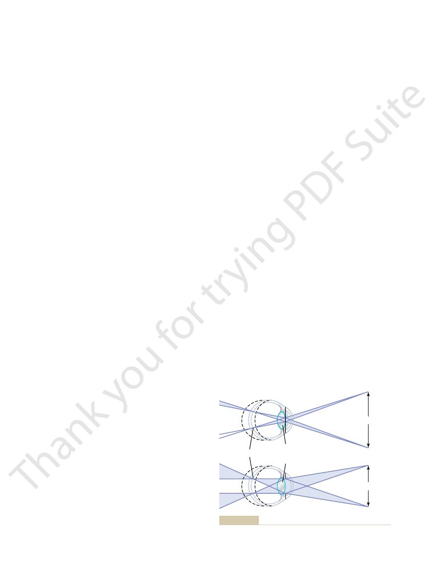
“Depth of Focus” of the Lens System Increases with Decreas-
–11 shows two eyes that
are exactly alike except for the diameters of the pupil-
front of each of these two eyes are two small point
ing a “
”
system has far greater depth of focus than the bottom
the focal plane or the lens strength can change con-
siderably from normal and the image will still remain
Lens
Lens
Focal point
Point sources of light
Point sources of light
pupillary apertures on
Effect of small
Figure 49–11
(top) and large (bottom)
“depth of focus.”
that is, by trying
is demonstrated in the lower diagram of Figure 49
using a convex lens in front of the eye. This correction
that is,
Conversely, in a person who has
upper diagram of Figure 49
diverge rays. Such correction is demonstrated in the
in front of the eye a concave spherical lens, which will
have too much refractive power, as in
concave lens diverge. If the refractive surfaces of the eye
focused clearly. A myopic person has a de
object comes still closer to the eye, the person can use
enough that its image can be focused. Then, when the
s eye, it
objects sharply on the retina. However, as an object
when the ciliary muscle is completely relaxed. A myopic
lens system of the eye.
12. This is usually due to too long an eyeball,
Figure 49
front of the retina, as shown in the bottom panel of
when the ciliary muscle is completely relaxed, the
ness,
In myopia, or
objects.
ciently to focus even distant objects, much less near
presbyopic,
the ciliary muscle has contracted to its limit. In old age,
much accommodative power left, and objects closer
accommodate for the distant objects, he or she still has
ing distant objects on the retina. If the person has used
accommodation, a farsighted person is capable of focus-
the strength of the lens. By using the mechanism of
abnormality, the ciliary muscle must contract to increase
by the time they reach the retina. To overcome this
12, parallel light rays are not bent
panel of Figure 49
that is too weak. In this condition, as seen in the middle
eyeball that is too short or, occasionally, a lens system
farsightedness,
Hyperopia, which is also
to focus objects at close range, the eye must contract its
objects clearly with its ciliary muscle relaxed. However,
This means that the emmetropic eye can see all distant
when the ciliary muscle is completely relaxed.
emmetropic,
eye is considered to be normal, or
12, the
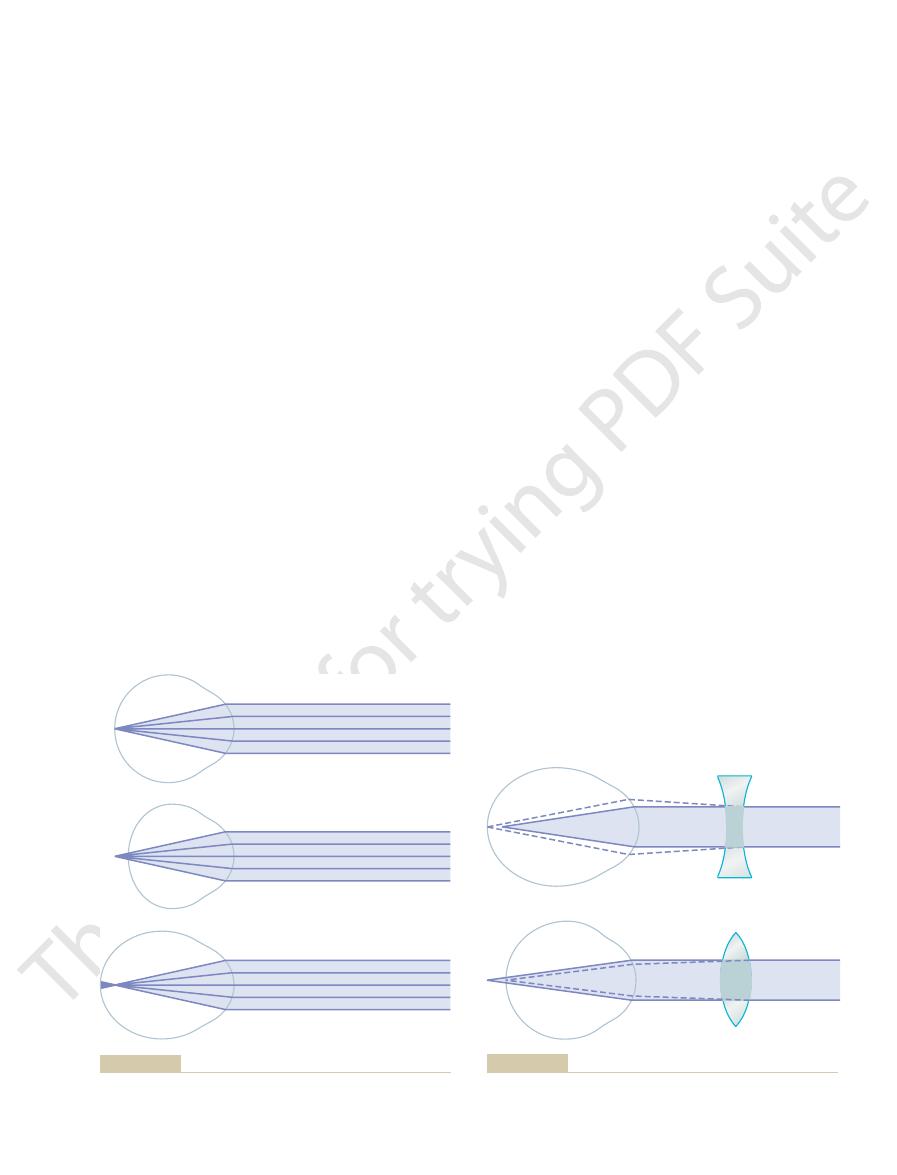
As shown in Figure 49
rays are always in focus, as explained earlier.
through the center of the lens, and the central most
with a very small aperture, almost all the rays pass
pupil is extremely small. The reason for this is that,
The greatest possible depth of focus occurs when the
ring.
depth of focus, moving the retina only
nearly in sharp focus, whereas when a lens system has
The Eye: I. Optics of Vision
Chapter 49
619
a “shallow”
slightly away from the focal plane causes extreme blur-
Errors of Refraction
Emmetropia (Normal Vision).
–
“
” if par-
allel light rays from distant objects are in sharp focus on
the retina
ciliary muscle and thereby provide appropriate degrees
of accommodation.
Hyperopia (Farsightedness).
known as “
” is usually due to either an
–
sufficiently by the relaxed lens system to come to focus
only a small amount of strength in the ciliary muscle to
and closer to the eye can also be focused sharply until
when the lens becomes “
” a farsighted
person is often unable to accommodate the lens suffi-
Myopia (Nearsightedness).
“nearsighted-
”
light rays coming from distant objects are focused in
–
but it can result from too much refractive power in the
No mechanism exists by which the eye can decrease
the strength of its lens to less than that which exists
person has no mechanism by which to focus distant
moves nearer to the person’
finally gets close
the mechanism of accommodation to keep the image
finite limiting
“far point” for clear vision.
Correction of Myopia and Hyperopia by Use of Lenses.
It will be recalled that light rays passing through a
myopia, this
excessive refractive power can be neutralized by placing
–13.
hyperopia—
someone who has too weak a lens system—the abnor-
mal vision can be corrected by adding refractive power
–13.
One usually determines the strength of the concave
or convex lens needed for clear vision by “trial and
error”—
first a strong lens and then a
Emmetropia
Hyperopia
Myopia
retina in hyperopia, and in front of the retina in myopia.
Parallel light rays focus on the retina in emmetropia, behind the
Figure 49–12
Correction of myopia with a concave lens, and correction of hyper-
Figure 49–13
opia with a convex lens.
the anterior surface of the cornea. The reason for this is
eye surface.
snugly against the anterior surface of the cornea. These
the appropriate axis.
spherical correction and the cylindrical correction at
this has been accomplished, the examiner directs the
patient sees all the crossed bars with equal clarity. When
are placed in line with the out-of-focus bars, until the
lenses, the axes of which
to the bars that are fuzzy. Once this axis is found, the
cylindrical component of the optical system is parallel
set of bars at right angles to the sharp bars. It can be
lenses in front of the astigmatic eye, a strength of lens
and horizontal axes. After placing various spherical
horizontal, and some at various angles to the vertical
15. Some of these parallel bars are vertical, some
use of parallel black bars of the type shown in Figure
system of an eye. One of these methods is based on the
of the abnormal cylindrical component of the lens
There are several methods for determining the axis
of the required cylindrical lens must be
in the remaining plane. To do this, both the
cylindrical lens is used to correct the remaining error
the two planes of the astigmatic lens. Then an additional
astigmatism, the usual procedure is to
and placed at right angles to each other. To correct for
made up of two cylindrical lenses of different strengths
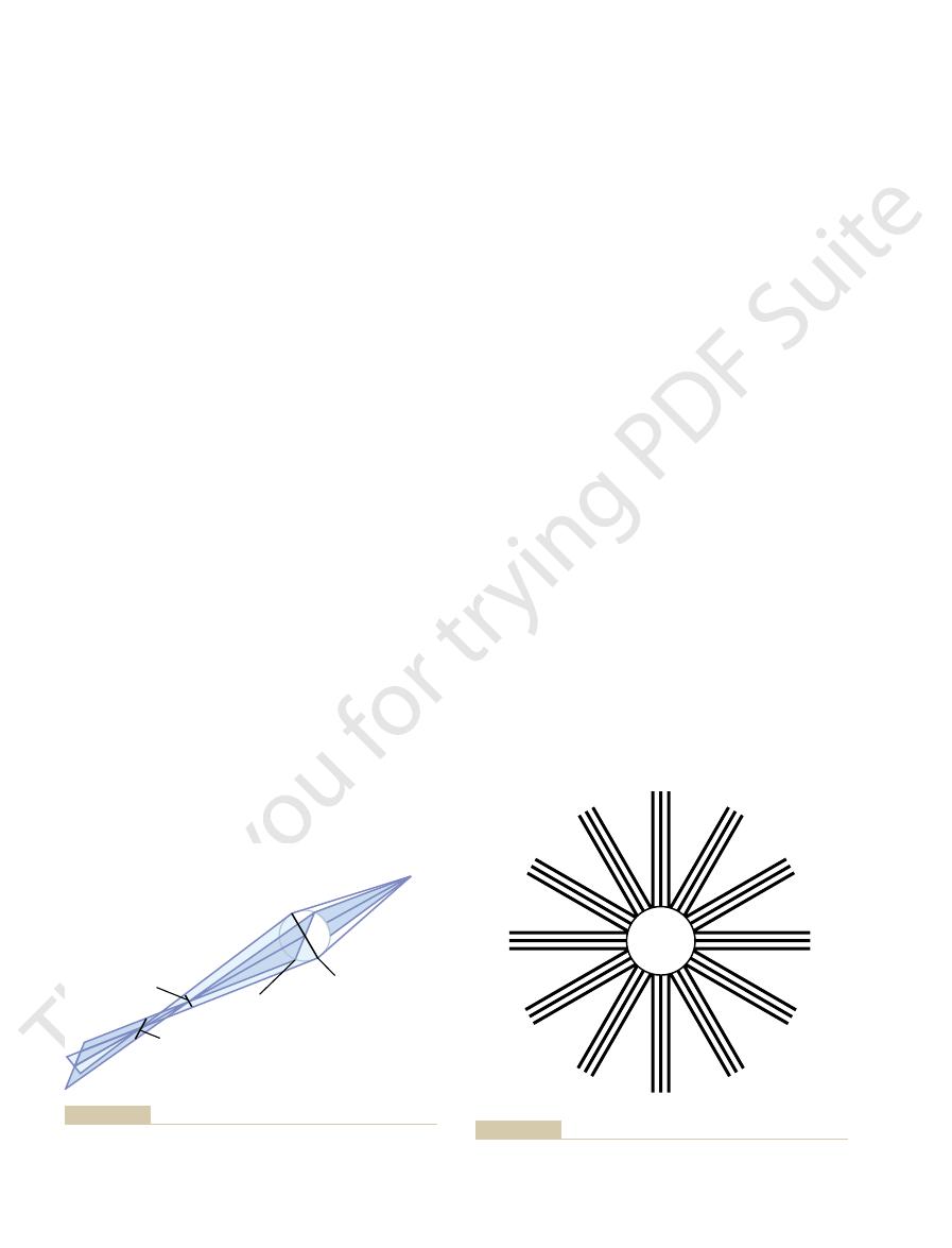
Correction of Astigmatism with a Cylindrical Lens.
person with astigmatism never sees in sharp focus.
accommodation. Thus, without the aid of glasses, a
mately equally in both planes; therefore, in astigmatism,
dation, the curvature of the eye lens changes approxi-
compensate for astigmatism because, during accommo-
The accommodative power of the eye can never
passing through the other plane.
all come to a common focal point, because the light rays
much as the light rays in vertical plane BD. It is obvious
plane, indicated by plane AC, are not bent nearly as
direction. By contrast, the light rays in the horizontal
vertical plane, indicated by plane BD, are refracted
through an oblong, astigmatic lens. The light rays in the
14, which shows rays
is demonstrated in Figure 49
striking the peripheral portions of the other plane. This
plane, light rays striking the peripheral portions of the
through the short axis.
sidewise to the incoming light. The degree of curvature
in one plane of the eye. An example of an astigmatic
tance from that of the plane at right angles. This most
The Nervous System: B. The Special Senses
620
Unit X
stronger or weaker lens until the one that gives the best
visual acuity is found.
Astigmatism
Astigmatism is a refractive error of the eye that causes
the visual image in one plane to focus at a different dis-
often results from too great a curvature of the cornea
lens would be a lens surface like that of an egg lying
in the plane through the long axis of the egg is not
nearly as great as the degree of curvature in the plane
Because the curvature of the astigmatic lens along
one plane is less than the curvature along the other
lens in one plane are not bent nearly as much as the rays
–
of light originating from a point source and passing
greatly by the astigmatic lens because of the greater cur-
vature in the vertical direction than in the horizontal
that light rays passing through an astigmatic lens do not
passing through one plane focus far in front of those
each of the two planes requires a different degree of
One
may consider an astigmatic eye as having a lens system
find a spherical
lens by trial and error that corrects the focus in one of
axis and
the strength
determined.
49–
is usually found that causes sharp focus of one set of
parallel bars but does not correct the fuzziness of the
shown from the physical principles of optics discussed
earlier in this chapter that the axis of the out-of-focus
examiner tries progressively stronger and weaker posi-
tive or negative cylindrical
optician to grind a special lens combining both the
Correction of Optical Abnormalities by Use of Contact Lenses
Glass or plastic contact lenses can be inserted that fit
lenses are held in place by a thin layer of tear fluid that
fills the space between the contact lens and the anterior
A special feature of the contact lens is that it nullifies
almost entirely the refraction that normally occurs at
A
B
C
D
Focal line
for plane BD
Focal line
for plane AC
Plane BD
(more refractive
power)
Plane AC
(less refractive
power)
Point source
of light
Figure 49–14
Astigmatism, demonstrating that light rays focus at one focal dis-
tance in one focal plane (plane AC) and at another focal distance
in the plane at a right angle (plane BD).
12
11
10
9
8
7
6
5
4
3
2
1
tations for determining the axis of astigmatism.
Chart composed of parallel black bars at different angular orien-
Figure 49–15
the retina, (2) the phenomenon of moving parallax,
means: (1) the sizes of the images of known objects on
of an Object from the Eye—
Determination of Distance
normal visual acuity.
expresses the ratio of two distances, which is also the
vision. In other words, the clinical method for express-
able to see at 200 feet, the person is said to have 20/200
that is, normal vision. If the
she should be able to see at 20 feet, the person is said
placed 20 feet away from the person being tested. If
The chart for
Clinical Method for Stating Visual Acuity.
Chapter 51.
more peripheral parts of the retina, as discussed in
10-fold as the periphery is approached. This is caused
becomes progressively poorer, decreasing more than
eld. Outside this foveal area, the visual acuity
micrometers) in diameter, which means that maximum
The fovea is less than 0.5 millimeter (less than 500
as two points instead of one. This means that a person
seconds between them, they can usually be recognized
seconds of arc. That is, when light rays from two sepa-
The normal visual acuity of the human eye for dis-
discrimination between points is also shown in Figure
slightly greater than the width of a foveal cone. This
much as 2 micrometers apart on the retina, which is
center point and shaded edges, a person can normally
Nevertheless, because the spot of light has a bright
is about 1.5 micrometers,
the central part of the retina, where vision
fovea
The average diameter of the cones in the
Figure 49
the edges, as shown by the two-point images in
olution of the normal eye optical system. The spot is
eter of about 11 micrometers, even with maximal res-
perfect, such a retinal spot ordinarily has a total diam-
However, because the lens system of the eye is never
focused on the retina, should be in
Theoretically, light from a distant point source, when
Visual Acuity
removed lens.
lens in front of the eye; usually, however, an arti
power, which must be replaced by a powerful convex
is done, the eye loses a large portion of its refractive
be corrected by surgical removal of the lens. When this
greatly that it seriously impairs vision, the condition can
When a cataract has obscured light transmission so
bers.
bers become denatured. Later, these same proteins
cataract formation, the proteins in some of the lens
or opaque area or areas in the lens. In the early stage of
that occurs mainly in older people. A cataract is a cloudy
the image, in addition to correcting the focus.
person sees through the lens, whereas lenses placed 1
eld of clear vision than glasses do, and (2) the
The contact lens has several other advantages as well,
however, the corneal refraction is neutralized, and
the vision satisfactorily; when a contact lens is used,
contact lens, the bulging cornea causes such severe
Without the
keratoconus.
such as those who have an odd-shaped, bulging
This is especially important in people whose eye refrac-
major role. Thus, the refraction of this surface of the
Instead, the outer surface of the contact lens plays the
cornea, so that the anterior surface of the cornea no
The Eye: I. Optics of Vision
Chapter 49
621
that the tears between the contact lens and the cornea
have a refractive index almost equal to that of the
longer plays a significant role in the eye’s optical system.
contact lens substitutes for the cornea’s usual refraction.
tive errors are caused by an abnormally shaped cornea,
cornea—a condition called
abnormality of vision that almost no glasses can correct
normal refraction by the outer surface of the contact
lens is substituted.
including (1) the lens turns with the eye and gives a
broader fi
contact lens has little effect on the size of the object the
centimeter or so in front of the eye do affect the size of
Cataracts
“Cataracts” are an especially common eye abnormality
fi
coagulate to form opaque areas in place of the normal
transparent protein fi
ficial
plastic lens is implanted in the eye in place of the
finitely small.
brightest in its center and shades off gradually toward
–16.
of
the retina—
is most highly developed—
which is one seventh the diameter of the spot of light.
distinguish two separate points if their centers lie as
49–16.
criminating between point sources of light is about 25
rate points strike the eye with an angle of at least 25
with normal visual acuity looking at two bright pin-
point spots of light 10 meters away can barely distin-
guish the spots as separate entities when they are 1.5
to 2 millimeters apart.
visual acuity occurs in less than 2 degrees of the visual
fi
by the connection of more and more rods and cones
to each optic nerve fiber in the nonfoveal,
testing eyes usually consists of letters of different sizes
the person can see well the letters of a size that he or
to have 20/20 vision—
person can see only letters that he or she should be
ing visual acuity is to use a mathematical fraction that
ratio of one’s visual acuity to that of a person with
“Depth Perception”
A person normally perceives distance by three major
2
µ
m
17 mm
1 mm
10 meters
Maximum visual acuity for two point sources of light.
Figure 49–16
pupil, and the observer sees into the subject
observed eye. Thus, the retina is illuminated through the
18, that light from a bulb is re
Figure 49
front of the observed eye in such a manner, as shown in
other. To illuminate the retina of the observed eye, an
means for illuminating the retina to be examined. Then,
To make an ophthalmoscope, one need only devise a
other.
focused on the retina of the observer, provided the two
made to emit light, the image of his or her retina will be
of the observing eye. Thus, if the retina of one person is
tance behind the lens. Any spot of light on the retina of
an emmetropic eye of another person, they focus again
the lens system. Then, when these parallel rays pass into
lens system, they are parallel with one another because
the lens system of the eye. After passing through the
emmetropic eye,
18 and can be explained as follows.
in Figure 49
principles are simple. The basic components are shown
appears to be a relatively complicated instrument, its
retina with clarity. Although the ophthalmoscope
The ophthalmoscope is an instrument through which an
than a person who has only one eye. However, stere-
when objects are nearby
present all the time when both eyes are being used. It
front of the eyes. This gives a type of parallax that is
17, which shows the images of a
strated in Figure 49
of the two retinas. This type of parallax is demon-
eye but on the right side of the retina of the right eye,
other. For instance, an object 1 inch in front of the nose
more than 2 inches to one side of the other eye, the
even though only one eye is used.
bly. Thus, by using this mechanism of moving parallax,
way across the retinas, whereas the image of an object
inch in front of the eye, the image moves almost all the
almost completely stationary. For instance, by moving
retinas, while the images of distant objects remain
moves his or her head to one side or the other, the
she perceives no moving parallax, but when the person
into the distance with the eyes completely still, he or
is that of moving parallax. If an individual looks off
retina. One does not consciously think about the size,
feet tall, one can determine how far away the person
and (3) the phenomenon of stereopsis. This ability to
The Nervous System: B. The Special Senses
622
Unit X
determine distance is called depth perception.
Determination of Distance by Sizes of Retinal Images of Known
Objects.
If one knows that a person being viewed is 6
is simply by the size of the person’s image on the
but the brain has learned to calculate automatically
from image sizes the distances of objects when the
dimensions are known.
Determination of Distance by Moving Parallax.
Another
important means by which the eyes determine distance
images of close-by objects move rapidly across the
the head 1 inch to the side when the object is only 1
200 feet away from the eyes does not move percepti-
one can tell the relative distances of different objects
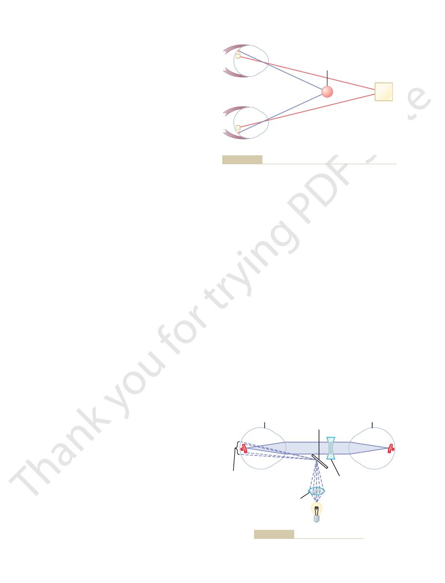
Determination of Distance by Stereopsis—Binocular Vision.
Another method by which one perceives parallax is
that of “binocular vision.” Because one eye is a little
images on the two retinas are different from each
forms an image on the left side of the retina of the left
whereas a small object 20 feet in front of the nose has
its image at closely corresponding points in the centers
–
red spot and a yellow square actually reversed on the
two retinas because they are at different distances in
is almost entirely this binocular parallax (or stereop-
sis) that gives a person with two eyes far greater ability
to judge relative distances
opsis is virtually useless for depth perception at dis-
tances beyond 50 to 200 feet.
Ophthalmoscope
observer can look into another person’s eye and see the
–
If a bright spot of light is on the retina of an
light rays from this spot diverge toward
the retina is located one focal length distance behind
to a point focus on the retina of the second person,
because his or her retina is also one focal length dis-
the observed eye projects to a focal spot on the retina
eyes are emmetropic and are simply looking into each
the reflected light from that retina can be seen by the
observer simply by putting the two eyes close to each
angulated mirror or a segment of a prism is placed in
–
flected into the
’s pupil by
looking over the edge of the mirror or prism or through
an appropriately designed prism.
1. Size of image
2. Stereopsis
Object of known
distance and size
Unknown
object
1. Size of image
2. Stereopsis
Object of known
distance and size
Unknown
object
and (2) as a result of stereopsis.
Perception of distance (1) by the size of the image on the retina
Figure 49–17
Mirror
Corrective lens in
turret (– 4 diopters
for normal eyes)
Illuminate retina
showing blood
vessel
Observer’s eye
Observed eye
Collimating lens
Figure 49–18
Optical system of the ophthalmoscope.
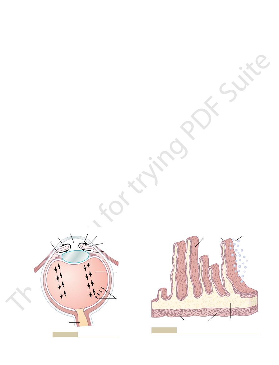
From here, the
into the anterior chamber of the eye.
ows, as shown in Figure 49
After aqueous humor is formed by the ciliary processes,
from the Eye
Outflow of Aqueous Humor
glucose.
diffusion; they include amino acids, ascorbic acid, and
of the eye. In addition, several nutrients are transported
spaces, and the resulting solution washes from the
to maintain electrical neutrality. Then all these ions
into the spaces between the epithelial cells. The sodium
secretion by the epithelium of the ciliary processes.
cells, and immediately beneath them is a highly vascu-
small size of the ciliary body. The surfaces of these
a large area, considering the
centimeters in each eye
19. Because of their folded architecture, the total
uid chambers of the eye can be seen in Figure
20, and their relation to
processes is shown in Figure 49
attach to the eyeball. A cross section of these ciliary
ciliary processes,
of 2 to 3 microliters each minute.
by the Ciliary Body
Formation of Aqueous Humor
reabsorbed. The balance between formation and reab-
flow
slowly in the vitreous humor, but there is little
molecules. Both water and dissolved substances can
vitreous body,
humor, sometimes called the
uid, whereas the vitreous
posterior surface of the lens and the retina. The aqueous
vitreous humor,
of the lens, and
aqueous humor,
Figure 49
The eye is
of each eye. To correct for this, it is necessary that
a lens of appropriate strength. In normal young adults,
observed retina. The usual ophthalmoscope has a series
is abnormal, it is necessary to correct the refractive
with completely emmetropic eyes. If the refractive
The Eye: I. Optics of Vision
Chapter 49
623
It is clear that these principles apply only to people
power of either the observed eye or the observer’s eye
power for the observer to see a sharp image of the
of very small lenses mounted on a turret so that the
turret can be rotated from one lens to another until the
correction for abnormal refraction is made by selecting
natural accommodative reflexes occur that cause an
approximate +2-diopter increase in strength of the lens
the lens turret be rotated to approximately –4-diopter
correction.
Fluid System of the Eye—
Intraocular Fluid
filled with intraocular fluid, which maintains
sufficient pressure in the eyeball to keep it distended.
–19 demonstrates that this fluid can be divided
into two portions—
which lies in front
which is between the
humor is a freely flowing fl
is a gelati-
nous mass held together by a fine fibrillar network com-
posed primarily of greatly elongated proteoglycan
diffuse
of fluid.
Aqueous humor is continually being formed and
sorption of aqueous humor regulates the total volume
and pressure of the intraocular fluid.
Aqueous humor is formed in the eye at an average rate
Essentially all of it is
secreted by the
which are linear folds
projecting from the ciliary body into the space behind
the iris where the lens ligaments and ciliary muscle
–
the fl
49–
surface area of the ciliary processes is about 6 square
—
processes are covered by highly secretory epithelial
lar area.
Aqueous humor is formed almost entirely as an active
Secretion begins with active transport of sodium ions
ions pull chloride and bicarbonate ions along with them
together cause osmosis of water from the blood capil-
laries lying below into the same epithelial intercellular
spaces of the ciliary processes into the anterior chamber
across the epithelium by active transport or facilitated
it first fl
–19, through the pupil
fluid
flows anterior to the lens and into the angle between the
Vitreous
humor
Aqueous humor Iris
Flow of fluid
Optic nerve
Lens
Formation
of aqueous
humor
Spaces of Fontana
Canal of Schlemm
Ciliary body
Diffusion of
fluid and other
constituents
Filtration and
diffusion at
retinal vessels
Formation and flow of fluid in the eye.
Figure 49–19
Formation
of aqueous
humor
Vascular layer
Ciliary muscle
Ciliary processes
surfaces.
Anatomy of the ciliary processes. Aqueous humor is formed on
Figure 49–20
days or even hours. As the pressure rises, the axons of
loss of vision when maintained for long periods.
70 mm Hg. Pressures above 25 to 30 mm Hg can cause
pathologically high, sometimes rising acutely to 60 to
of the most common causes of blindness. It is a disease
from the aqueous humor, thereby helping to maintain a
capable of phagocytizing proteins and small particles
The surface of the iris and other surfaces of the eye
phagocytic system keeps the trabecular spaces cleaned.
lar substances that can then be absorbed. Thus, this
trabecular plates are large numbers of phagocytic cells.
explained subsequently. However, on the surfaces of the
anterior chamber, sometimes causing
anterior chamber to the canal of Schlemm; this debris
or during intraocular infection, the debris is likely to
aqueous humor, as occurs after hemorrhage into the eye
When large amounts of debris are present in the
Mechanism for Cleansing the Trabecular Spaces and Intraocular
mally remains at about this level of 15 mm Hg.
uid from the ciliary body. The pressure nor-
sure rises. At about 15 mm Hg in the normal eye, the
minute openings of only 2 to 3 micrometers. The rate of
the wall of the canal of Schlemm. These trabeculae have
This out
15 mm Hg. The level of this pressure is determined
2 mm Hg of its normal level, which averages about
remains constant in the normal eye, usually within
and this is calibrated in terms of intraocular pressure.
placement is recorded on the scale of the tonometer,
plunger to be displaced inward. The amount of dis-
plunger, causing the part of the cornea beneath the
cornea. A small force is then applied to a central
cornea of the eye is anesthetized with a local anesthetic,
22. The
the principle of which is shown in Figure 49
tonometer,
s eye to measure intraocular pressure, this
Tonometry.
15 mm Hg, with a range from 12 to 20 mm Hg.
The average normal intraocular pressure is about
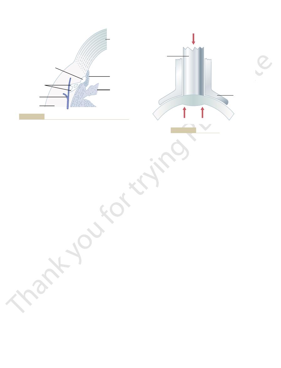
aqueous veins.
only aqueous humor, and they are called
than with blood.The small veins that lead from the canal
blood vessel, so much aqueous humor normally
ticulate matter up to the size of red blood cells, can pass
that even large protein molecules, as well as small par-
around the eye. Its endothelial membrane is so porous
canal of Schlemm. The canal of Schlemm is a thin-
angle, showing that the spaces between the trabeculae
empties into extraocular veins. Figure 49
canal of Schlemm,
ulae,
cornea and the iris,
The Nervous System: B. The Special Senses
624
Unit X
then through a meshwork of trabec-
finally entering the
which
–21 demon-
strates the anatomical structures at this iridocorneal
extend all the way from the anterior chamber to the
walled vein that extends circumferentially all the way
from the anterior chamber into the canal of Schlemm.
Even though the canal of Schlemm is actually a venous
flows
into it that it is filled only with aqueous humor rather
of Schlemm to the larger veins of the eye usually contain
Intraocular Pressure
Because it is impractical to pass a needle into
a patient’
pressure is measured clinically by using a “
”
–
and the footplate of the tonometer is placed on the
Regulation of Intraocular Pressure.
Intraocular pressure
±
mainly by the resistance to outflow of aqueous humor
from the anterior chamber into the canal of Schlemm.
flow resistance results from the meshwork of
trabeculae through which the fluid must percolate on its
way from the lateral angles of the anterior chamber to
fluid flow into the canal increases markedly as the pres-
amount of fluid leaving the eye by way of the canal of
Schlemm usually averages 2.5
ml/min and equals the
inflow of fl
Fluid.
accumulate in the trabecular spaces leading from the
can prevent adequate reabsorption of fluid from the
“glaucoma,” as
Immediately outside the canal of Schlemm is a layer of
interstitial gel that contains large numbers of reticu-
loendothelial cells that have an extremely high capacity
for engulfing debris and digesting it into small molecu-
behind the iris are covered with an epithelium that is
clear fluid.
“Glaucoma,” a Principal Cause of Blindness.
Glaucoma is one
of the eye in which the intraocular pressure becomes
Extremely high pressures can cause blindness within
Cornea
Sclera
Trabeculae
Iris
Blood veins
Canal of Schlemm
Aqueous veins
of aqueous humor from the eyeball into the conjunctival veins.
Anatomy of the iridocorneal angle, showing the system for outflow
Figure 49–21
Intraocular pressure
Pressure applied
Central plunger
Footplate
Principles of the tonometer.
Figure 49–22

Lancet 363:1711, 2004.
Weinreb RN, Khaw PT: Primary open-angle glaucoma.
ments. Cambridge: Cambridge University Press, 1997.
Smith G, Atchison DA: The Eye and Visual Optical Instru-
cance. Clin Exp Optom 86:3, 2003.
Smith G: The optical properties of the crystalline lens and
Curr Opin Ophthalmol 15:119, 2004.
Schwartz K, Budenz D: Current management of glaucoma.
biology of myopia. Clin Exp Optom 86:295, 2003.
Schaeffel F, Simon P, Feldkaemper M, et al: Molecular
Curr Biol 14:R279, 2004.
Shea J, Walsh V: Visual awareness: the eye
Retin Eye Res 22:307, 2003.
ment and pathological complications of myopia. Prog
McBrien NA, Gentle A: Role of the sclera in the develop-
the normal lens. Physiol Rev 77:21, 1997.
Mathias RT, Rae JL, Baldo GJ: Physiological properties of
lens capsule. Prog Retin Eye Res 22:749, 2003.
Krag S, Andreassen TT: Mechanical properties of the human
BMJ 328:97, 2004.
1: diagnosis.
Khaw PT, Shah P, Elkington AR: Glaucoma
Motility and Binocular Vision. St Louis: CV Mosby,
Guyton DL: Sights and Sounds in Ophthalmology: Ocular
rst century. Clin Exp Ophthalmol 32:305, 2004.
Gilmartin B: Myopia: precedents for research in the twenty-
surgery. Curr Opin Ophthalmol 11:207, 2000.
Farr AK, Guyton DL: Strabismus after retinal detachment
Ophthalmol 15:16, 2004.
Doane JF: Accommodating intraocular lenses. Curr Opin
examination. Semin Neurol 23:63, 2003.
Corbett JJ: The bedside and of
visual impairment in the world today. JAMA 290:2057,
Congdon NG, Friedman DS, Lietman T: Important causes of
on vision. Physiol Rev 75:323, 1995.
Buisseret P: In
reduce the pressure.
absorption of aqueous humor. When drug therapy fails,
especially in older individuals,
increase in intraocular pressure. In chronic conditions,
ammation, white blood cells and tissue debris
at the iridocorneal junction. For instance, in acute
In most cases of glaucoma, the abnormally high pres-
artery, which enters the eyeball at the optic disc, also
bers. It is possible that compression of the retinal
bers, which eventually causes death of the involved
the brain. The result is lack of appropriate nutrition of
ow of cytoplasm from the retinal neu-
eyeball at the optic disc. This compression is believed to
The Eye: I. Optics of Vision
Chapter 49
625
the optic nerve are compressed where they leave the
block axonal fl
ronal cell bodies into the optic nerve fibers leading to
the fi
fi
adds to the neuronal damage by reducing nutrition to
the retina.
sure results from increased resistance to fluid outflow
through the trabecular spaces into the canal of Schlemm
eye infl
can block these trabecular spaces and cause an acute
fibrous occlusion of the
trabecular spaces appears to be the likely culprit.
Glaucoma can sometimes be treated by placing drops
in the eye that contain a drug that diffuses into the
eyeball and reduces the secretion or increases the
operative techniques to open the spaces of the trabec-
ulae or to make channels to allow fluid to flow directly
from the fluid space of the eyeball into the subconjunc-
tival space outside the eyeball can often effectively
References
fluence of extraocular muscle proprioception
2003.
fice neuro-ophthalmology
fi
1989.
—
O’
fields have it?
their signifi