
electrode is surrounded by a thin plastic membrane. In the space between the
in blood, a miniature glass
When the glass electrode is used to measure CO
explained in Chapter 30; that is,
solution until an equilibrium state is established. In this equilibrium state, the
bonate is exposed to carbon dioxide gas, the carbon dioxide dissolves in the
in the following way: When a weak solution of sodium bicar-
direct measure of pH, and this is generally read directly from a voltmeter scale,
purpose are miniaturized. The voltage generated by the glass electrode is a
the type used in all chemical laboratories. However, the electrodes used for this
minutes, using no more than a few drops of blood. They are the following.
respiratory distress or acute abnormalities of acid-base balance. Several simple
, and pH. It is often important to make these
, CO
Study of Blood Gases and Blood pH
clinical pulmonary physiologist. Some other interesting tools are described here.
space. This array of measurements is only part of the armamentarium of the
tional residual capacity, dead space, physiologic shunt, and physiologic dead
ing respiratory abnormalities, including measuring vital capacity, tidal air, func-
In the previous few chapters, we have discussed a number of methods for study-
Useful Methods for Studying Respiratory Abnormalities
a diagnosis of “respiratory insufficiency.”
entirely different for these diseases, so it is no longer satisfactory simply to make
between the lungs and tissues. Therapy is often
inadequate ventilation. Others result from abnor-
exchange. Some respiratory diseases result from
Oxygen Therapy
Pathophysiology, Diagnosis,
Respiratory Insufficiency—
C
H
A
P
T
E
R
4
2
524
Diagnosis and treatment of most respiratory disor-
ders depend heavily on understanding the basic
physiologic principles of respiration and gas
malities of diffusion through the pulmonary mem-
brane or abnormal blood transport of gases
Among the most fundamental of all tests of pulmonary performance are deter-
minations of the blood Po
2
2
measurements rapidly as an aid in determining appropriate therapy for acute
and rapid methods have been developed to make these measurements within
Determination of Blood pH.
Blood pH is measured using a glass pH electrode of
or it is recorded on a chart.
Determination of Blood CO
2
.
A glass electrode pH meter can also be used to deter-
mine blood CO
2
pH of the solution is a function of the carbon dioxide and bicarbonate ion con-
centrations in accordance with the Henderson-Hasselbalch equation that is
2
electrode and plastic membrane is a solution of sodium bicarbonate of known
pH
HCO
CO
3
2
=
-
lo
+
g
6.1
capacity (TLC) and reduced residual volume (RV).
constricted lungs and partial airway obstruction. Note
ow-volume curve, along with two additional
Figure 42
Abnormalities of the Maximum Expiratory Flow-Volume Curve.
pressure, thus progressively reducing the maximum
structures are relaxed, so that the bronchi and bron-
elements; however, as the lung becomes smaller, these
The main reason for this is that in the enlarged lung
ow rate also becomes less.
Note also that as the lung volume becomes smaller,
ow rate that he or she can achieve.
tional expiratory effort the person exerts, this is still
than 400 L/min. But regardless of how much addi-
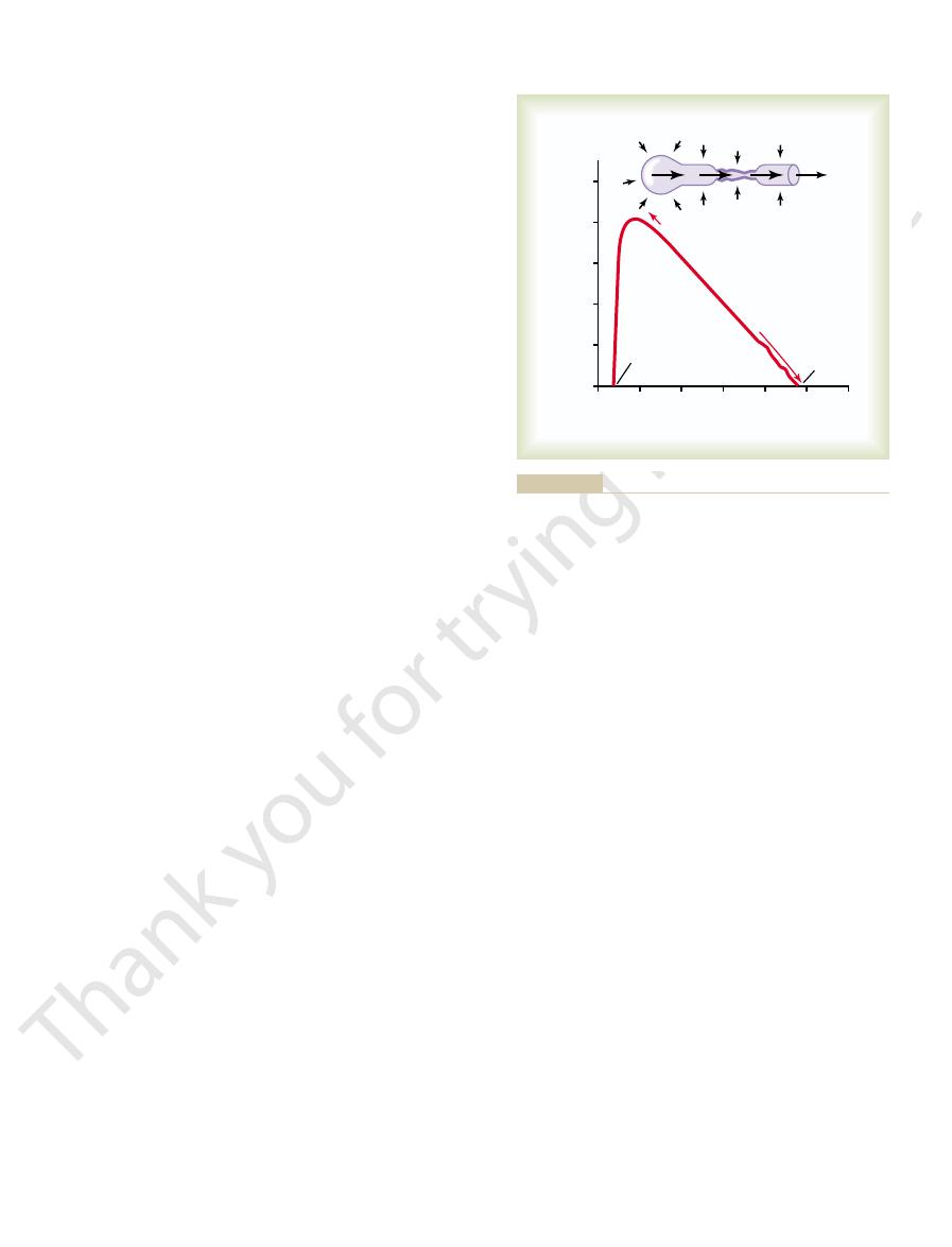
maximum expiratory airflow
can expire at no greater rate. Note that the person
ow. The curve
Figure 42
Therefore, beyond a critical degree of expiratory force,
ow.
amount, thus preventing further increase in
alveolar pressure, but it also increases the degree of
will oppose movement of air to the exterior. Once
to collapse the bronchioles at the same time, which
the alveoli toward the bronchioles, but it also tends
fore, not only does this pressure force air from
outsides of both the alveoli and the bronchioles.There-
ways caused by compressing the chest cage.The arrows
Figure 42
Figure 42
These principles can be understood by referring to
large volume of air than when they are almost empty.
ow. The maximum expiratory
with greatly increased additional force. This is the
follows: When a person expires with great force, the
, which can be de
maximum expiratory flow
culty in breathing. This has led to the concept called
during expiration, sometimes causing tremendous dif-
In many respiratory diseases, particularly in asthma,
almost moment by moment at the bedside.
so using a single, droplet-size sample of blood. Thus,
are built into the same apparatus, and all
, and Po
the electrode.
limeter is used, and this is separated from the blood
as well). In practice, a negative platinum
deposit on the electrode. Furthermore, the rate of
different from the voltage of the solution, oxygen will
. Electric current is made to
The concentration of oxygen
the pH is measured by the glass electrode, and the CO
solution. Only a drop or so of blood is required. Next,
surface of the plastic membrane, allowing carbon
concentration. Blood is then superfused onto the outer
Respiratory Insufficiency—Pathophysiology, Diagnosis, Oxygen Therapy
Chapter 42
525
dioxide to diffuse from the blood into the bicarbonate
2
is calculated by use of the above formula.
Determination of Blood PO
2
.
in a fluid can be measured by a technique called
polarography
flow
between a small negative electrode and the solution.
If the voltage of the electrode is more than
-0.6 volt
current flow through the electrode will be directly pro-
portional to the concentration of oxygen (and there-
fore to PO
2
electrode with a surface area of about 1 square mil-
by a thin plastic membrane that allows diffusion of
oxygen but not diffusion of proteins or other sub-
stances that will “poison”
Often all three of the measuring devices for pH,
CO
2
2
these measurements can be made within a minute or
changes in the blood gases and pH can be followed
Measurement of Maximum
Expiratory Flow
the resistance to airflow becomes especially great
fi
fined as
expiratory airflow reaches a maximum flow beyond
which the flow cannot be increased any more even
maximum expiratory fl
flow is much greater when the lungs are filled with a
–1.
–1A shows the effect of increased pressure
applied to the outsides of the alveoli and air passage-
indicate that the same pressure compresses the
the bronchioles have almost completely collapsed,
further expiratory force can still greatly increase the
bronchiolar collapse and airway resistance by an equal
fl
a maximum expiratory flow has been reached.
–1B shows the effect of different degrees
of lung collapse (and therefore of bronchiolar collapse
as well) on the maximum expiratory fl
recorded in this section shows the maximum expira-
tory flow at all levels of lung volume after a healthy
person first inhales as much air as possible and then
expires with maximum expiratory effort until he or she
quickly reaches a
of more
the maximum fl
the maximum expiratory fl
the bronchi and bronchioles are held open partially by
way of elastic pull on their outsides by lung structural
chioles are collapsed more easily by external chest
expiratory flow rate as well.
–2 shows the normal maximum expiratory
fl
flow-
volume curves recorded in two types of lung diseases:
that the constricted lungs have both reduced total lung
Total lung
6
0
1
2
3
A
B
Residual
volume
capacity
Maximum expiratory flow
4
5
Expiratory air flow (L/min)
Lung volume (liters)
0
500
400
300
200
100
becomes smaller.
decreasing maximum expiratory air flow as the lung volume
lung volume on the maximum expiratory air flow, showing
Effect of
ratory effort, an effect that limits expiratory flow rate.
Collapse of the respiratory passageway during maximum expi-
Figure 42–1
A,
B,
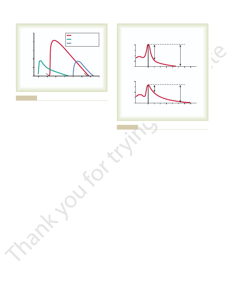
excess air in the lungs. However, this term is usually
The term
Abnormalities
of Specific Pulmonary
obstruction, as often occurs in acute asthma, this can
decreased to only 47 per cent. In serious airway
that, with airway obstruction, this value
/FVC%) is 80 per cent. However, note in Figure
), the percentage of the FVC that is
(see Figure 42
) with the normal. In the normal person
rst second. Therefore, it is customary to compare
, especially during
these persons can expire each second
however, a
in basic lung volumes in the two persons. There is,
greatly different, indicating only a moderate difference
tion. The total volume changes of the FVCs are not
Now, study the difference between the two records
gure.
FVC, as shown in the
and as completely as possible. The total distance of the
to the total lung capacity, then exhales into the
the FVC maneuver, the person
person with partial airway obstruction. In performing
person with normal lungs and in Figure 42
. Such a record is shown in Figure 42
and one that is also simple, is to make a record on a
Forced Expiratory Volume
Forced Expiratory Vital Capacity and
. Serious airway obstruction also
The classic disease that causes severe airway
normal airways, the maximum expiratory
2. Also, because of the obstruction of the
in Figure 42
both the TLC and the RV, as shown by the green curve
Over a period of months or years, this effect increases
lung easily but then becomes trapped in the lungs.
expands the alveoli. Therefore, air tends to enter the
to cause expiration. By contrast, the extra negative
the closing tendency of the airways is greatly increased
, it is usually
airway obstruction
, and
kyphosis
eases that constrict the chest cage, such as
, and dis-
lung itself, such as
cannot rise to equal that of the normal curve. Con-
sible expiratory effort, the maximal expiratory
normal maximum volume, even with the greatest pos-
Furthermore, because the lung cannot expand to a
526
Unit VII
Respiration
flow
stricted lung diseases include fibrotic diseases of the
tuberculosis and silicosis
,
scoliosis
fibrotic pleurisy.
In diseases with
much more difficult to expire than to inspire because
by the extra positive pressure required in the chest
pleural pressure that occurs during inspiration actually
“pulls” the airways open at the same time that it
–
airways and because they collapse more easily than
flow rate is
greatly reduced.
obstruction is asthma
occurs in some stages of emphysema.
Another exceedingly useful clinical pulmonary test,
spirometer of the forced expiratory vital capacity
(FVC)
–3A for a
–3B for a
first inspires maximally
spirometer with maximum expiratory effort as rapidly
downslope of the lung volume record represents the
fi
(1) for normal lungs and (2) for partial airway obstruc-
major difference in the amounts of air that
the fi
the recorded forced expiratory volume during the first
second (FEV
1
–3A
expired in the first second divided by the total FVC
(FEV
1
42–3B
decrease to less than 20 per cent.
Physiologic Peculiarities
Chronic Pulmonary Emphysema
pulmonary emphysema literally means
6
5
4
3
2
1
0
7
TLC
RV
Expiratory air flow (L/min)
Lung volume (liters)
0
500
Normal
Airway obstruction
Constricted lungs
400
300
200
100
curve. TLC, total lung capacity; RV, residual volume.
airway obstruction—-on the maximum expiratory flow-volume
Effect of two respiratory abnormalities—constricted lungs and
Figure 42–2
0
1
2
3
4
5
6
7
0
1
2
3
4
5
6
7
Maximum
inspiration
FEV
1
FVC
FEV
1
/FVC%
= 80%
Lung volume change (liters)
AIRWAY OBSTRUCTION
Seconds
NORMAL
4
3
2
1
0
A
FEV
1
FVC
FEV
1
/FVC%
= 47%
4
3
2
1
0
B
(The “zero” on the volume scale is residual volume.)
in a person with partial airway obstruction.
Recordings during the forced vital capacity maneuver:
Figure 42–3
A, in a
healthy person and B,
This disease begins with infection in the alveoli; the
, caused most frequently by
5. A common type of pneumonia is
uid and blood cells, as shown in Figure
The term
smoking.
all these effects is severe, prolonged, devastating
alveoli plus loss of alveolar walls. The net result of
many years. The person develops both hypoxia and
frequently causes right-sided heart failure.
. This in
markedly, causing
through which blood can pass. As a result, the
4. Loss of large portions of the alveolar walls also
same lungs.
in wasted ventilation, both effects occurring in the
), resulting
in poor aeration of the blood, and very high V
), resulting
, with a very low
ventilated. This often causes
ventilated, while other portions are poorly
in some parts of the lungs than in other parts,
3. The obstructive process is frequently much worse
of the lung, which
2. The marked loss of alveolar walls greatly
alveoli but also compresses the bronchioles,
breathing. It is especially dif
increases airway
1. The bronchiolar obstruction
struction versus lung parenchymal destruction.Among
extremely varied, depending on the severity of the
The physiologic effects of chronic emphysema are
emphysematous lung is that shown in Figures
. Therefore, the
destruction of as much as 50 to 80 per cent of the
combined with the lung infection, causes
and overstretching them. This,
cult to expire, thus causing
3. The obstruction of the airways makes it especially
airways.
chronic obstruction
2. The infection, excess mucus, and in
alveolar macrophages occurs, so that they become
exacerbates the condition. Too, inhibition of the
of excess mucus secretion occurs, which further
easily out of the passageways. Also, stimulation
nicotine. As a result, mucus cannot be moved
the respiratory epithelium, an effect caused by
airways, including partial paralysis of the cilia of
bronchioles. The chronic infection seriously
, caused by inhaling smoke or
process of the lungs caused by many years of smoking.
Respiratory Insufficiency—Pathophysiology, Diagnosis, Oxygen Therapy
Chapter 42
527
used to describe complex obstructive and destructive
It results from the following major pathophysiologic
changes in the lungs:
1. Chronic infection
other substances that irritate the bronchi and
deranges the normal protective mechanisms of the
less effective in combating infection.
flammatory
edema of the bronchiolar epithelium together
cause
of many of the smaller
diffi
entrapment of
air in the alveoli
marked
alveolar walls
final picture of the
42–4 (top) and 42–5.
disease and the relative degrees of bronchiolar ob-
the different abnormalities are the following:
resistance and results in greatly increased work of
ficult for the person
to move air through the bronchioles during
expiration because the compressive force on the
outside of the lung not only compresses the
which further increases their resistance during
expiration.
decreases the diffusing capacity
reduces the ability of the lungs to oxygenate the
blood and remove carbon dioxide from the blood.
so that some portions of the lungs are well
extremely abnormal
ventilation-perfusion ratios
V
.
a/Q
.
in some parts (physiologic shunt
.
a/Q
.
in other parts (physiologic dead space
decreases the number of pulmonary capillaries
pulmonary vascular resistance often increases
pulmonary hypertension
turn overloads the right side of the heart and
Chronic emphysema usually progresses slowly over
hypercapnia because of hypoventilation of many
air
hunger that can last for years until the hypoxia and
hypercapnia cause death—a high penalty to pay for
Pneumonia
pneumonia includes any inflammatory con-
dition of the lung in which some or all of the alveoli
are filled with fl
42–
bacterial pneu-
monia
pneumococci.
pulmonary membrane becomes inflamed and highly
porous so that fluid and even red and white blood cells
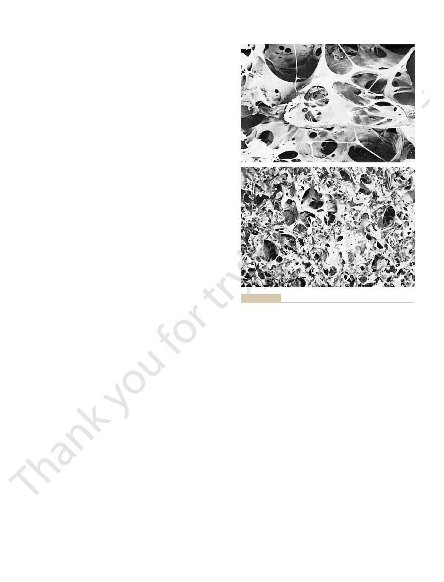
and the Department of Anatomy, The Medical College of
emphysema. (Reproduced with permission of Patricia Delaney
bottom figure
) with the normal
top figure
Figure 42–4
Contrast of the emphysematous lung (
lung (
), showing extensive alveolar destruction in
Wisconsin.)
monary vessels of the collapsed lung. This resistance
resistance to blood flow
7. Collapse of the lung tissue not
shown in Figure 42
The effects on overall pulmonary function caused by
lung.
atelectatic, a condition called
uid. This almost always is the
illaries into the alveoli, thus causing the alveoli to
the alveoli, which pull
brotic tissue and cannot collapse, absorption of air
the alveoli. However, if the lung is rigid because of
is pliable enough, this will lead simply to collapse of
owing in the pulmonary capillaries. If the lung tissue
object such as a tumor. The air entrapped beyond the
The airway obstruction type of
Airway Obstruction.
in localized areas of a lung or in an entire lung. Its most
Atelectasis means collapse of the alveoli. It can occur
about 78 per cent, which is far below normal.
cent saturated.Therefore, the average saturation of the
97 per cent saturated with oxygen, whereas that
ventilation-perfusion ratio in pneumonia, showing that
Figure 42
hypoxemia
perfusion ratio. Both these effects cause
This results in two major pulmonary abnormalities: (1)
ow through the lung continues normally.
to only one lung, with alveolar ventilation reduced
stages, the pneumonia process might well be localized
lungs change in different stages of the disease. In early
In pneumonia, the gas exchange functions of the
uid and cellular debris.
lung, become
of the lungs, sometimes whole lobes or even a whole
virus from alveolus to alveolus. Eventually, large areas
uid and cells,
leak out of the blood into the alveoli.Thus, the infected
528
Unit VII
Respiration
alveoli become progressively filled with fl
and the infection spreads by extension of bacteria or
“consolidated,” which means that they
are filled with fl
while blood fl
reduction in the total available surface area of the res-
piratory membrane and (2) decreased ventilation-
(low blood oxygen) and hypercapnia (high blood
carbon dioxide).
–6 shows the effect of the decreased
the blood passing through the aerated lung becomes
passing through the unaerated lung is about 60 per
blood pumped by the left heart into the aorta is only
Atelectasis
common causes are (1) total obstruction of the airway
or (2) lack of surfactant in the fluids lining the alveoli.
atelectasis usually results from (1) blockage of many
small bronchi with mucus or (2) obstruction of a major
bronchus by either a large mucus plug or some solid
block is absorbed within minutes to hours by the blood
fl
fi
from the alveoli creates very negative pressures within
fluid out of the pulmonary cap-
fill
completely with edema fl
effect that occurs when an entire lung becomes
massive collapse of the
massive collapse (atelectasis) of an entire lung are
–
only occludes the alveoli but also almost always
increases the
through the pul-
Normal
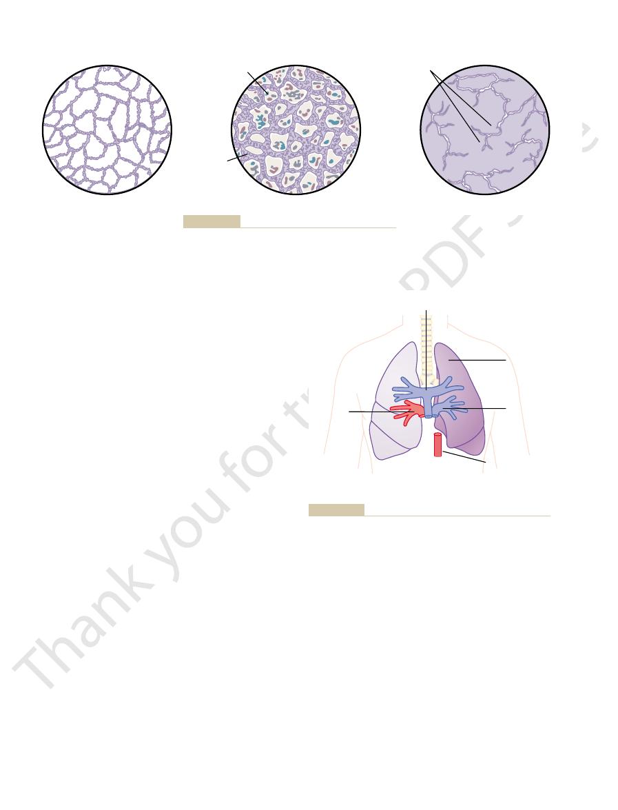
Pneumonia
Emphysema
Fluid and blood cells
Confluent alveoli
Edema
Figure 42–5
Lung alveolar changes in pneumonia and emphysema.
Mean = 78%
Pneumonia
Pulmonary arterial blood
60% saturated with O
2
Left
pulmonary
vein 60%
saturated
Right
pulmonary
vein 97%
saturated
Aorta:
Blood
1
/
2
= 97%
1
/
2
= 60%
pulmonary artery, the right and left pulmonary veins, and the aorta.
Effect of pneumonia on percentage saturation of oxygen in the
Figure 42–6

years, the chest cage becomes permanently enlarged,
expiring air from the lungs. Also, over a period of
The
this chapter.
air hunger,
dyspnea, or
ratory volume. Also, all of this together results in
ing. Clinical measurements show (1) greatly reduced
expiration. That is, the asthmatic person often can
occluded, further occlusion resulting from the external
the outsides of the bronchioles. Because the bronchi-
than during inspiration in asthma, caused by bronchi-
As discussed earlier in this chapter, the bronchiolar
Therefore, the airway resistance increases greatly.
and (2) spasm of the bronchiolar smooth muscle.
secretion of thick mucus into the bronchiolar lumens,
edema in the walls of the small bronchioles, as well as
substance of anaphylaxis, are to produce (1) localized
effects of all these factors, especially the slow-reacting
. The combined
, and (d)
chemotactic factor
(which is a mixture of leukotrienes), (c)
slow-reacting substance of anaphylaxis
, (b)
several different substances. Among them are (a)
IgE antibodies), the pollen reacts with the mast cell
sensitive (that is, to which the person has developed
with the bronchioles and small bronchi. When the asth-
antibodies are mainly attached to mast cells
rst place, as explained in Chapter 34. In asthma,
mally large amounts of IgE antibodies, and these anti-
typical allergic person has a tendency to form abnor-
of asthma is believed to occur in the following way: The
The allergic reaction that occurs in the allergic type
the air, such as irritants in smog.
pollens. In older people, the cause is almost always
allergic hypersensitivity, especially sensitivity to plant
younger than age 30 years, the asthma is caused by
stances in the air. In about 70 per cent of patients
The usual cause of asthma is contractile hypersensi-
at some time in life.
cult breathing. It occurs in 3 to 5 per cent of all people
smooth muscle in the bronchioles, which partially
atelectatic.
explained in Chapter 37, many of these infants die of
uid. As
This causes a serious tendency for the lungs of these
babies, the quantity of surfactant secreted by the
), which often occurs in newborn premature
However, in a number of conditions, such as in
plays a major role in preventing alveolar collapse.
tension in the alveoli 2- to 10-fold, which normally
alveoli. The surfactant in turn decreases the surface
discussed in Chapter 37. It was pointed out that the
The secre-
an entire lung.
promised, so that the aortic blood has only mild
through the unaerated lung. As a result, the overall
situation shown in Figure 42
lated lung and therefore becomes well aerated. In the
nately, most of the blood is routed through the venti-
through the atelectatic lung becomes slight. Fortu-
Because of the vascular constriction, blood
tion, as explained in Chapter 38.
volume of the lung decreases. In addition, hypoxia in
itself, which compresses and folds the vessels as the
Respiratory Insufficiency—Pathophysiology, Diagnosis, Oxygen Therapy
Chapter 42
529
increase occurs partially because of the lung collapse
the collapsed alveoli causes additional vasoconstric-
flow
–7, five sixths of the blood
passes through the aerated lung and only one sixth
ventilation-perfusion ratio is only moderately com-
oxygen desaturation despite total loss of ventilation in
Lack of “Surfactant” as a Cause of Lung Collapse.
tion and function of surfactant in the alveoli were
surfactant is secreted by special alveolar epithelial
cells into the fluids that coat the inside surface of the
hyaline
membrane disease (also called respiratory distress syn-
drome
alveoli is so greatly depressed that the surface tension
of the alveolar fluid becomes several times normal.
babies to collapse or to become filled with fl
suffocation when large portions of the lungs become
Asthma
Asthma is characterized by spastic contraction of the
obstructs the bronchioles and causes extremely diffi-
tivity of the bronchioles in response to foreign sub-
hypersensitivity to nonallergenic types of irritants in
bodies cause allergic reactions when they react with the
specific antigens that have caused them to develop in
the fi
these
that
are present in the lung interstitium in close association
matic person breathes in pollen to which he or she is
–
attached antibodies and causes the mast cells to release
histamine
eosinophilic
bradykinin
diameter becomes more reduced during expiration
olar collapse during expiratory effort that compresses
oles of the asthmatic lungs are already partially
pressure creates especially severe obstruction during
inspire quite adequately but has great difficulty expir-
maximum expiratory rate and (2) reduced timed expi-
“
” which is discussed later in
functional residual capacity and residual volume
of the lung become especially increased during the
acute asthmatic attack because of the difficulty in
causing a “barrel chest,” and both the functional resid-
ual capacity and lung residual volume become perma-
nently increased.
= 91%
Atelectasis
Pulmonary arterial blood
60% saturated with O
2
Left
pulmonary
vein 60%
saturated-
flow
1
/
5
normal
Right
pulmonary
vein 97%
saturated
Aorta:
Blood
5
/
6
= 97%
1
/
6
= 60%
Mean saturation
Effect of atelectasis on aortic blood oxygen saturation.
Figure 42–7

value of about 100 mm Hg to as high as 600 mm Hg.
hypoventilation hypoxia, because oxygen therapy can
, essentially the same result occurs as in
hypoxia caused by impaired alveolar membrane
cial. (However, this provides no
normal air. Therefore, here again oxygen therapy can
per cent oxygen can move
, a person breathing 100
hypoventilation hypoxia
effective therapy.
inspired gases and, therefore, provide 100 per cent
, oxygen therapy can com-
atmospheric hypoxia
valuable.
when oxygen therapy will be of value and, if so, how
different types of hypoxia, one can readily decide
intranasal tube.
a mask, or (3) administering oxygen through an
with oxygen, (2) allowing the patient to breathe either
s head in a
of Hypoxia
Oxygen Therapy in Different Types
high-altitude physiology.
(2) reduced work capacity of the muscles. These effects
mental activity, sometimes culminating in coma, and
can cause death of cells throughout the body, but in
Hypoxia, if severe enough,
Effects of Hypoxia on the Body.
, in which several
system can lead to this type of hypoxia. A special
tissue cellular oxidative
cannot use oxygen even when plenty is available. Also,
cyanide
cytochrome oxidase
, in which the action of the enzyme
The classic
Inadequate Tissue Capability to Use Oxygen.
needs further elaboration: this is the hypoxia caused
self-evident from the discussions earlier in the chapter.
This classi
ciency, or
oxygen, because of toxicity, vitamin de
b. Diminished cellular metabolic capacity for using
a. Poisoning of cellular oxidation enzymes
5. Inadequate tissue capability of using oxygen
d. Tissue edema
cerebral, coronary vessels)
ciency (peripheral,
c. Localized circulatory de
ciency
b. General circulatory de
a. Anemia or abnormal hemoglobin
4. Inadequate oxygen transport to the tissues by the
3. Venous-to-arterial shunts (
c. Diminished respiratory membrane diffusion
b. Abnormal alveolar ventilation-perfusion ratio
a. Hypoventilation caused by increased airway
2. Pulmonary disease
b. Hypoventilation (neuromuscular disorders)
ciency of oxygen in the atmosphere
a. De
1. Inadequate oxygenation of the blood in the lungs
principles of oxygen therapy. The following is a
types of hypoxia; then we can discuss the physiologic
Therefore, it is important to understand the different
value; and, at still other times, it is of almost no value.
therapy is of great value; other times, it is of moderate
of bodywide cellular hypoxia. Sometimes, oxygen
sion of oxygen and carbon dioxide.
in the lungs, further reducing overall pulmonary diffu-
increased thickness of the respiratory membrane
These effects cause (1) increased
as reduced total amount of functional lung tissue.
brosis throughout the lungs, as well
Thus, tuberculosis in its late stages is characterized
large abscess cavities.
bacilli spread throughout the lungs, often causing
untreated, the walling-off process fails and tubercle
cent of all people who develop tuberculosis, if
extension of the infection. However, in about 3 per
. This walling-off process helps to limit further
tissue reaction in the lungs, including (1) invasion of
In tuberculosis, the tubercle bacilli cause a peculiar
Tuberculosis
530
Unit VII
Respiration
the infected tissue by macrophages and (2) “walling
off” of the lesion by fibrous tissue to form the so-called
tubercle
transmission of the tubercle bacilli in the lungs and
therefore is part of the protective process against
extreme destruction of lung tissue with formation of
by many areas of fi
“work” on the part
of the respiratory muscles to cause pulmonary venti-
lation and reduced vital capacity and breathing capac-
ity; (2) reduced total respiratory membrane surface area
and
,
causing progressively diminished pulmonary diffusing
capacity; and (3) abnormal ventilation-perfusion ratio
Hypoxia and Oxygen Therapy
Almost any of the conditions discussed in the past
few sections of this chapter can cause serious degrees
descriptive classification of the causes of hypoxia:
because of extrinsic reasons
fi
resistance or decreased pulmonary compliance
(including either increased physiologic dead
space or increased physiologic shunt)
“right-to-left” cardiac
shunts)
blood
fi
fi
fi
other factors
fication of the types of hypoxia is mainly
Only one of the types of hypoxia in the classification
by inadequate capability of the body’s tissue cells to
use oxygen.
cause of inability of the tissues to use oxygen is cyanide
poisoning
is completely blocked by the
—to such an extent that the tissues simply
deficiencies of some of the
enzymes or of other elements in the tissue oxidative
example occurs in the disease beriberi
important steps in tissue utilization of oxygen and
formation of carbon dioxide are compromised because
of vitamin B deficiency.
less severe degrees it causes principally (1) depressed
are specifically discussed in Chapter 43 in relation to
Oxygen can be administered by (1) placing the
patient’
“tent” that contains air fortified
pure oxygen or high concentrations of oxygen from
Recalling the basic physiologic principles of the
In
pletely correct the depressed oxygen level in the
In
five times as much oxygen
into the alveoli with each breath as when breathing
be extremely benefi
benefit for the excess blood carbon dioxide also caused
by the hypoventilation.)
In
diffusion
increase the Po
2
in the lung alveoli from the normal

circle: (1) more carbon dioxide, (2) further decrease in
piration rather than stimulate it, thus causing a vicious
to 120 to 150 mm Hg. At these higher levels of P
becomes lethargic and sometimes even semicomatose.
rises to 80 to 100 mm Hg, the person
severe.
, becomes
air hunger,
can, and
75 mm Hg, an otherwise normal person by then is
When the alveolar P
oxygen, so that the resulting tissue hypercapnia is
hypoxia. However, the transport capacity of the blood
decreases carbon dioxide removal from the tissues,
ciency, diminished
Hypercapnia then occurs along with the hypoxia. And
atmosphere is affected as much as is oxygen transfer.
Conversely, in hypoxia caused by hypoventilation,
rapidly as oxygen. If hypercapnia does begin to occur,
the pulmonary membrane or through the tissues,
or use of oxygen by the tissues. Therefore, it is readily
poisoning of the oxidative
, or
too little oxygen in the air
The reasons for this are the following.
hypoventilation
is caused by
hypercapnia. However, hypercapnia usually occurs
rst thought, that any respi-
One might suspect, on
uids.
Hypercapnia
conditions.
frequently to cyanosis, even under otherwise normal
, the great excess of avail-
Conversely, in a person with excess red blood cells, as
almost never becomes cyanotic
genated hemoglobin in each 100 milliliters of blood. A
nite cyanosis appears whenever the
In general, de
laries. This deoxygenated hemoglobin has an intense
bin in the skin blood vessels, especially in the capil-
means blueness of the skin, and its
The term
Therefore, oxygen therapy is of hardly any measura-
tissues. Instead, the tissue metabolic enzyme system is
, there is abnormality neither
quate tissue use of oxygen
hypoxia caused by inade-
altered. This small amount of extra oxygen may be the
extra oxygen, between 7 and 30 per cent, can be
cient. Even so, a small amount of
alveoli. The problem instead is that one or more of the
, oxygen therapy is of much less value
transport of oxygen
hypoxia caused by anemia, abnormal hemoglobin
as would occur with no therapy.
8, which shows that the
demonstrated in Figure 42
an increase of more than 800 per cent. This highly ben-
normal value of 60 mm Hg to as high as 560 mm Hg,
This raises the oxygen pressure gradient for diffusion
Respiratory Insufficiency—Pathophysiology, Diagnosis, Oxygen Therapy
Chapter 42
531
of oxygen from the alveoli to the blood from the
eficial effect of oxygen therapy in diffusion hypoxia is
–
pulmonary blood in this patient with pulmonary
edema picks up oxygen three to four times as rapidly
In
, circulatory deficiency, or physio-
logic shunt
because normal oxygen is already available in the
mechanisms for transporting oxygen from the lungs to
the tissues is defi
trans-
ported in the dissolved state in the blood when alveo-
lar oxygen is increased to maximum even though the
amount transported by the hemoglobin is hardly
difference between life and death.
In the different types of
of oxygen pickup by the lungs nor of transport to the
simply incapable of using the oxygen that is delivered.
ble benefit.
Cyanosis
cyanosis
cause is excessive amounts of deoxygenated hemoglo-
dark blue-purple color that is transmitted through the
skin.
fi
arterial blood contains more than 5 grams of deoxy-
person with anemia
because there is not enough hemoglobin for 5 grams
to be deoxygenated in 100 milliliters of arterial blood.
occurs in polycythemia vera
able hemoglobin that can become deoxygenated leads
Hypercapnia means excess carbon dioxide in the body
fl
fi
ratory condition that causes hypoxia would also cause
in association with hypoxia only when the hypoxia
or circulatory deficiency.
Hypoxia caused by
, too
little hemoglobin
enzymes has to do only with the availability of oxygen
understandable that hypercapnia is not a concomitant
of these types of hypoxia.
In hypoxia resulting from poor diffusion through
serious hypercapnia usually does not occur at the same
time because carbon dioxide diffuses 20 times as
this immediately stimulates pulmonary ventilation,
which corrects the hypercapnia but not necessarily the
hypoxia.
carbon dioxide transfer between the alveoli and the
in circulatory defi
flow of blood
resulting in tissue hypercapnia in addition to tissue
for carbon dioxide is more than three times that for
much less than the tissue hypoxia.
co
2
rises above about 60 to
breathing about as rapidly and deeply as he or she
“
” also called dyspnea
If the Pco
2
Anesthesia and death can result when the Pco
2
rises
co
2
,
the excess carbon dioxide now begins to depress res-
Arterial end
Venous end
P
O
2
in alveoli and blood (mm Hg)
Blood in pulmonary capillary
0
100
Capillary blood
Alveolar P
O
2
with tent therapy
Normal alveolar P
O
2
Pulmonary edema
+
O
2
therapy
Pulmonary edema with no
therapy
200
300
monary edema with and without oxygen tent therapy.
Absorption of oxygen into the pulmonary capillary blood in pul-
Figure 42–8
30 mm Hg positive pressure in the lungs can cause
sometimes to lethal levels. For instance, continuous
peripheral veins becomes impeded. As a result, use
greater than pressure everywhere else in the body.
respirator, the pressure inside the lungs becomes
tive pressure by a resuscitator, or when the pressure
When air is forced into the lungs under posi-
Effect of the Resuscitator and the Tank Respirator on Venous
5 cm H
negative pressures. Ordinarily these pressures are
outward, negative pressure causes inspiration. Check
body and causes expiration; as the diaphragm moves
moves inward, positive pressure develops around the
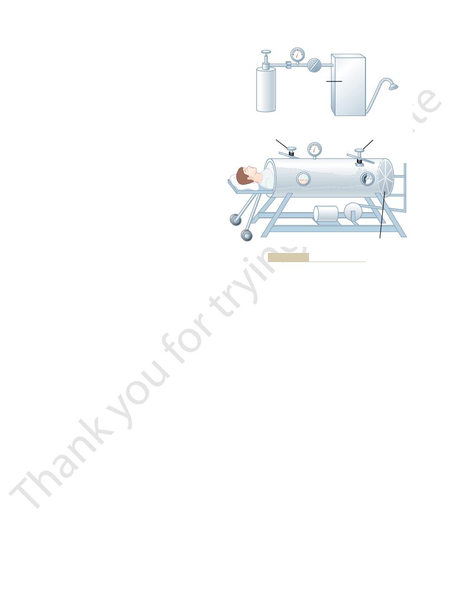
the pressure inside the tank. As the leather diaphragm
head a motor-driven leather diaphragm moves back
collar. At the end of the tank opposite the patient
Figure 42
Tank Respirator (the “Iron-Lung”).
that are commonly set at 12 to 15 cm H
usage was at one time greatly decried. However, resus-
lungs because of excessive positive pressure. Their
the lungs during the remainder of the cycle.
during the positive-pressure cycle of the resuscitator
cheal tube. This apparatus forces air through the mask
and, with some machines, negative pressure as well;
consists of a tank supply of oxygen or air; a
ciples of operation. The resuscitator shown in Figure
are available, and each has its own characteristic prin-
Resuscitator.
Artificial Respiration
small or crowded rooms.
cient quantity of air, such as on entering
dyspnea. This feeling is greatly enhanced in people
. For instance, almost
of an abnormal state of mind. This is called
Finally, the person
breathe forcefully. In these instances, the forceful
normality of the respiratory gases, the person has to
uids are normal, but to attain this
times, however, the levels of both carbon dioxide and
uids. At
piratory muscles to provide adequate ventilation; and
hypercapnia and, to a much less extent, hypoxia; (2)
uids, especially
ment of the sensation of dyspnea. They are (1) abnor-
ity to ventilate enough to satisfy the demand for air. A
respiration, (3) then more carbon dioxide, and so
532
Unit VII
Respiration
forth—culminating rapidly in a respiratory death.
Dyspnea
Dyspnea means mental anguish associated with inabil-
common synonym is air hunger.
At least three factors often enter into the develop-
mality of respiratory gases in the body fl
the amount of work that must be performed by the res-
(3) state of mind.
A person becomes very dyspneic especially from
excess buildup of carbon dioxide in the body fl
oxygen in the body fl
activity of the respiratory muscles frequently gives the
person a sensation of dyspnea.
’s respiratory functions may be
normal and still dyspnea may be experienced because
neurogenic
dyspnea or emotional dyspnea
anyone momentarily thinking about the act of breath-
ing may suddenly start taking breaths a little more
deeply than ordinarily because of a feeling of mild
who have a psychological fear of not being able to
receive a suffi
Many types of respiratory resuscitators
42–9A
mechanism for applying intermittent positive pressure
and a mask that fits over the face of the patient or a
connector for joining the equipment to an endotra-
or endotracheal tube into the lungs of the patient
and then usually allows the air to flow passively out of
Earlier resuscitators often caused damage to the
citators now have adjustable positive-pressure limits
2
O pressure for
normal lungs (but sometimes much higher for non-
compliant lungs).
–9B shows the
tank respirator with a patient’s body inside the tank
and the head protruding through a flexible but airtight
’s
and forth with sufficient excursion to raise and lower
valves on the respirator control the positive and
adjusted so that the negative pressure that causes
inspiration falls to
-10 to -20 cm H
2
O and the positive
pressure rises to 0 to
+
2
O.
Return.
around the patient’s body is reduced by the tank
Flow of blood into the chest and heart from the
of excessive pressures with either the resuscitator or
the tank respirator can reduce the cardiac output—
exposure for more than a few minutes to greater than
death because of inadequate venous return to the
heart.
Positive
pressure
valve
Negative
pressure
valve
Mechanism
for applying
positive and
negative
pressure
A
B
Leather diaphragm
Tank respirator.
Resuscitator.
Figure 42–9
A,
B,

ed. Nat Rev Genet 5:376,
Wills-Karp M, Ewart SL: Time to draw breath: asthma-
in lung function and disease. N Engl J Med 347:2141,
Whitsett JA, Weaver TE: Hydrophobic surfactant proteins
cott Williams & Wilkins, 2001.
Philadelphia: Lippin-
Integrated, Case-Based Approach
West JB: Pulmonary Physiology and Pathophysiology: An
asthma. Clin Sci (Lond) 103:201, 2002.
Wardlaw AJ, Brightling CE, Green R, et al: New insights
c review. JAMA 290:2301, 2003.
disease: scienti
Sin DD, McAlister FA, Man SF, Anthonisen NR: Contem-
Mol Physiol 286:L715, 2004.
ammation: for better or worse? Am J Physiol Lung Cell
brosis, gene therapy, and lung
Schwiebert LM: Cystic
review. Chest 125:1081, 2004.
Rodrigo GJ, Rodrigo C, Hall JB: Acute asthma in adults: a
L1313, 2001.
for asthma. Am J Physiol Lung Cell Mol Physiol 281:
and airway smooth muscle cell interactions: implications
Page S, Ammit AJ, Black JL, Armour CL: Human mast cell
N Engl J Med 345:1257, 2001.
Naureckas ET, Solway J: Clinical practice. Mild asthma.
muscle function and training. Sports Med 34:117, 2004.
obstructive pulmonary disease: the role of respiratory
McConnell AK, Romer LM: Dyspnoea in health and
ogy 8:432, 2003.
and functional properties in health and disease. Respirol-
Knight DA, Holgate ST: The airway epithelium: structural
Sci 326:174, 2003.
as sampled by the expired breath condensate. Am J Med
Dwyer TM: Cigarette smoke-induced airway in
Physiol Lung Cell Mol Physiol 287:L24, 2004.
ammation. Am J
of HO-1: protection against airway in
Carter EP, Garat C, Imamura M: Continual emerging roles
Annu Rev Physiol 63:471, 2001.
Cardoso WV: Molecular regulation of lung development.
L887, 2004.
lung disease. Am J Physiol Lung Cell Mol Physiol 286:
Basu S, Fenton MJ: Toll-like receptors: function and roles in
asthma? Ann Intern Med 139:359, 2003.
Barnes PJ, Adcock IM: How do corticosteroids work in
cine. Philadelphia: Mosby, 2002.
Albert R, Spiro S, Jett J: Comprehensive Respiratory Medi-
Respiratory Insufficiency—Pathophysiology, Diagnosis, Oxygen Therapy
Chapter 42
533
References
fl
flammation
fi
infl
porary management of chronic obstructive pulmonary
fi
into the relationship between airway inflammation and
.
2002.
susceptibility genes are identifi
2004.