
pumping, as we shall see later in this and subsequent chapters.
the volume of these vessels. This can
veins,
The innervation of the large vessels, particularly of the
flow through the tissues.
The innervation of the
the capillaries, precapillary sphincters, and metar-
of sympathetic nerve fibers to the blood vessels, demonstrating that in most
Figure 18–2 shows distribution
Sympathetic Innervation of the Blood Vessels.
The precise pathways of these fibers in the spinal cord and in the sympathetic
distributed to the vasculature of the peripheral areas.
on the right side of Figure 18–1, and (2) almost immediately into peripheral por-
innervate mainly the vasculature of the internal viscera and the heart, as shown
by two routes to the circulation: (1) through specific
one of which lies on each side of the vertebral column. Next, they pass
chain,
or two lumbar spinal nerves. They then pass immediately into a
nervous control of the circulation. Sympathetic vasomotor nerve fibers leave
Figure 18–1 shows the anatomy of sympathetic
Sympathetic Nervous System.
see later in the chapter.
also contributes specifically to regulation of heart function, as we shall
. The
teristics, as follows.
sion, we need to present still other specific anatomical and functional charac-
60, and this subject was also introduced in Chapter 17. For our present discus-
. The total function of this system is presented in Chapter
The nervous system controls the circulation almost entirely through the
arterial pressure.
activity by the heart, and, especially, providing very rapid control of systemic
ing blood flow to different areas of the body, increasing or decreasing pumping
tion has more global functions, such as redistribut-
tissue blood flow control mechanisms. We shall see
As discussed in Chapter 17, adjustment of blood
Circulation
Nervous Regulation of the
of Arterial Pressure
Circulation, and Rapid Control
C
H
A
P
T
E
R
1
8
204
Nervous Regulation of the
flow tissue by tissue is mainly the function of local
in this chapter that nervous control of the circula-
auto-
nomic nervous system
Autonomic Nervous System
By far the most important part of the autonomic nervous system for regulating
the circulation is the sympathetic nervous system
parasympathetic nervous
system
the spinal cord through all the thoracic spinal nerves and through the first one
sympathetic
sympathetic nerves that
tions of the spinal nerves
chains are discussed more fully in Chapter 60.
tissues all the vessels except
terioles are innervated.
small arteries and arterioles allows sympathetic stim-
ulation to increase resistance to blood flow and thereby to decrease rate of blood
makes it possi-
ble for sympathetic stimulation to decrease
push blood into the heart and thereby play a major role in regulation of heart
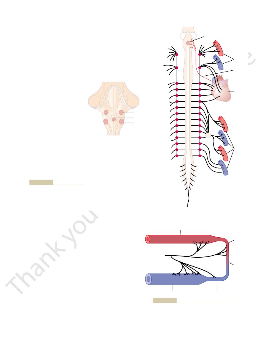
The effects of parasympathetic stimulation on
shown in Figure 18–1 by the dashed red line
nerves,
regulation of the circulation. Its most important circu-
trointestinal actions, it plays only a minor role in
functions of the body, such as control of multiple gas-
and volume of pumping.
markedly increases the activity of the heart, both
shown in Figure 18–1 and also discussed in Chapter 9.
sympathetic fibers also go directly to the heart, as
sympathetic nerve fibers supplying the blood vessels,
Nervous Regulation of the Circulation, and Rapid Control of Arterial Pressure
Chapter 18
205
Sympathetic Nerve Fibers to the Heart.
In addition to
It should be recalled that sympathetic stimulation
increasing the heart rate and enhancing its strength
Parasympathetic Control of Heart Function, Especially Heart
Rate.
Although the parasympathetic nervous system is
exceedingly important for many other autonomic
latory effect is to control heart rate by way of parasym-
pathetic nerve fibers to the heart in the vagus
from the brain medulla directly to the heart.
heart function were discussed in detail in Chapter 9.
Vasoconstrictor
Sympathetic chain
Cardioinhibitor
Vasodilator
Blood
vessels
Vasomotor center
Blood
vessels
Heart
Vagus
to the heart.
shown by the red dashed line is a
of the circulation. Also
control
Figure 18–1
Anatomy of sympathetic nervous
vagus nerve that carries parasym-
pathetic signals
Arteries
Sympathetic
vasoconstriction
Arterioles
Capillaries
Venules
Veins
Sympathetic innervation of the systemic circulation.
Figure 18–2
need to decrease heart pumping, the
heart rate and contractility. Conversely, when there is
activity. The
amount of vascular constriction, it also controls heart
At the
Control of Heart Activity by the Vasomotor Center.
1 to 3 minutes, until the norepinephrine was destroyed.
vessels once again became constricted, and the arterial
was transported in the blood to all blood vessels, the
fibers throughout the body). As this injected hormone
throughout the body. A few minutes later, a small
result, the arterial pressure fell from 100 to 50 mm Hg,
impulses from the spinal cord to the periphery. As a
This blocked all transmission of sympathetic nerve
constrictor tone. In the experiment of this figure,
Figure 18–4 demonstrates the significance of vaso-
partial state of contraction in the blood vessels, called
. These impulses normally maintain a
second. This continual firing is called
entire body, causing continuous slow firing of these
normal conditions, the vasoconstrictor area of the
mally Caused by Sympathetic Vasoconstrictor Tone.
Continuous Partial Constriction of the Blood Vessels Is Nor-
pressure, which we describe later in this chapter.
control of many circulatory functions. An example
the vasomotor center, thus providing “reflex”
, and output signals from
medulla and lower pons. The neurons of this area
3. A
medulla. The fibers from these neurons project
2. A
fibers to all levels of the spinal cord, where they
anterolateral portions of the upper medulla. The
1. A
center, as follows:
center is still unclear, experiments have made it
body.
to virtually all arteries, arterioles, and veins of the
. This center transmits
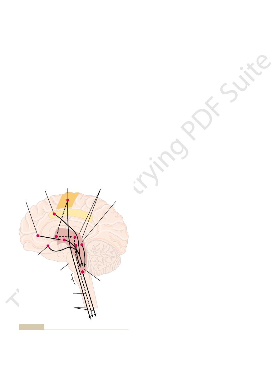
of the pons, shown in Figures 18–1 and 18–3, is an area
Vasomotor Center in the Brain and Its Control of the Vasocon-
intestines, spleen, and skin but much less potent in
strictor effect is especially powerful in the kidneys,
some tissues than others. This sympathetic vasocon-
essentially all segments of the circulation, but more to
fibers. The vasoconstrictor fibers are distributed to
The sympathetic nerves carry tremendous numbers of
Its Control by the Central Nervous System
Sympathetic Vasoconstrictor System and
heart muscle contractility.
Principally, parasympathetic stimulation causes a
206
Unit IV
The Circulation
marked decrease in heart rate and a slight decrease in
vasoconstrictor nerve fibers and only a few vasodilator
skeletal muscle and the brain.
strictor System.
Located bilaterally mainly in the retic-
ular substance of the medulla and of the lower third
called the vasomotor center
parasympathetic impulses through the vagus nerves to
the heart and transmits sympathetic impulses through
the spinal cord and peripheral sympathetic nerves
Although the total organization of the vasomotor
possible to identify certain important areas in this
vasoconstrictor area located bilaterally in the
neurons originating in this area distribute their
excite preganglionic vasoconstrictor neurons of the
sympathetic nervous system.
vasodilator area located bilaterally in the
anterolateral portions of the lower half of the
upward to the vasoconstrictor area just described;
they inhibit the vasoconstrictor activity of this area,
thus causing vasodilation.
sensory area located bilaterally in the tractus
solitarius in the posterolateral portions of the
receive sensory nerve signals from the circulatory
system mainly through the vagus and
glossopharyngeal nerves
this sensory area then help to control activities of
both the vasoconstrictor and vasodilator areas of
is the baroreceptor reflex for controlling arterial
Under
vasomotor center transmits signals continuously to
the sympathetic vasoconstrictor nerve fibers over the
fibers at a rate of about one half to two impulses per
sympathetic vaso-
constrictor tone
vasomotor tone.
total spinal anesthesia was administered to an animal.
demonstrating the effect of losing vasoconstrictor tone
amount of the hormone norepinephrine was injected
into the blood (norepinephrine is the principal
vasoconstrictor hormonal substance secreted at the
endings of the sympathetic vasoconstrictor nerve
pressure rose to a level even greater than normal for
same time that the vasomotor center is controlling the
lateral portions of the vasomotor center
transmit excitatory impulses through the sympathetic
nerve fibers to the heart when there is need to increase
medial portion of
Reticular
substance
Motor
Cingulate
Orbital
Temporal
Pons
Medulla
VASOMOTOR
CENTER
VASOCONSTRICTOR
VASODILATOR
Mesencephalon
tion of the circulation. The dashed lines represent inhibitory
Areas of the brain that play important roles in the nervous regula-
Figure 18–3
pathways.
at their endings, although in primates, the vasodilator
not norepinephrine,
acetylcholine,
dilator fibers release
constrictor fibers. In lower animals such as the cat, these
The sympathetic nerves to skeletal
Nervous System.
Vasodilator
dilates rather than constricts certain vessels, as dis-
a “beta” adrenergic receptor stimulatory effect, which
to cause vasoconstriction, but in an occasional tissue
where they act directly on all blood vessels, usually
carried in the blood stream to all parts of the body,
. These two hormones are
are transmitted to the blood vessels. They cause the
Adrenal Medullae and Their Relation to the Sympathetic Vaso-
vasoconstriction, as discussed in Chapter 60.
alpha adren-
rine. Norepinephrine acts directly on the
The substance secreted at the endings of the
Norepinephrine—The Sympathetic Vasoconstrictor Transmitter
stimulus. Thus, widespread basal areas of the brain can
motor center, depending on the precise portions of
, and the
, the
, the
, the
Also, stimulation of the
hypothalamus and thence to the vasomotor center.
for instance, excites the vasomotor center
inhibit the vasomotor center. Stimulation of the
thalamus cause mainly excitation, whereas the
motor center. The
The
of the reticular substance cause excitation, whereas
in Figure 18–3 by the rose-colored area. In general, the
vasomotor center. This reticular substance is shown
Control of the Vasomotor Center by Higher Nervous Centers.
either increase or decrease heart activity. Heart rate
contractility. Therefore, the vasomotor center can
, which then
Nervous Regulation of the Circulation, and Rapid Control of Arterial Pressure
Chapter 18
207
the vasomotor center sends signals to the adjacent
dorsal motor nuclei of the vagus nerves
transmit parasympathetic impulses through the vagus
nerves to the heart to decrease heart rate and heart
and strength of heart contraction ordinarily increase
when vasoconstriction occurs and ordinarily decrease
when vasoconstriction is inhibited.
Large numbers of small neurons located throughout
the reticular substance of the pons, mesencephalon,
and diencephalon can either excite or inhibit the
neurons in the more lateral and superior portions
the more medial and inferior portions cause inhibition.
hypothalamus plays a special role in controlling
the vasoconstrictor system because it can exert either
powerful excitatory or inhibitory effects on the vaso-
posterolateral portions of the hypo-
anterior
portion can cause either mild excitation or inhibition,
depending on the precise part of the anterior hypo-
thalamus stimulated.
Many parts of the cerebral cortex can also excite or
motor
cortex,
because of impulses transmitted downward into the
anterior temporal lobe
orbital areas of the frontal cortex, the anterior part of
the cingulate gyrus
amygdala
septum
hippocampus can all either excite or inhibit the vaso-
these areas that are stimulated and on the intensity of
have profound effects on cardiovascular function.
Substance.
vasoconstrictor nerves is almost entirely norepineph-
ergic receptors of the vascular smooth muscle to cause
constrictor System.
Sympathetic impulses are transmit-
ted to the adrenal medullae at the same time that they
medullae to secrete both epinephrine and norepineph-
rine into the circulating blood
epinephrine causes vasodilation because it also has
cussed in Chapter 60.
Sympathetic
System and its Control by the Central
muscles carry sympathetic vasodilator fibers as well as
0
5
10
15
20
25
Arterial pressure (mm Hg)
Seconds
150
125
100
75
50
25
0
Total spinal
anesthesia
Injection of norepinephrine
resulting from loss of “vasomotor
marked decrease in pressure
the arterial pressure, showing
Effect of total spinal anesthesia on
Figure 18–4
tone.”

from danger.
, and it
few seconds. This is called the
instance, during extreme fright, the arterial pressure
cise, a similar rise in pressure can also occur. For
activity.
center. These increase the arterial pressure instanta-
ing system of the brain stem is also activated, which
vated to cause exercise, most of the reticular activat-
results mainly from the following effect: At the same
The increase in arterial pressure during exercise
rises about 30 to 40 per cent, which increases blood
exercise. In most heavy exercise, the arterial pressure
in Chapter 17. Additional increase results from simul-
increased metabolism of the muscle cells, as explained
blood flow. Part of this increase results from local
heavy exercise, the muscles require greatly increased
in pressure that occurs during muscle exercise. During
Types of Stress
Increase in Arterial Pressure
one half normal within 10 to 40 seconds. Therefore,
two times normal within 5 to 10 seconds. Conversely,
arterial pressure is its rapidity of response, beginning
Rapidity of Nervous Control of Arterial Pressure.
to the acute rise in arterial pressure.
under normal conditions. This contributes still more
blood. During strong sympathetic stimulation, the
force of the heart muscle, this, too, increasing the
addition, sympathetic nervous signals have a
increasing to as great as three times normal. In
increase in the heart rate, the rate sometimes
cardiac pumping.
autonomic nervous system, further enhancing
the heart itself is directly stimulated by the
3. Finally,
pressure.
quantities of blood. This, too, increases the arterial
volume of blood in the heart chambers. The stretch
blood vessels toward the heart, thus increasing the
This displaces blood out of the large peripheral
pressure.
peripheral resistance, thereby increasing the arterial
This greatly increases the total
each of which helps to increase arterial pressure. They
heart. Thus, three major changes occur simultaneously,
together. At the same time, there is reciprocal inhibi-
increases in arterial pressure. For this purpose, the
Arterial Pressure
in Rapid Control of
Role of the Nervous System
nerves of the muscles.
nerves, and also through the spinal cord to the
centers of the medulla, to the heart through the vagus
The pathway probably then goes to the vasodilatory
vasovagal syncope.
causes the person to lose consciousness. This overall
falls rapidly, which reduces blood flow to the brain and
to slow the heart rate markedly. The arterial pressure
becomes activated, and at the same time, the vagal car-
fainting. In this case, the muscle vasodilator system
Emotional Fainting—Vasovagal Syncope.
before the muscles require increased nutrients.
anticipatory increase in blood flow
that at the onset of exercise, the sympathetic vasodila-
response to their needs. Yet some experiments suggest
Possible Unimportance of the Sympathetic Vasodilator System.
anterior hypothalamus.
Figure 18–3. The principal area of the brain controlling
The pathway for central nervous system control of
vasculature.
208
Unit IV
The Circulation
effect is believed to be caused by epinephrine exciting
specific beta adrenergic receptors in the muscle
the vasodilator system is shown by the dashed lines in
this system is the
It is doubtful that the sympathetic vasodilator system
plays an important role in the control of the circulation
in the human being because complete block of the sym-
pathetic nerves to the muscles hardly affects the ability
of these muscles to control their own blood flow in
tor system might cause initial vasodilation in skeletal
muscles to allow
even
A particularly
interesting vasodilatory reaction occurs in people who
experience intense emotional disturbances that cause
dioinhibitory center transmits strong signals to the heart
effect is called
Emotional fainting
begins with disturbing thoughts in the cerebral cortex.
center of the anterior hypothalamus next to the vagal
sympa-
thetic vasodilator
One of the most important functions of nervous
control of the circulation is its capability to cause rapid
entire vasoconstrictor and cardioaccelerator functions
of the sympathetic nervous system are stimulated
tion of parasympathetic vagal inhibitory signals to the
are as follows:
1. Almost all arterioles of the systemic circulation
are constricted.
2. The veins especially (but the other large vessels of
the circulation as well) are strongly constricted.
of the heart then causes the heart to beat with far
greater force and therefore to pump increased
Much of this is caused by an
significant direct effect to increase contractile
capability of the heart to pump larger volumes of
heart can pump about two times as much blood as
An espe-
cially important characteristic of nervous control of
within seconds and often increasing the pressure to
sudden inhibition of nervous cardiovascular stimula-
tion can decrease the arterial pressure to as little as
nervous control of arterial pressure is by far the most
rapid of all our mechanisms for pressure control.
During Muscle Exercise and Other
An important example of the ability of the nervous
system to increase the arterial pressure is the increase
vasodilation of the muscle vasculature caused by
taneous elevation of arterial pressure caused by sym-
pathetic stimulation of the overall circulation during
flow almost an additional twofold.
time that the motor areas of the brain become acti-
includes greatly increased stimulation of the vasocon-
strictor and cardioacceleratory areas of the vasomotor
neously to keep pace with the increase in muscle
In many other types of stress besides muscle exer-
sometimes rises to as high as double normal within a
alarm reaction
provides an excess of arterial pressure that can imme-
diately supply blood to any or all muscles of the body
that might need to respond instantly to cause flight
toward normal. Thus, the baroreceptor feedback
of arterial pressure, around 100 mm Hg, even a slight
about 30 mm Hg higher.
except that they operate, in general, at pressure levels
about 180 mm Hg. The responses of the aortic barore-
50 to 60 mm Hg, but above these levels, they respond
sinus nerve. Note that the carotid sinus baroreceptors
the rate of impulse transmission in a Hering’s carotid
Figure 18–6
“aortic baroreceptors” in the arch of the aorta are
medullary area of the brain stem. Signals from the
the high neck, and then to the
Hering’s nerves
baroreceptors” are transmitted through very small
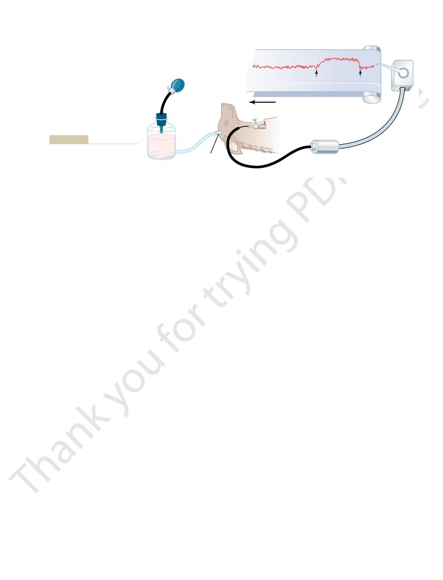
Figure 18–5 shows that signals from the “carotid
carotid sinus,
tion, an area known as the
regions; but, as shown in Figure 18–5, baroreceptors
stretched. A few baroreceptors are located in the wall
lie in the walls of the arteries; they are stimulated when
Physiologic Anatomy of the Baroreceptors and Their Innerva-
the central nervous system. “Feedback” signals are
arteries. A rise in arterial pressure stretches the
, located
Basically, this reflex is initiated by stretch receptors,
System—Baroreceptor Reflexes
The Baroreceptor Arterial Pressure Control
which we explain in the following sections.
negative feedback reflex mechanisms
tain the arterial pressure at or near normal. Almost
sure, there are multiple subconscious special nervous
Normal Arterial Pressure
Nervous Regulation of the Circulation, and Rapid Control of Arterial Pressure
Chapter 18
209
Reflex Mechanisms for Maintaining
Aside from the exercise and stress functions of the
autonomic nervous system to increase arterial pres-
control mechanisms that operate all the time to main-
all of these are
,
By far the best known of the nervous mechanisms for
arterial pressure control is the baroreceptor reflex.
called either baroreceptors or pressoreceptors
at specific points in the walls of several large systemic
baroreceptors and causes them to transmit signals into
then sent back through the autonomic nervous system
to the circulation to reduce arterial pressure down-
ward toward the normal level.
tion.
Baroreceptors are spray-type nerve endings that
of almost every large artery of the thoracic and neck
are extremely abundant in (1) the wall of each inter-
nal carotid artery slightly above the carotid bifurca-
and (2) the
wall of the aortic arch.
to the glossopharyngeal nerves in
tractus solitarius in the
transmitted through the vagus nerves also to the same
tractus solitarius of the medulla.
Response of the Baroreceptors to Pressure.
shows the effect of different arterial pressure levels on
are not stimulated at all by pressures between 0 and
progressively more rapidly and reach a maximum at
ceptors are similar to those of the carotid receptors
Note especially that in the normal operating range
change in pressure causes a strong change in the
baroreflex signal to readjust arterial pressure back
mechanism functions most effectively in the pressure
range where it is most needed.
Glossopharyngeal nerve
Hering’s nerve
Vagus nerve
Aortic baroreceptors
Carotid body
Carotid sinus
The baroreceptor system for controlling arterial pressure.
Figure 18–5
0
160
244
Number of impulses from carotid
sinus nerves per second
80
D
I
D
P
= maximum
Arterial blood pressure (mm Hg)
P, change in arterial blood pressure in mm Hg.
I, change in carotid sinus nerve impulses per second;
pressure.
Activation of the baroreceptors at different levels of arterial
Figure 18–6
D
D
blood pressure regulation has been controversial. One
arterial pressure, their importance in long-term
Are the Baroreceptors Important in Long-Term Regulation of
In summary, a primary purpose of the arterial
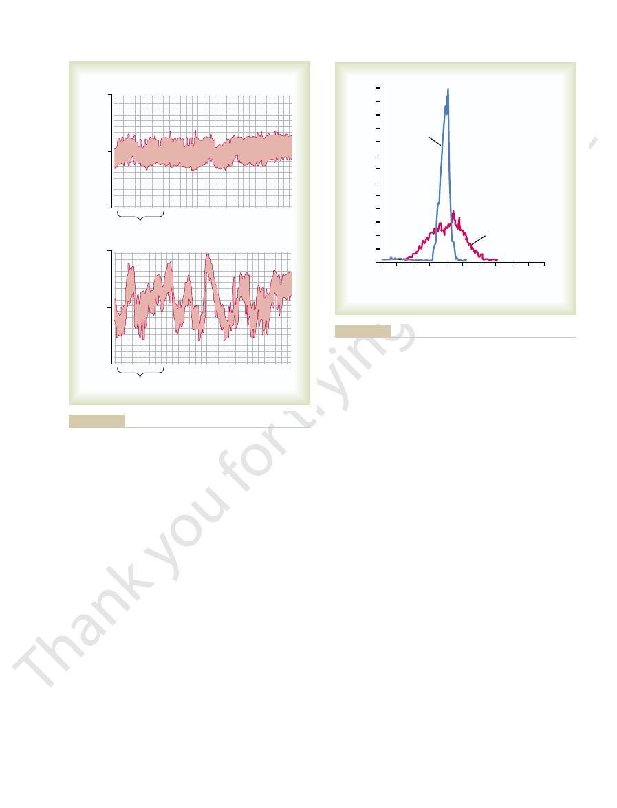
Hg. Thus, one can see the extreme variability of pres-
falling to as low as 50 mm Hg or rising to over 160 mm
that the pressure range increased 2.5-fold, frequently
became the broad, low curve of the figure, showing
the baroreceptors, the frequency distribution curve
exactly 100 mm Hg. Conversely, after denervation of
115 mm Hg—indeed, during most of the day at almost
the day within a narrow range between 85 and
both the normal dog and the denervated dog. Note
Figure 18–9 shows the frequency distributions of the
ment, eating, defecation, and noises.
events of the day, such as lying down, standing, excite-
aorta had been removed. Note the extreme variability
from a normal dog, and the lower record shows an
function of the baroreceptors. The upper record in this
Figure 18–8 shows the importance of this buffer
, and the nerves
sure, it is called a
Function of the Baroreceptor
the head and upper body.
the body. This minimizes the decrease in pressure in
cause loss of consciousness. However, the falling pres-
to fall, and marked reduction of this pressure could
been lying down. Immediately on standing, the arterial
The ability of the baroreceptors to maintain
another minute.
the pressure in the carotid sinuses to rise, and the
carotids are occluded. Removal of the occlusion allows
than usual, causing the aortic arterial pressure to rise
their inhibitory effect on the vasomotor center. The
result, the baroreceptors become inactive and lose
arteries. This reduces the carotid sinus pressure; as a
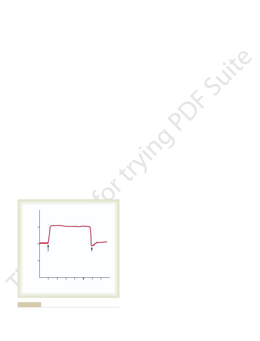
Figure 18–7 shows a typical reflex change in arterial
sure has opposite effects, reflexly causing the pressure
and a decrease in cardiac output. Conversely, low pres-
Therefore, exci-
The net effects are (1)
parasympathetic center.
ius of the medulla, secondary signals
Circulatory Reflex Initiated by the Baroreceptors.
when the pressure is stationary at 150 mm Hg.
Hg but at that moment is rising rapidly, the rate of
sure. That is, if the mean arterial pressure is 150 mm
rapidly changing pressure
respond much more to a
more, the baroreceptors
systole and decreases again during diastole. Further-
changes in arterial pressure; in fact, the rate of impulse
The baroreceptors respond extremely rapidly to
210
Unit IV
The Circulation
firing increases in the fraction of a second during each
than to a stationary pres-
impulse transmission may be as much as twice that
After the
baroreceptor signals have entered the tractus solitar-
inhibit the vaso-
constrictor center of the medulla and excite the vagal
vasodi-
lation of the veins and arterioles throughout the
peripheral circulatory system and (2) decreased heart
rate and strength of heart contraction.
tation of the baroreceptors by high pressure in the
arteries reflexly causes the arterial pressure to decrease
because of both a decrease in peripheral resistance
to rise back toward normal.
pressure caused by occluding the two common carotid
vasomotor center then becomes much more active
and remain elevated during the 10 minutes that the
carotid sinus reflex now causes the aortic pressure to
fall immediately to slightly below normal as a momen-
tary overcompensation and then return to normal in
Function of the Baroreceptors During Changes in Body
Posture.
relatively constant arterial pressure in the upper body
is important when a person stands up after having
pressure in the head and upper part of the body tends
sure at the baroreceptors elicits an immediate reflex,
resulting in strong sympathetic discharge throughout
Pressure “Buffer”
Control System.
Because the baroreceptor system
opposes either increases or decreases in arterial pres-
pressure buffer system
from the baroreceptors are called buffer nerves.
figure shows an arterial pressure recording for 2 hours
arterial pressure recording from a dog whose barore-
ceptor nerves from both the carotid sinuses and the
of pressure in the denervated dog caused by simple
mean arterial pressures recorded for a 24-hour day in
that when the baroreceptors were functioning nor-
mally the mean arterial pressure remained throughout
sure in the absence of the arterial baroreceptor
system.
baroreceptor system is to reduce the minute by minute
variation in arterial pressure to about one third that
which would occur were the baroreceptor system not
present.
Arterial Pressure?
Although the arterial baroreceptors
provide powerful moment-to-moment control of
reason that the baroreceptors have been considered
10
12
14
0
2
4
6
8
150
Both common
carotids clamped
Carotids released
100
50
Arterial pressure (mm Hg)
0
Minutes
caused by clamping both common carotids (after the two vagus
Typical carotid sinus reflex effect on aortic arterial pressure
Figure 18–7
nerves have been cut).
receptors excite nerve fibers that, along with the
adjacent to the aorta). The chemo-
each common carotid artery, and usually one to three
carotid bodies,
chemoreceptor organs
gen ion excess. They are located in several small
tive to oxygen lack, carbon dioxide excess, and hydro-
chemoreceptors
The
ate the response.
that chemoreceptors, instead of stretch receptors, initi-
chemoreceptor reflex
cussed in Chapters 19 and 29.
its associated nervous and hormonal mechanisms), dis-
interaction with additional systems, principally the
toward normal. Thus, long-term regulation of mean
blood volume, which helps to restore arterial pressure
kidneys. This, in turn, causes a gradual decrease in
sure, the baroreceptor reflexes may mediate decreases
For example, with prolonged increases in arterial pres-
influencing sympathetic nerve activity of the kidneys.
long-term blood pressure regulation, especially by
however, have suggested that the baroreceptors do
longer than a few days at a time. Experimental studies,
their potency as a control system for correcting dis-
This “resetting” of the baroreceptors may attenuate
impulses, but gradually, over 1 to 2 days, the rate of
low level, the baroreceptors at first transmit no
Conversely, when the arterial pressure falls to a very
the mean arterial pressure still remains at 160 mm Hg.
days, at the end of which time the rate of firing will
the rate of firing diminishes considerably; then it
are at first transmitted. During the next few minutes,
160 mm Hg, a very high rate of baroreceptor impulses
pressure rises from the normal value of 100 mm Hg to
level to which they are exposed. That is, if the arterial
Nervous Regulation of the Circulation, and Rapid Control of Arterial Pressure
Chapter 18
211
by some physiologists to be relatively unimportant in
chronic regulation of arterial pressure chronically is
that they tend to reset in 1 to 2 days to the pressure
diminishes much more slowly during the next 1 to 2
have returned to nearly normal despite the fact that
baroreceptor firing returns toward the control level.
turbances that tend to change arterial pressure for
not completely reset and may therefore contribute to
in renal sympathetic nerve activity that promote
increased excretion of sodium and water by the
arterial pressure by the baroreceptors requires
renal–body fluid–pressure control system (along with
Control of Arterial Pressure by the Carotid and Aortic
Chemoreceptors—Effect of Oxygen Lack on Arterial Pressure.
Closely associated with the baroreceptor pressure
control system is a
that operates in
much the same way as the baroreceptor reflex except
are chemosensitive cells sensi-
about 2 millimeters in size (two
one of which lies in the bifurcation of
aortic bodies
NORMAL
24
DENERVATED
Time (min)
Arterial pressure (mm Hg)
200
100
0
200
100
0
24
1973. By permission of the American Heart Association, Inc.)
blood pressure and other variables in dogs. Circ Res 32:564,
Guyton AC: Role of baroreceptor reflex in daily control of arterial
had been denervated. (Redrawn from Cowley AW Jr, Liard JF,
several weeks after the baroreceptors
Two-hour records of arterial pressure in a normal dog
Figure 18–8
(above) and
in the same dog (below)
0
50
100
150
200
Percentage of occurrence
250
0
1
2
3
4
5
6
Mean arterial pressure (mm Hg)
Normal
Denervated
tion, Inc.)
Res 32:564, 1973. By permission of the American Heart Associa-
control of arterial blood pressure and other variables in dogs. Circ
AW Jr, Liard JP, Guyton AC: Role of baroreceptor reflex in daily
the baroreceptors had been denervated. (Redrawn from Cowley
period in a normal dog and in the same dog several weeks after
Frequency distribution curves of the arterial pressure for a 24-hour
Figure 18–9

The kidneys, for instance, often
tion caused by intense cerebral ischemia is often so great
as 250 mm Hg.
activity is tremendous: it can elevate the mean arterial
The magnitude of the ischemic effect on vasomotor
ischemic response.
central nervous system ischemic response,
elevation in arterial pressure. This arterial pressure ele-
center, also contribute to the marked stimulation and
It is possible that other factors, such as buildup of
pathetic vasomotor nervous control areas in the brain’s
blood flow to the vasomotor center, the local con-
from the brain stem vasomotor center: at low levels of
pump. This effect is believed to be caused by failure of
excited. When this occurs, the systemic arterial pressure
cerebral ischemia—
severely enough to cause nutritional deficiency—that is,
the brain. However, when blood flow to the vasomotor
chemoreceptors, and the low-pressure receptors, all of
by reflexes that originate in the baroreceptors, the
s Vasomotor
Control of Arterial
prevent damming of blood in the veins, atria, and pul-
strength of heart contraction. Thus, this reflex helps
Then efferent signals are transmitted back through
The stretch receptors of the atria that elicit
as 15 per cent. An additional 40 to 60 per cent increase
the sinus node: it was pointed out in Chapter 10 that
as 75 per cent. A small part of this increase is caused by
heart rate, sometimes increasing the heart rate as much
is discussed again in Chapter 29, along with other mech-
greater arterial pressure. This volume reflex mechanism
heart to greater cardiac output and leads, therefore, to
normal. (We will also see in Chapter 19 that atrial
from the tubules. Combination of these two effects—
fluid into the kidney tubules. The diminution of antidi-
pressure to rise, with resultant increase in filtration of
antidiuretic hormone. The decreased afferent arteriolar
of the afferent arterioles in the kidneys. And still
Atrial Reflexes That Activate the Kidneys—The “Volume Reflex.”
potent for control of arterial pressure.
volume, and they elicit reflexes parallel to the barore-
cannot detect the systemic arterial pressure, they do
Thus, one can see that even though the low-pressure
sure rises about 100 mm Hg.
also are denervated, the pres-
low-pressure receptors
denervated, the pressure rises about 40 mm Hg. If
rises only about 15 mm Hg. With the
into a dog with allreceptors intact, the arterial pressure
example, if 300 milliliters of blood suddenly are infused
in response to changes in blood volume. To give an
role, especially in minimizing arterial pressure changes
arteries. These low-pressure receptors play an important
They are similar to
low-pressure receptors.
Pressure and Other Circulatory Factors.
Atrial and Pulmonary Artery Reflexes That Help Regulate Arterial
The chemoreceptors are discussed in much more
pressure.
Therefore, it is at the lower pressures that this reflex
troller until the arterial pressure falls below 80 mm Hg.
pressure back toward normal. However, this chemo-
the vasomotor center, and this elevates the arterial
The signals transmitted from the chemoreceptors
below a critical level, the chemoreceptors become stim-
with arterial blood. Whenever the arterial pressure falls
abundant blood flow through a small nutrient artery, so
baroreceptor fibers, pass through Hering’s nerves and
212
Unit IV
The Circulation
the vagus nerves into the vasomotor center of the brain
stem.
Each carotid or aortic body is supplied with an
that the chemoreceptors are always in close contact
ulated because diminished blood flow causes decreased
oxygen as well as excess buildup of carbon dioxide and
hydrogen ions that are not removed by the slowly
flowing blood.
excite
receptor reflex is not a powerful arterial pressure con-
becomes important to help prevent still further fall in
detail in Chapter 41 in relation to respiratory control,
in which they play a far more important role than in
pressure control.
Both the atria and
the pulmonary arteries have in their walls stretch recep-
tors called
the baroreceptor stretch receptors of the large systemic
arterial barorecep-
tors
the
receptors in the pulmonary artery and in the atria
detect simultaneous increases in pressure in the low-
pressure areas of the circulation caused by increase in
ceptor reflexes to make the total reflex system more
Stretch of the atria also causes significant reflex dilation
other signals are transmitted simultaneously from the
atria to the hypothalamus to decrease secretion of
resistance in the kidneys causes the glomerular capillary
uretic hormone diminishes the reabsorption of water
increase in glomerular filtration and decrease in reab-
sorption of the fluid—increases fluid loss by the kidneys
and reduces an increased blood volume back toward
stretch caused by increased blood volume also elicits a
hormonal effect on the kidneys—release of atrial natri-
uretic peptide that adds still further to the excretion of
fluid in the urine and return of blood volume toward
normal.)
All these mechanisms that tend to return the blood
volume back toward normal after a volume overload act
indirectly as pressure controllers as well as blood
volume controllers because excess volume drives the
anisms of blood volume control.
Atrial Reflex Control of Heart Rate (the Bainbridge Reflex).
An
increase in atrial pressure also causes an increase in
a direct effect of the increased atrial volume to stretch
such direct stretch can increase the heart rate as much
in rate is caused by a nervous reflex called the Bain-
bridge reflex.
the Bainbridge reflex transmit their afferent signals
through the vagus nerves to the medulla of the brain.
vagal and sympathetic nerves to increase heart rate and
monary circulation.
Central Nervous System Ischemic
Response—
Pressure by the Brain’
Center in Response to Diminished
Brain Blood Flow
Most nervous control of blood pressure is achieved
which are located in the peripheral circulation outside
center in the lower brain stem becomes decreased
to cause
the vasoconstrictor and
cardioaccelerator neurons in the vasomotor center
respond directly to the ischemia and become strongly
often rises to a level as high as the heart can possibly
the slowly flowing blood to carry carbon dioxide away
centration of carbon dioxide increases greatly and
has an extremely potent effect in stimulating the sym-
medulla.
lactic acid and other acidic substances in the vasomotor
vation in response to cerebral ischemia is known as the
or simply CNS
pressure for as long as 10 minutes sometimes to as high
The degree of sympathetic vasoconstric-
that some of the peripheral vessels become totally or
almost totally occluded.
the vessels in the muscles and in the abdomen. The
exercise tightens the muscles, thereby compressing
blood vessels throughout the body. Even anticipation of
skeletal muscles contract during exercise, they compress
When the
normal skeletal muscles.
The abdominal compression reflex is probably much
an increase in both cardiac output and arterial pressure.
. The resulting effect on
for the heart to pump. This overall response is called the
result, increased quantities of blood are made available
abdominal vascular reservoirs toward the heart. As a
the abdomen, helping to translocate blood out of the
muscles. This compresses all the venous reservoirs of
tal muscles of the body, particularly to the abdominal
chemoreceptor reflex is elicited, nerve signals are trans-
When a baroreceptor or
responses are the following.
system, at least two conditions in which the skeletal
Cardiac Output and
Control of Arterial Pressure
Special Features of Nervous
rises high enough to compress the cerebral arteries.
into the cranial vault around the brain. The Cushing
typical Cushing reaction is shown in Figure 18–10,
ing blood to begin again to flow through the brain. A
higher than the cerebrospinal fluid pressure, thus allow-
the brain to relieve the brain ischemia. Ordinarily, the
pressure, blood will flow once again into the vessels of
the arterial pressure to rise. When the arterial pressure
brain. This initiates a CNS ischemic response that causes
pressure, it compresses the whole brain as well as the
the brain in the cranial vault. For instance, when the
The so-called
the “last ditch stand” pressure control mechanism.
pressure whenever blood flow to the brain decreases dan-
very powerfully to prevent further decrease in arterial
arterial pressure. Instead, it operates principally as an
stimulation at a pressure of 15 to 20 mm Hg. Therefore,
60 mm Hg and below, reaching its greatest degree of
the arterial pressure falls far below normal, down to
ischemic response, it does not become significant until
of the most powerful of all the activators of the sympa-
the CNS ischemic response is one
discharge. Therefore,
Nervous Regulation of the Circulation, and Rapid Control of Arterial Pressure
Chapter 18
213
entirely cease their production of urine because of renal
arteriolar constriction in response to the sympathetic
thetic vasoconstrictor system.
Importance of the CNS Ischemic Response as a Regulator of Arte-
rial Pressure.
Despite the powerful nature of the CNS
it is not one of the normal mechanisms for regulating
emergency pressure control system that acts rapidly and
gerously close to the lethal level. It is sometimes called
Cushing Reaction.
Cushing reaction is a
special type of CNS ischemic response that results from
increased pressure of the cerebrospinal fluid around
cerebrospinal fluid pressure rises to equal the arterial
arteries in the brain and cuts off the blood supply to the
has risen to a level higher than the cerebrospinal fluid
blood pressure comes to a new equilibrium level slightly
caused in this instance by pumping fluid under pressure
reaction helps protect the vital centers of the brain from
loss of nutrition if ever the cerebrospinal fluid pressure
Role of the Skeletal Nerves and
Skeletal Muscles in Increasing
Arterial Pressure
Although most rapidly acting nervous control of the cir-
culation is effected through the autonomic nervous
nerves and muscles also play major roles in circulatory
Abdominal Compression Reflex.
mitted simultaneously through skeletal nerves to skele-
abdominal compression reflex
the circulation is the same as that caused by sympathetic
vasoconstrictor impulses when they constrict the veins:
more important than has been realized in the past
because it is well known that people whose skeletal
muscles have been paralyzed are considerably more
prone to hypotensive episodes than are people with
Increased Cardiac Output and Arterial Pressure Caused by
Skeletal Muscle Contraction During Exercise.
Connector to
subarachnoid
space
Pressure
bottle
Arterial
pressure
transducer
Pen
recorder
CSF pressure
reduced
CSF pressure
raised
Zero
pressure
Moving paper
Arterial
pressure
brospinal fluid (CSF) pressure.
resulting from increased cere-
rapid rise in arterial pressure
Cushing reaction,” showing a
Figure 18–10
“

side instead of following a straight course.
guiding mechanism, the plane will oscillate from side to
systems. For instance, if the feedback “gain” is too great
The vasomotor waves are of considerable theoretical
sure receptor and the subsequent pressure response.
oscillate if the intensity of “feedback” is strong enough
Thus, any reflex pressure control mechanism can
repeated itself cyclically as long as the cerebrospinal
ischemia was relieved and again the pressure fell. This
then initiated another rise in pressure. Again the
value, causing brain ischemia once again. The ischemia
pathetic nervous system became inactive. As a result,
high value, the brain ischemia was relieved and the sym-
200 mm Hg. When the arterial pressure rose to such a
160 mm Hg, which compressed the cerebral vessels and
ment, the cerebrospinal fluid pressure was raised to
ischemic pressure control mechanism. In this experi-
Figure 18–11
The record in
control becomes weaker.
circulation becomes powerful, whereas baroreceptor
because in this low range, chemoreceptor control of the
arterial pressure is in the range of 40 to 80 mm Hg
taneously with the baroreceptor reflex. It probably plays
same type of waves. This reflex usually oscillates simul-
chemoreceptor reflex
The
another cycle, and the oscillation continues on and on.
few seconds later. This high pressure then initiates
response is not instantaneous, and it is delayed until a
again, elevating the pressure to a high value. The
lowers the pressure a few seconds later. The decreased
That is, a high pressure excites the baroreceptors;
much less intense than shown in the figure. They are
in experimental pressure recordings, although usually
The vasomotor waves of Figure 18–11
Oscillation of the Baroreceptor and Chemoreceptor Reflexes.
some of which are the following.
of one or more nervous pressure control mechanisms,
The cause of vasomotor waves is “reflex oscillation”
showing the cyclical rise and fall in arterial pressure.
waves.” Such records are demonstrated in Figure 18–11,
These waves are called
each cycle varies from 26 seconds in the anesthetized
more slowly than the respiratory waves. The duration of
great as 10 to 40 mm Hg at times—that rise and fall
piration, some much larger waves are also noted—as
Waves
Vasomotor
20 mm Hg with each respiratory cycle.
ration, the blood pressure can rise and fall as much as
remainder of the respiratory cycle. During deep respi-
waves, the net result during normal respiration is usually
receptors.
3. The pressure changes caused in the thoracic vessels
decreases the cardiac output and arterial pressure.
This reduces the quantity of blood returning to the
2. Every time a person inspires, the pressure in the
the vasomotor center with each respiratory cycle.
respiratory center of the medulla “spill over” into
1. Many of the “breathing signals” that arise in the
some of which are reflex in nature, as follows:
sure. The waves result from several different effects,
manner, causing
usually rises and falls 4 to 6 mm Hg in a wavelike
With each cycle of respiration, the arterial pressure
Respiratory Waves in the
100 mm Hg up to 130 to 160 mm Hg.
exercise, an increase usually from a normal mean of
The increase in cardiac output in turn is an essential
cardiac output that sometimes occurs in heavy exercise.
increase the cardiac output. This is an essential effect in
eral vessels into the heart and lungs and, therefore, to
214
Unit IV
The Circulation
resulting effect is to translocate blood from the periph-
helping to cause the fivefold to sevenfold increase in
ingredient in increasing the arterial pressure during
Arterial Pressure
respiratory waves in the arterial pres-
thoracic cavity becomes more negative than usual,
causing the blood vessels in the chest to expand.
left side of the heart and thereby momentarily
by respiration can excite vascular and atrial stretch
Although it is difficult to analyze the exact relations
of all these factors in causing the respiratory pressure
an increase in arterial pressure during the early part
of expiration and a decrease in pressure during the
Arterial Pressure “
”
—Oscillation of Pressure
Reflex Control Systems
Often while recording arterial pressure from an animal,
in addition to the small pressure waves caused by res-
dog to 7 to 10 seconds in the unanesthetized human.
vasomotor waves or “Mayer
B are often seen
caused mainly by oscillation of the baroreceptor reflex.
this then inhibits the sympathetic nervous system and
pressure in turn reduces the baroreceptor stimulation
and allows the vasomotor center to become active once
can also oscillate to give the
the major role in causing vasomotor waves when the
Oscillation of the CNS Ischemic Response.
A resulted from oscillation of the CNS
initiated a CNS ischemic pressure response up to
the arterial pressure fell rapidly back to a much lower
fluid pressure remained elevated.
and if there is a delay between excitation of the pres-
importance because they show that the nervous reflexes
that control arterial pressure obey the same principles
as those applicable to mechanical and electrical control
in the guiding mechanism of an automatic pilot for an
airplane and there is also delay in response time of the
Pressure (mm Hg)
200
160
120
80
40
0
A
B
100
60
Vasomotor waves caused by baroreceptor reflex
response.
Vasomotor waves caused by oscillation of the CNS ischemic
Figure 18–11
A,
B,
oscillation.

N Y Acad Sci 940:431, 2001.
angiotensin II, nitric oxide, and exercise training. Ann
of sympathetic outflow in heart failure. The roles of
Zucker IH, Wang W, Pliquett RU, et al: The regulation
Physiol 282:R1044, 2002.
rogenic hypertension. Am J Physiol Regul Integr Comp
Thrasher TN: Unloading arterial baroreceptors causes neu-
Hypertens Rep 5:262, 2003 .
tension: role of the rostral ventrolateral medulla. Curr
Sved AF, Ito S, Sved JC: Brainstem mechanisms of hyper-
Am J Physiol Regul Integr Comp Physiol 281:R683, 2001.
Morrison SF: Differential control of sympathetic outflow.
News Physiol Sci 16:266, 2001.
Mifflin SW:What does the brain know about blood pressure?
Am J Physiol Regul Integr Comp Physiol 286:R1, 2004.
nerve activity: is there a role for arterial baroreceptors?
Malpas SC: What sets the long-term level of sympathetic
term blood pressure regulation. Am J Hypertens 14:147S,
Lohmeier TE: The sympathetic nervous system and long-
hypertension. Hypertension 39:550, 2002.
Lohmeier TE, Lohmeier JR, Warren S, et al: Sustained acti-
system. Ann N Y Acad Sci 940:338, 2001.
Krieger EM, Da Silva GJ, Negrao CE: Effects of exercise
Circulation 105:2518, 2002.
tension, orthostatic tachycardia, and malignant vagotonia.
of baroreflex failure: hypertensive crisis, volatile hyper-
Ketch T, Biaggioni I, Robertson R, Robertson D: Four faces
tens 14:103S, 2001.
of leptin and sympathetic nervous system. Am J Hyper-
Hall JE, Hildebrandt DA, Kuo J: Obesity hypertension: role
phia: WB Saunders Co, 1980.
Guyton AC: Arterial Pressure and Hypertension. Philadel-
Intern Med 137:753, 2002.
clinical disorders of the autonomic nervous system. Ann
Goldstein DS, Robertson D, Esler M, et al: Dysautonomias:
Physiol 284:R259, 2003.
brain: new perspectives. Am J Physiol Regul Integr Comp
Felder RB, Francis J, Zhang ZH, et al: Heart failure and the
Ann N Y Acad Sci 940:500, 2001.
control of sympathetic outflow in human heart failure.
Floras JS: Arterial baroreceptor and cardiopulmonary reflex
practice. Acta Physiol Scand 177:275, 2003.
humans: translation from pathophysiology into clinical
Esler M, Lambert G, Brunner-La Rocca HP, et al: Sympa-
and arterial pressure. Ann N Y Acad Sci 940:324, 2001.
DiCarlo SE, Bishop VS: Central baroreflex resetting as a
940:395, 2001.
nerves in control of renal function. Ann N Y Acad Sci
DiBona GF: Peripheral and central interactions between
50:61, 1974.
tion in angiotensin II–induced hypertension. Circulation
Cowley AW Jr, Guyton AC: Baroreceptor reflex contribu-
medial hypothalamus. Neuroscience 126:229, 2004.
Cao WH, Fan W, Morrison SF: Medullary pathways mediat-
84: 169, 2004.
roendocrine control of body fluid metabolism. Physiol Rev
Antunes-Rodrigues J, De Castro M, Elias LLK, et al: Neu-
Nervous Regulation of the Circulation, and Rapid Control of Arterial Pressure
Chapter 18
215
References
ing specific sympathetic responses to activation of dorso-
the renin-angiotensin system and the renal sympathetic
means of increasing and decreasing sympathetic outflow
thetic nerve activity and neurotransmitter release in
training on baroreflex control of the cardiovascular
vation of the central baroreceptor pathway in angiotensin
2001.