more than required to maintain full tissue oxygenation but no more than this.
delivery of oxygen, the blood flow is always controlled at a level only slightly
no less. For instance, in tissues for which the most important requirement is
lated at the minimal level that will supply the tissue’s requirements— no more,
require many times more blood flow than the heart can pump.
ity of the tissue is little or great? The answer is equally simple: To do this would
tissue of the body, always enough to supply the tissue’s needs whether the activ-
tion: Why not simply allow a very large blood flow all the time through every
Importance of Blood Flow Control by the Local Tissues.
min/100 g of muscle).
as high as 16,000 ml/min in the body’s total muscle vascular bed (or 80 ml/
increase more than 60-fold and the blood flow as much as 20-fold, increasing to
4 ml/min/100 g. Yet, during heavy exercise, muscle metabolic activity can
bolic activity of the muscles is very low, and so also is the blood flow, only
between 30 and 40 per cent of the total body mass. In the resting state, the meta-
of the body, only a total of 750 ml/min, even though the muscles constitute
Conversely, most surprising is the low blood flow to all the
tion of cleansing the blood of waste products.
This extreme amount of flow is required for the kidneys to perform their func-
of 1350 ml/min in the liver, which is 95 ml/min/100 g of liver tissue.
large blood flows in some organs—for example, several hundred milliliters per
Note in Table 17–1 the very
Variations in Blood Flow in Different Tissues and Organs.
We shall see that most of these factors exert extreme degrees of local blood
allows the kidneys to excrete the waste products of the body.
perature. Also, delivery of adequate quantities of blood plasma to the kidneys
Certain organs have special requirements. For instance, blood flow to the skin
6. Transport of various hormones and other substances to the different tissues
5. Maintenance of proper concentrations of other ions in the tissues
4. Removal of hydrogen ions from the tissues
3. Removal of carbon dioxide from the tissues
2. Delivery of other nutrients, such as glucose, amino acids, and fatty acids
1. Delivery of oxygen to the tissues
to this is manyfold, including the following:
What are some of the specific needs of the tissues for blood flow? The answer
bolic needs.
Local Control of Blood
Blood Flow by the Tissues
C
H
A
P
T
E
R
1
7
195
Local and Humoral Control of
Flow in Response to
Tissue Needs
One of the most fundamental principles of circula-
tory function is the ability of each tissue to control
its own local blood flow in proportion to its meta-
determines heat loss from the body and in this way helps to control body tem-
flow control.
minute per 100 grams of thyroid or adrenal gland tissue and a total blood flow
Also note the extremely large blood flow through the kidneys—1100 ml/min.
inactive muscles
One might ask the simple ques-
Experiments have shown that the blood flow to each tissue usually is regu-

mainly in response to oxygen deficiency. For instance,
, and
histamine, potassium ions
carbon dioxide
suggested are
metarterioles, and arterioles to cause dilation. Some of
through the tissues to the precapillary sphincters,
The
nutrients to a tissue, the greater the rate of formation
ing to this theory, the greater the rate of metabolism
lation—Possible Special Role of Adenosine.
Vasodilator Theory for Acute Local Blood Flow Regu-
lack theory
oxygen
They are (1) the
olism changes or the availability of oxygen changes.
There are two basic theories for the regulation of
effect of oxygen deficiency to increase blood flow.
much as sevenfold, thus demonstrating the extreme
Total cyanide poisoning of oxygen usage by a local
supply of oxygen to the tissues.
blood, thus almost maintaining an exact constant
increases almost enough, but not quite enough, to
leg increases about threefold; that is, the blood flow
per cent of normal, the blood flow through an isolated
tissues increases markedly. Figure 17–2 shows that as
tissues to use oxygen), the blood flow through the
cyanide poisoning (which poisons the ability of the
ability of hemoglobin to transport oxygen), or (4) in
tude at the top of a high mountain, (2) in pneumonia,
oxygen to the tissues decreases, such as (1) at high alti-
nutrients is oxygen. Whenever the availability of
Acute Local Blood Flow Regulation When Oxygen Availability
a local tissue, such as in a skeletal muscle. Note that an
Figure
Effect of Tissue Metabolism on Local Blood Flow.
Acute Control of Local Blood Flow
tissues.
needs of the tissues. These changes come about as a
months. In general, these long-term changes provide
changes in flow over a period of days, weeks, or even
however, means slow, controlled
tenance of appropriate local tissue blood flow.
metarterioles, and precapillary sphincters, occurring
vasodilation or vasoconstriction of the arterioles,
phases: (1) acute control and (2) long-term control.
Flow Control
deficiency, and yet the workload on the heart is kept
By controlling local blood flow in such an exact way,
196
Unit IV
The Circulation
the tissues almost never suffer from oxygen nutritional
at a minimum.
Mechanisms of Blood
Local blood flow control can be divided into two
Acute control is achieved by rapid changes in local
within seconds to minutes to provide very rapid main-
Long-term control,
even better control of the flow in proportion to the
result of an increase or decrease in the physical sizes
and numbers of actual blood vessels supplying the
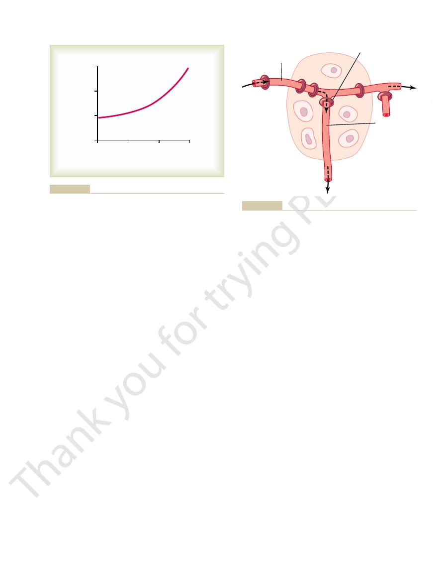
17–1 shows the approximate quantitative acute effect
on blood flow of increasing the rate of metabolism in
increase in metabolism up to eight times normal
increases the blood flow acutely about fourfold.
Changes.
One of the most necessary of the metabolic
(3) in carbon monoxide poisoning (which poisons the
the arterial oxygen saturation decreases to about 25
make up for the decreased amount of oxygen in the
tissue area can cause local blood flow to increase as
local blood flow when either the rate of tissue metab-
vasodilator theory and (2) the
.
Accord-
or the less the availability of oxygen or some other
of vasodilator substances in the tissue cells.
vasodilator substances then are believed to diffuse
the different vasodilator substances that have been
adenosine,
, adenosine
phosphate compounds,
hydrogen tons.
Most of the vasodilator theories assume that the
vasodilator substance is released from the tissue
Blood Flow to Different Organs and Tissues Under Basal
Table 17–1
Total
100.0
5000
Other tissues
3.5
175
1.3
Adrenal glands
0.5
25
300
Thyroid gland
1
50
160
Skin (cool weather)
6
300
3
Bone
5
250
3
Muscle (inactive state)
15
750
4
Arterial
(6)
300
Portal
(21)
1050
Liver
27
1350
95
Kidneys
22
1100
360
Bronchi
2
100
25
Heart
4
200
70
Brain
14
700
50
Per cent
ml/min
ml/min/100 g
Conditions
Based mainly on data compiled by Dr. L. A. Sapirstein.
Blood flow (x normal)
0
0
2
3
4
7
6
5
4
3
2
1
8
Rate of metabolism (x normal)
1
Normal level
Effect of increasing rate of metabolism on tissue blood flow.
Figure 17–1
The cyclical opening and closing is called
oles open and close cyclically several times per minute,
nutrition. The precapillary sphincters and metarteri-
open or completely closed. The number of precapillary
scope—for example, in a bat’s wing—one sees that the
muscle fibers. Observing such a tissue under a micro-
single sidearm capillary and its surrounding tissue. At
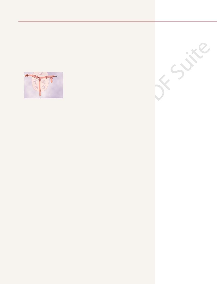
shows a tissue unit, consisting of a metarteriole with a
could operate is shown in Figure 17–3. This figure
local blood vessels, and this, too, would cause local
and therefore naturally dilate. Also, increased utiliza-
absence of adequate oxygen, it is reasonable to
to cause vascular muscle contraction. Therefore, in the
oxygen are involved). Oxygen (and other nutrients
nutrient lack theory
the oxygen lack theory or, more accurately, the
favor still another theory, which can be called either
Oxygen Lack Theory for Local Blood Flow Control.
tors could increase the blood flow sufficiently.
flow. But a combination of several different vasodila-
has been the following: It has been difficult to prove
well as in the heart. The problem with the different
Although research evidence is less clear, many phys-
heart muscle cells to cause coronary vasodilation, pro-
(ATP), which (3) increases the release of adenosine. It
utilization of oxygen, followed by (1) decreased
increases an extra amount, this, too, causes increased
more active than normal and the heart’s metabolism
back to normal. Also, whenever the heart becomes
becomes too little, and this causes enough local vasodi-
example, minute quantities of adenosine are released
vasodilators for controlling local blood flow. For
sible, or partially responsible, for the local blood flow
between the tissue cells; these substances then cause
Local and Humoral Control of Blood Flow by the Tissues
Chapter 17
197
experiments have shown that decreased availability of
oxygen can cause both adenosine and lactic acid (con-
taining hydrogen ions) to be released into the spaces
intense acute vasodilation and therefore are respon-
regulation.
Many physiologists have suggested that the sub-
stance adenosine is the most important of the local
from heart muscle cells when coronary blood flow
lation in the heart to return coronary blood flow
oxygen concentration in the heart muscle cells with (2)
consequent degradation of adenosine triphosphate
is believed that much of this adenosine leaks out of the
viding increased coronary blood flow to supply the
increased nutrient demands of the active heart.
iologists also have suggested that the same adenosine
mechanism is the most important controller of blood
flow in skeletal muscle and many other tissues as
vasodilator theories of local blood flow regulation
that sufficient quantities of any single vasodilator sub-
stance (including adenosine) are indeed formed in the
tissues to cause all the measured increase in blood
Although the vasodilator theory is widely accepted,
several critical facts have made other physiologists
(because other nutrients besides
as well) is required as one of the metabolic nutrients
believe that the blood vessels simply would relax
tion of oxygen in the tissues as a result of increased
metabolism theoretically could decrease the availabil-
ity of oxygen to the smooth muscle fibers in the
vasodilation.
A mechanism by which the oxygen lack theory
the origin of the capillary is a precapillary sphincter,
and around the metarteriole are several other smooth
precapillary sphincters are normally either completely
sphincters that are open at any given time is roughly
proportional to the requirements of the tissue for
with the duration of the open phases being propor-
tional to the metabolic needs of the tissues for oxygen.
vasomotion.
Let us explain how oxygen concentration in the
local tissue could regulate blood flow through the area.
Because smooth muscle requires oxygen to remain
50
25
Blood flow (x normal)
75
100
0
2
3
Arterial oxygen saturation (per cent)
1
through an isolated dog leg.
Effect of decreasing arterial oxygen saturation on blood flow
Figure 17–2
Precapillary sphincter
Metarteriole
Sidearm capillary
for controlling capillary blood flow.
precapillary sphincter
sidearm capillary
passing through
metarteriole
back control of blood flow, showing a
Diagram of a tissue unit area for explanation of acute local feed-
Figure 17–3
the tissue and a
with its
“acute” curve in Figure 17–4. Note that between an
flow.” After autoregulation has occurred, the local
flow toward normal is called “
the arterial pressure is kept elevated. This return of
tissues returns almost to the normal level, even though
within less than a minute, the blood flow in most
pressure causes immediate rise in blood flow. But,
In any tissue of the body, an acute increase in arterial
Arterial Pressure Changes from Normal—
“Autoregulation” of Blood Flow When the
as 20-fold during intense exercise.
pointed out earlier, active hyperemia in skeletal
required to sustain its new level of function. As
and, therefore, to increase local blood flow. In this way,
stances. The result is to dilate the local blood vessels
The increase in local metabolism causes the cells
control, one can easily understand this
flow through the tissue increases. Here again, by
brain during rapid mental activity, the rate of blood
gland during a hypersecretory period, or even the
active, such as an exercising muscle, a gastrointestinal
When any tissue becomes highly
ents to the tissues.
during the period of occlusion. This mechanism
lar occlusion, the extra blood flow during the reactive
that cause vasodilation. After short periods of vascu-
that is, lack of flow sets into motion all of those factors
local “metabolic” blood flow regulation mechanism;
blood flow has been stopped for an hour or more. This
normal; this increased flow will continue for a few
and then is unblocked, blood flow through the tissue
When the blood supply to a tissue
examples of metabolic control of local blood flow are
metabolic needs of the tissues. Two additional special
nisms” because all of them function in response to the
local blood flow control are called “metabolic mecha-
The mechanisms that we have described thus far for
Control of Local Blood Flow
deficiency of these vitamins might lead to diminished
ATP in the tissue cells, one can well understand how
disease, the peripheral vascular blood flow everywhere
. In this
, and
lation occurs in the vitamin deficiency disease
has not been studied adequately. In addition, vasodi-
amino acids or fatty acids, are deficient, although this
this same effect occurs when other nutrients, such as
cause local tissue vasodilation. Also, it is possible that
Under special conditions, it has been
Local Blood Flow.
mechanisms.
response to the metabolic needs of the tissues.
oxygen lack theory
or an
Thus, on the basis of available data, either a
the cycle again.
enough, the sphincters would open once more to begin
cells consume the excess oxygen. But when the excess
a certain level, the precapillary and metarteriole
increase in oxygen concentration. Consequently, when
contracted, one might assume that the strength of con-
198
Unit IV
The Circulation
traction of the sphincters would increase with an
the oxygen concentration in the tissue rises above
sphincters presumably would close until the tissue
oxygen is gone and the oxygen concentration falls low
vasodilator substance theory
could explain acute local blood flow regulation in
Probably the truth lies in a combination of the two
Possible Role of Other Nutrients Besides Oxygen in Control of
shown that lack of glucose in the perfusing blood can
beriberi,
in which the patient has deficiencies of the vitamin B
substances thiamine, niacin
riboflavin
in the body often increases twofold to threefold.
Because these vitamins all are needed for oxygen-
induced phosphorylation that is required to produce
smooth muscle contractile ability and therefore also
local vasodilation.
Special Examples of Acute “Metabolic”
reactive hyperemia and active hyperemia.
Reactive Hyperemia.
is blocked for a few seconds to as long an hour or more
usually increases immediately to four to seven times
seconds if the block has lasted only a few seconds but
sometimes continues for as long as many hours if the
phenomenon is called reactive hyperemia.
Reactive hyperemia is another manifestation of the
hyperemia phase lasts long enough to repay almost
exactly the tissue oxygen deficit that has accrued
emphasizes the close connection between local blood
flow regulation and delivery of oxygen and other nutri-
Active Hyperemia.
simply applying the basic principles of local blood flow
active hyper-
emia.
to devour tissue fluid nutrients extremely rapidly and
also to release large quantities of vasodilator sub-
the active tissue receives the additional nutrients
muscle can increase local muscle blood flow as much
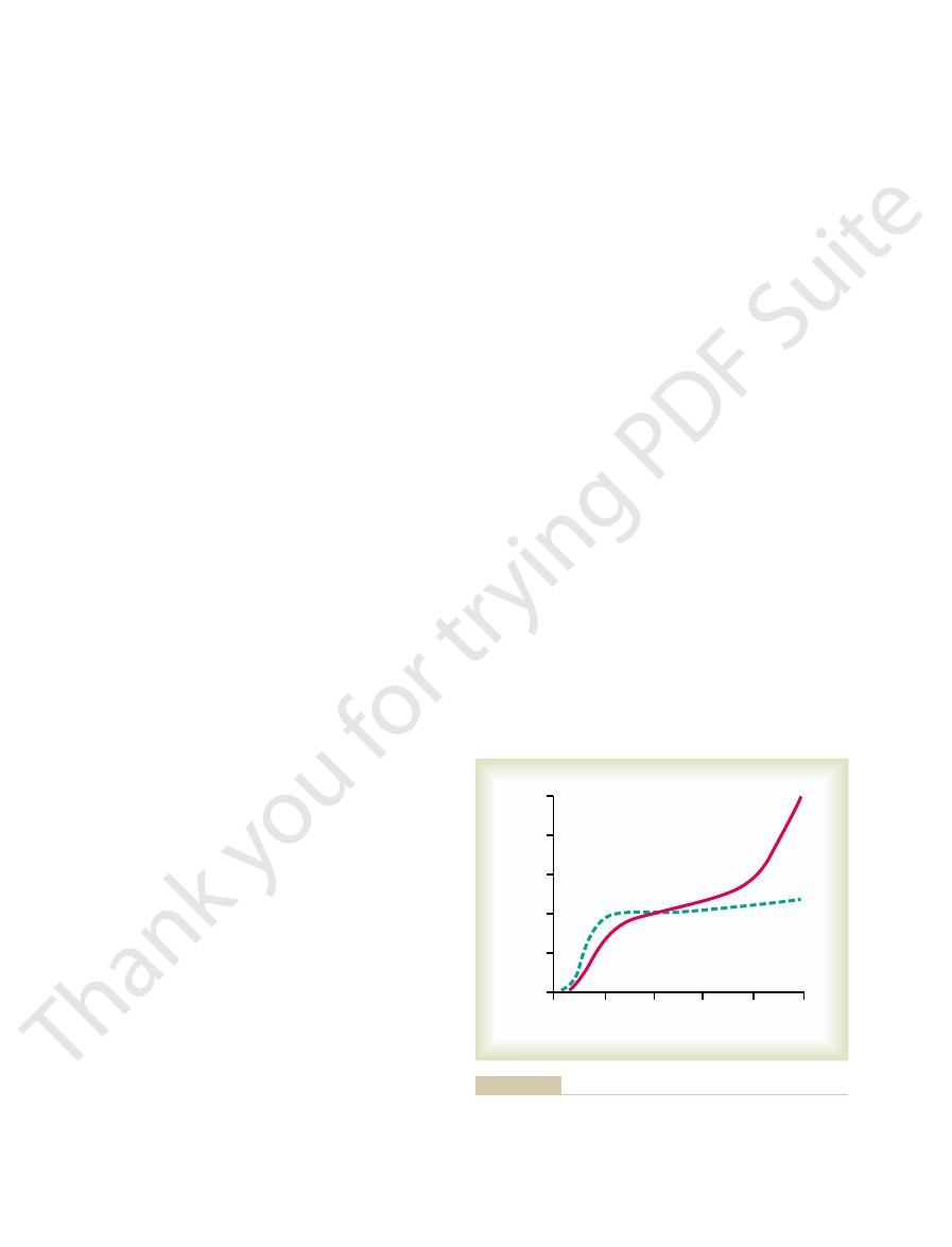
“Metabolic” and “Myogenic” Mechanisms
autoregulation of blood
blood flow in most body tissues will be related to arte-
rial pressure approximately in accord with the solid
Blood flow (x normal)
100
50
0
0.5
0
1.0
1.5
2.0
2.5
150
200
250
Arterial pressure (mm Hg)
Acute
Long-term
curve shows the effect if the arterial pressure is raised extremely
sure is raised over a period of a few minutes. The dashed green
a muscle. The solid red curve shows the effect if the arterial pres-
Effect of different levels of arterial pressure on blood flow through
Figure 17–4
slowly over a period of many weeks.

response, the effectiveness of local blood flow control
lar blood flow increases downstream. Without such a
nitric oxide then relaxes the blood vessels. This is for-
nificant increase in the release of nitric oxide. The
against the vascular walls. This stress contorts the
blood of only 6 seconds. Rapid flow of blood through
, which has a half-life in the
nitric oxide
, which is composed
traction of the arterial wall. The most important of
released, can affect the degree of relaxation or con-
arteries synthesize several substances that, when
The endothelial cells lining the arterioles and small
the larger arteries as well. This mechanism is the
vascular portion of the circulation increases, this sec-
upstream. Yet, when blood flow through a micro-
vessels, not the intermediate and larger arteries back
or tissue cell oxygen deficiency can reach only these
The local mechanisms for controlling tissue blood flow
When Microvascular Blood Flow Increases—
Mechanism for Dilating Upstream Arteries
. This special mechanism for cerebral
dioxide concentration and hydrogen ion
ions from the brain tissues. This is important
prominent roles. An increase of either or both of
tissue oxygen concentration, the concentrations of
, in addition to control of blood flow by
2. In the
filtration rate back to or near to normal. The
constriction of the afferent arterioles, in this way
glomerulus into the tubular system, appropriate
When too
juxtaglomerular apparatus.
This is located where the distal tubule lies adjacent
tubuloglomerular feedback,
, blood flow control is vested mainly
1. In the
throughout this text in relation to specific organs, but
operate in a few special areas. They all are discussed
tissues of the body, distinctly different mechanisms
Control in Specific Tissues
blood flow.
increased, such as during vigorous muscle exercise,
tissue. Indeed metabolic factors appear to override
pressure is increased. However, the importance of
The myogenic mechanism may be important in pre-
vascular wall or to the ion channels themselves.
teins that are tethered to cytoskeleton elements of the
cular ion channels are still uncertain, but likely involve
lar contraction. The precise mechanisms by which
to contract. Changes in vascular pressure may also
from the extracellular fluid into the cells, causing them
ization, which then rapidly increases calcium ion entry
veins, and even lymphatic vessels. Myogenic contrac-
rioles but can also be observed in arteries, venules,
or hormonal influences. It is most pronounced in arte-
The myogenic response is inherent to vascular
relaxes and allows increased flow.
stretch of the vessel is less, so that the smooth muscle
normal. Conversely, at low pressures, the degree of
stretches the vessel, this in turn causes reactive vascu-
vessel wall to contract for a few seconds. Therefore, it
explains the phenomenon of autoregulation. This
, however, suggests that still
The
pressure.
ents to the tissues. These nutrients (especially oxygen)
arterial pressure becomes too great, the excess flow
lation discussed in previous sections. Thus, when the
The
myogenic theory.
to explain this acute autoregulation mechanism. They
For almost a century, two views have been proposed
arterial pressure of about 70 mm Hg and 175 mm Hg,
Local and Humoral Control of Blood Flow by the Tissues
Chapter 17
199
the blood flow increases only 30 per cent even though
the arterial pressure increases 150 per cent.
have been called (1) the metabolic theory and (2) the
metabolic theory can be understood easily by
applying the basic principles of local blood flow regu-
provides too much oxygen and too many other nutri-
then cause the blood vessels to constrict and the flow
to return nearly to normal despite the increased
myogenic theory
another mechanism not related to tissue metabolism
theory is based on the observation that sudden stretch
of small blood vessels causes the smooth muscle of the
has been proposed that when high arterial pressure
lar constriction that reduces blood flow nearly back to
smooth muscle and can occur in the absence of neural
tion is initiated by stretch-induced vascular depolar-
open or close other ion channels that influence vascu-
changes in pressure cause opening or closing of vas-
mechanical effects of pressure on extracellular pro-
venting excessive stretch of blood vessel when blood
the myogenic mechanism in blood flow regulation
is unclear because this pressure sensing mechanism
cannot directly detect changes in blood flow in the
the myogenic mechanism in circumstances where the
metabolic demands of the tissues are significantly
which can cause dramatic increases in skeletal muscle
Special Mechanisms for Acute Blood Flow
Although the general mechanisms for local blood flow
control discussed thus far are present in almost all
two notable ones are as follows:
kidneys
in a mechanism called
in which the composition of the fluid in the early
distal tubule is detected by an epithelial structure
of the distal tubule itself called the macula densa.
to the afferent and efferent arterioles at the
nephron
much fluid filters from the blood through the
feedback signals from the macula densa cause
reducing both renal blood flow and glomerular
details of this mechanism are discussed in
Chapter 26.
brain
carbon dioxide and hydrogen ions play very
these dilates the cerebral vessels and allows rapid
washout of the excess carbon dioxide or hydrogen
because the level of excitability of the brain itself is
highly dependent on exact control of both carbon
concentration
blood flow control is presented in Chapter 61.
The Endothelium-Derived Relaxing Factor
(Nitric Oxide)
can dilate only the very small arteries and arterioles in
each tissue because tissue cell vasodilator substances
ondarily entrains another mechanism that does dilate
following:
these is a vasodilator substance called endothelium-
derived relaxing factor (EDRF)
principally of
the arteries and arterioles causes shear stress on the
endothelial cells because of viscous drag of the blood
endothelial cells in the direction of flow and causes sig-
tunate because it increases the diameters of the
upstream arterial blood vessels whenever microvascu-

growth of new vessels.
larger vessels. Thus, angiogenesis explains the manner
the wall, so that some of the new vessels eventually
great enough, smooth muscle cells eventually invade
loop through which blood begins to flow. If the flow is
fold over into a tube. Next, the tube connects with
The cells in each cord continue to divide and rapidly
directed toward the source of the angiogenic factor.
cells at the point of sprouting. This is followed by rapid
to sprout from other small vessels. The first step is dis-
vessel growth in the same way. They cause new vessels
tissue oxygen or other nutrients, or both, that leads to
equate blood supply. Presumably, it is deficiency of
, each
, and
(VEGF), fibroblast growth factor
vascular endothelial growth factor
small peptides. Three of those that have been best
blood vessels have been found, almost all of which are
Factor in Formation of New Blood Vessels
Importance of Vascular Endothelial Growth
and this eventually causes blindness. (This condition is
grow out from the retina into the eye’s vitreous humor;
the sudden decrease in available oxygen; indeed, there
the infant is taken out of the oxygen tent, there is
the small vessels that already have formed. Then when
baby’s eyes and even causes degeneration of some of
The excess oxygen causes almost immediate cessation
babies put into oxygen tents for therapeutic purposes.
conductivity as is normally true.This same effect is also
high altitudes, where the atmospheric oxygen is low. A
also for long-term control. One example of this is
Role of Oxygen in Long-Term Regulation.
quently lags far behind the needs of the tissues.
blood flow, whereas in older tissues, vascularity fre-
older, so that in the neonate, the vascularity will adjust
the elderly person. Furthermore, the final degree of
well-established tissues. Therefore, the time required
tissue; however, it occurs much more slowly in old,
growth tissue, such as in scar tissue and cancerous
extremely young animals. It also occurs rapidly in new
This reconstruction occurs rapidly (within days) in
the tissue vasculature to meet the needs of the tissues.
Thus, there is actual physical reconstruction of
vascularity decreases.
vascularity increases; if the metabolism is decreased,
of the tissues. For instance, if the metabolism in a
The mechanism of long-term local blood flow regula-
Change in “Tissue Vascularity”
Mechanism of Long-Term Regulation—
increased quantities of oxygen and other nutrients, the
of a tissue change. Thus, if a tissue becomes chroni-
effect on the rate of local blood flow.
rial pressure between 50 and 250 mm Hg have little
tion has had time to occur, long-term changes in arte-
flow regulation. Note that once the long-term regula-
Figure 17–4 shows by the dashed green curve the
rial pressure remains at 150 mm Hg indefinitely, within
instance, in the aforementioned example, if the arte-
regulation gives far more complete regulation. For
in addition to the acute regulation. This long-term
However, over a period of hours, days, and weeks, a
cent increase in blood flow.
at the same time, it demonstrates that the regulation is
acute mechanisms for local blood flow regulation, but
nal control value. This illustrates the rapidity of the
Then, within 30 seconds to 2 minutes, the flow
increased from 100 to 150 mm Hg, the blood flow
For instance, when the arterial pressure suddenly is
the exact additional requirements of the tissues.
of these acute mechanisms, the blood flow usually
tions have changed. Yet, even after full activation
Thus far, most of the mechanisms for local blood flow
Long-Term Blood Flow Regulation
small arteries.
200
Unit IV
The Circulation
would be significantly decreased because a significant
part of the resistance to blood flow is in the upstream
regulation that we have discussed act within a few
seconds to a few minutes after the local tissue condi-
is adjusted only about three quarters of the way to
increases almost instantaneously about 100 per cent.
decreases back to about 15 per cent above the origi-
still incomplete because there remains an excess 15 per
long-term type of local blood flow regulation develops
a few weeks the blood flow through the tissues gradu-
ally reapproaches almost exactly the normal flow level.
extreme effectiveness of this long-term local blood
Long-term regulation of blood flow is especially
important when the long-term metabolic demands
cally overactive and therefore requires chronically
arterioles and capillary vessels usually increase both in
number and size within a few weeks to match the
needs of the tissue—unless the circulatory system has
become pathological or too old to respond.
tion is principally to change the amount of vascularity
given tissue is increased for a prolonged period,
for long-term regulation to take place may be only
a few days in the neonate or as long as months in
response is much better in younger tissues than in
to match almost exactly the needs of the tissue for
Oxygen is impor-
tant not only for acute control of local blood flow but
increased vascularity in tissues of animals that live at
second example is that fetal chicks hatched in low
oxygen have up to twice as much tissue blood vessel
dramatically demonstrated in premature human
of new vascular growth in the retina of the premature
explosive overgrowth of new vessels to make up for
is often so much overgrowth that the retinal vessels
called retrolental fibroplasia.)
A dozen or more factors that increase growth of new
characterized are
angiogenin
of which has been isolated from tissues that have inad-
formation of the vascular growth factors (also called
“angiogenic factors”).
Essentially all the angiogenic factors promote new
solution of the basement membrane of the endothelial
reproduction of new endothelial cells that stream
outward through the vessel wall in extended cords
another tube budding from another donor vessel
(another arteriole or venule) and forms a capillary
grow to be new arterioles or venules or perhaps even
in which metabolic factors in local tissues can cause

severely depressed. However, the real importance of
tissue area, the blood flow to that area can be
the small arterioles. If this occurs in an isolated
The effect of angiotensin II is to constrict powerfully
being 50 mm Hg or more.
constrictor substance. As little as
lation, thus providing a dual system of control: (1)
nephrine into the blood. These hormones then circu-
and contracts the veins and arterioles. In addition,
tissues release norepinephrine, which excites the heart
cise, the sympathetic nerve endings in the individual
When the sympathetic nervous system is stimulated
arteries during increased heart activity.)
vasodilation. (A special example of vasodilation
Vasoconstrictor Agents
following.
culatory effects. Among the most important of the
the blood throughout the entire body. Others are
such as hormones and ions. Some of these substances
Circulation
Humoral Control of the
als to develop that serious heart attacks occur.
ciency occurs too rapidly or too severely for collater-
oped rapidly enough to prevent myocardial damage. It
coronary vessels close. Yet most people do not know
of the coronary arteries. Almost all people by the age
The most important example of the development of
of weeks and months.
metabolic dilation, followed chronically by manifold
control, the acute control being rapid neurogenic and
needed during strenuous tissue activity. Thus, the
returns very near to normal, but the new channels
vessel. Under resting conditions, the blood flow usually
months thereafter, almost always forming multiple
The collateral vessels continue to grow for many
within a few days often all the tissue needs.
as much as half the tissue needs may be met, and
occurs within the ensuing hours, so that within 1 day
supply all the tissue needs. However, further opening
initial opening of collateral vessels, the blood flow
muscle fibers of the small vessels involved. After this
first minute or two, indicating that the dilation is
age to the vessel below. This dilation occurs within the
partial resupply of blood to the affected tissue. The
tissue of the body, a new vascular channel usually
When an artery or a vein is blocked in virtually any
Term Local Blood Flow Regulation
A Phenomenon of Long-
Development of Collateral
required extra flow.
vasodilatory stimuli, or other stimuli call forth the
propriate local stimuli such as oxygen lack, nerve
stricted, opening to allow extra flow only when ap-
However, after extra vascularity does develop, the
especially the required oxygen, so that the muscles
muscles would fail to receive the required nutrients,
every time that a person attempted heavy exercise, the
vascularity as required. Were it not for this capability,
ertheless, even this short need can cause enough
required for more than a few minutes each day. Nev-
blood flow. This great excess of flow may not be
flow need rather than by average need. For instance,
by Average Need.
Vascularity Is Determined by Maximum Blood Flow Need, Not
cular cells and disappearance of vessels. Therefore,
vessels, occasionally even causing dissolution of vas-
mones, have exactly the opposite effect on small blood
Certain other substances, such as some steroid hor-
Local and Humoral Control of Blood Flow by the Tissues
Chapter 17
201
blood vessels can also be made to disappear when not
needed.
An especially valuable characteristic
of long-term vascular control is that vascularity is
determined mainly by the maximum level of blood
during heavy exercise the need for whole body blood
flow often increases to six to eight times the resting
VEGF to be formed by the muscles to increase their
simply would fail to contract.
extra blood vessels normally remain mainly vasocon-
Circulation—
develops around the blockage and allows at least
first stage in this process is dilation of small vascular
loops that already connect the vessel above the block-
simply a neurogenic or metabolic relaxation of the
often is still less than one quarter that needed to
small collateral channels rather than one single large
seldom become large enough to supply the blood flow
development of collateral vessels follows the usual
principles of both acute and long-term local blood flow
growth and enlargement of new vessels over a period
collateral blood vessels occurs after thrombosis of one
of 60 years have had at least one of the smaller branch
that this has happened because collaterals have devel-
is in those other instances in which coronary insuffi-
Humoral control of the circulation means control by
substances secreted or absorbed into the body fluids—
are formed by special glands and transported in
formed in local tissue areas and cause only local cir-
humoral factors that affect circulatory function are the
Norepinephrine and Epinephrine.
Norepinephrine is an
especially powerful vasoconstrictor hormone; epi-
nephrine is less so and in some tissues even causes mild
caused by epinephrine occurs to dilate the coronary
in most or all parts of the body during stress or exer-
the sympathetic nerves to the adrenal medullae cause
these glands to secrete both norepinephrine and epi-
late to all areas of the body and cause almost the same
effects on the circulation as direct sympathetic stimu-
direct nerve stimulation and (2) indirect effects of
norepinephrine and/or epinephrine in the circulating
blood.
Angiotensin II.
Angiotensin II is another powerful vaso-
one millionth of a
gram can increase the arterial pressure of a human
angiotensin II is that it normally acts on many of the
arterioles of the body at the same time to increase the

(decrease in pH) causes dilation of the arterioles.
4. An
powerful vasodilation
3. An increase in
. This results from the ability of
2. An increase in
contraction, as discussed in Chapter 8.
. This results from the general
1. An increase in
circulation, but some specific effects are:
overall regulation
either dilate or constrict local blood vessels. Most of
Vascular Control by Ions and Other
edema. The local vasodilatory and edema-producing
to leak out of the circulation into the tissues, inducing
many pathological conditions, the intense arteriolar
of both fluid and plasma protein into the tissues. In
increase greatly capillary porosity, allowing leakage
the arterioles and, like bradykinin, has the ability to
inflamed or is the subject of an allergic reaction. Most
testinal glands.
fluids in inflamed tissues. It also is believed that
There is reason to believe that kinins play special
size.
through the arm as much as sixfold, and even smaller
1 microgram
instance, injection of
. For
also present in the body fluids.
angiotensin, as discussed in Chapter 19. The activated
, the same
or by
carboxypeptidase
. Once formed, bradykinin persists for only
tissues. As kallikrein becomes activated, it acts imme-
tion of the blood, by tissue inflammation, or by other
form. This inactive kallikrein is activated by macera-
, which
plasma or tissue fluids. A proteolytic enzyme of par-
The kinins are small polypeptides that are split away
fluids of some organs.
Vasodilator Agents
injury.
blood vessel damage, release of local endothelin and
tizing chemical into the blood vessel. After severe
release is damage to the endothelium, such as that
of all or most blood vessels. The usual stimulus for
tion. This substance is present in the endothelial cells
, which requires only
Vessels.
Endothelin—A Powerful Vasoconstrictor in Damaged Blood
to help control body fluid volume. That is why this
the blood (discussed in Chapter 28), and therefore
Vasopressin has a major function to increase greatly
60 mm Hg. In many instances, this can, by itself, bring
little role in vascular control. However, experiments
minute amounts of vasopressin are secreted, so that
effects on circulatory function. Yet, normally, only
nerve axons to the posterior pituitary gland, where it
most potent vascular constrictor substances. It is
a vasoconstrictor, thus making it one of the body’s
, is even more powerful than angiotensin II as
,
also called
Vasopressin
Vasopressin.
in the regulation of arterial pressure, as is discussed in
rial pressure. Thus, this hormone plays an integral role
, thereby increasing the arte-
total peripheral resistance
202
Unit IV
The Circulation
detail in Chapter 19.
antidiuretic
hormone
formed in nerve cells in the hypothalamus of the brain
(see Chapter 75) but is then transported downward by
is finally secreted into the blood.
It is clear that vasopressin could have enormous
most physiologists have thought that vasopressin plays
have shown that the concentration of circulating blood
vasopressin after severe hemorrhage can rise high
enough to increase the arterial pressure as much as
the arterial pressure almost back up to normal.
water reabsorption from the renal tubules back into
hormone is also called antidiuretic hormone.
Still another vasoconstrictor substance that
ranks along with angiotensin and vasopressin in its
vasoconstrictor capability is a large 21 amino acid
peptide called endothelin
nanogram quantities to cause powerful vasoconstric-
caused by crushing the tissues or injecting a trauma-
subsequent vasoconstriction helps to prevent exten-
sive bleeding from arteries as large as 5 millimeters in
diameter that might have been torn open by crushing
Bradykinin.
Several substances called kinins cause pow-
erful vasodilation when formed in the blood and tissue
by proteolytic enzymes from alpha2-globulins in the
ticular importance for this purpose is kallikrein
is present in the blood and tissue fluids in an inactive
similar chemical or physical effects on the blood or
diately on alpha2-globulin to release a kinin called
kallidin that then is converted by tissue enzymes into
bradykinin
a few minutes because it is inactivated by the enzyme
converting enzyme
enzyme that also plays an essential role in activating
kallikrein enzyme is destroyed by a kallikrein inhibitor
Bradykinin causes both powerful arteriolar
dilation and increased capillary permeability
of bradykinin into
the brachial artery of a person increases blood flow
amounts injected locally into tissues can cause marked
local edema resulting from increase in capillary pore
roles in regulating blood flow and capillary leakage of
bradykinin plays a normal role to help regulate blood
flow in the skin as well as in the salivary and gastroin-
Histamine.
Histamine is released in essentially every
tissue of the body if the tissue becomes damaged or
of the histamine is derived from mast cells in the
damaged tissues and from basophils in the blood.
Histamine has a powerful vasodilator effect on
dilation and increased capillary porosity produced
by histamine cause tremendous quantities of fluid
effects of histamine are especially prominent
during allergic reactions and are discussed in
Chapter 34.
Chemical Factors
Many different ions and other chemical factors can
them have little function in
of the
calcium ion concentration causes
vasoconstriction
effect of calcium to stimulate smooth muscle
potassium ion concentration causes
vasodilation
potassium ions to inhibit smooth muscle
contraction.
magnesium ion concentration
causes
because magnesium
ions inhibit smooth muscle contraction.
increase in hydrogen ion concentration

Annu Rev Physiol 65:501, 2003.
loglomerular feedback: adenosine, ATP, and nitric oxide.
Schnermann J, Levine DZ: Paracrine factors in tubu-
control of cardiovascular function. Physiol Rev 82:131,
Roman RJ: P-450 metabolites of arachidonic acid in the
diovascular disease. Circulation 108:2184, 2003.
Rich S, McLaughlin VV: Endothelin receptor blockers in car-
ical Society, 1984, p 627.
Physiology, Sec. 2, Vol. IV. Bethesda: American Physiolog-
exchange. In: Renkin EM, Michel CC (eds): Handbook of
Renkin EM: Control of microcirculation and blood-tissue
cytokines. Circulation 109:2487, 2004.
vasculogenesis for ischemic disease: Part I: angiogenic
Losordo DW, Dimmeler S: Therapeutic angiogenesis and
inhibitors. Nat Rev Cancer 2:727, 2002.
Kerbel R, Folkman J: Clinical translation of angiogenesis
Curr Hypertens Rep 5:144, 2003.
Iglarz M, Schiffrin EL: Role of endothelin-1 in hypertension.
Comp Physiol 282:R1280, 2002.
in the regulation of blood flow. Am J Physiol Regul Integr
Hester RL, Hammer LW: Venular-arteriolar communication
Sci 17:27, 2002.
in matching blood flow to metabolic activity. News Physiol
Harder DR, Zhang C, Gebremedhin D: Astrocytes function
nance of the kidney. J Am Soc Nephrol 10(Suppl 12):S258,
term arterial pressure regulation: the overriding domi-
Hall JE, Brands MW, Henegar JR: Angiotensin II and long-
regulation. Annu Rev Physiol 34:13, 1972.
Guyton AC, Coleman TG, Granger HJ: Circulation: overall
nervous system in the dog. Circ Res 25:379, 1969.
Granger HJ, Guyton AC: Autoregulation of the total sys-
its receptors. Nat Med 9:669, 2003.
Ferrara N, Gerber HP, LeCouter J: The biology of VEGF and
Erdös EG, Marcic BM: Kinins, receptors, kininases and
vascular myogenic response. Physiol Rev 79:387, 1999.
Davis MJ, Hill MA: Signaling mechanisms underlying the
Am J Physiol Regul Integr Comp Physiol 284:R1355, 2003.
NO production in the regulation of medullary blood flow.
Cowley AW Jr, Mori T, Mattson D, Zou AP: Role of renal
mechanisms. Ann N Y Acad Sci 979:111, 2002.
Chang L, Kaipainen A, Folkman J: Lymphangiogenesis new
Hypertens 11:177, 2002.
Campbell WB, Gauthier KM: What is new in endothelium-
J Physiol 259:R393, 1990.
vascular system: evidence for a metabolic hypothesis. Am
Adair TH, Gay WJ, Montani JP: Growth regulation of the
body.
sympathetic nervous vasoconstrictor system, to
indirect effect, transmitted through the
vasomotor center, has an extremely powerful
dioxide in the blood, acting on the brain
marked vasodilation in the brain. Also, carbon
causes moderate vasodilation in most tissues, but
increase in carbon dioxide concentration
6. An
, both of which cause
slight decrease in hydrogen ion
Conversely,
Local and Humoral Control of Blood Flow by the Tissues
Chapter 17
203
concentration causes arteriolar constriction.
5. Anions that have significant effects on blood
vessels are acetate and citrate
mild degrees of vasodilation.
cause widespread vasoconstriction throughout the
References
derived hyperpolarizing factors? Curr Opin Nephrol
inhibitors—where did they lead us? Biol Chem 382:43,
2001.
temic circulation following destruction of the central
1999.
2002.