Figure 3–6 shows the manner in which multiple numbers of nucleotides are
Organization of the Nucleotides to Form Two Strands of DNA Loosely Bound to Each Other.
DNA.
acid, and Figure 3–5 shows simple symbols for the four nucleotides that form
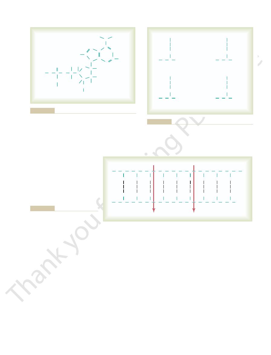
Figure 3–4 shows the chemical structure of deoxyadenylic
deoxycytidylic acids.
deoxyadenylic, deoxythymidylic, deoxyguanylic,
to form an acidic nucleotide. Four separate nucleotides are thus formed, one for
cule of phosphoric acid, one molecule of deoxyribose, and one of the four bases
The first stage in the formation of DNA is to combine one mole-
them, as illustrated in Figure 3–6.
molecule, and the nitrogenous bases lie between the two strands and connect
and deoxyribose form the two helical strands that are the backbone of the DNA
). The phosphoric acid
and two pyrimidines,
guanine,
(two purines,
deoxyribose,
involved in the formation of DNA. These include (1)
Figure 3–3 shows the basic chemical compounds
few paragraphs.
bound together in a regular pattern, details of which are explained in the next
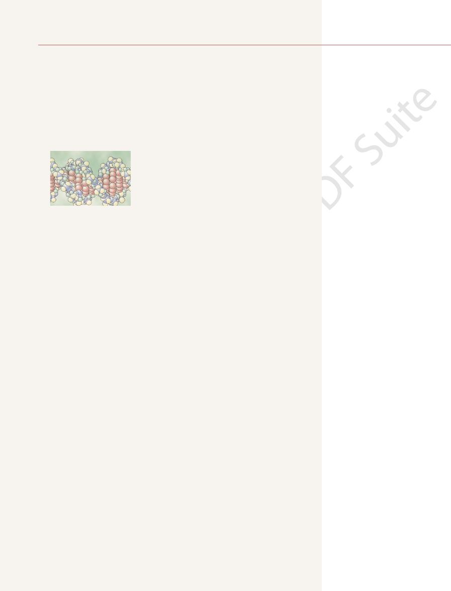
Figure 3–2. This molecule is composed of several simple chemical compounds
measured in the billions. A very short segment of such a molecule is shown in
long double-stranded helical molecules of DNA having molecular weights
In the cell nucleus, large numbers of genes are attached end on end in extremely
gen, and adenosine triphosphate (ATP).
cell, and they promote synthesis of all the cell chemicals, such as lipids, glyco-
instance, enzymes promote all the oxidative reactions that supply energy to the
that catalyze the different chemical reactions in the cells. For
lar organelles discussed in Chapter 2. However, by far the majority of the pro-
various lipids and carbohydrates, form the structures of the various intracellu-
which, in association with
structural proteins,
cally possible to form a very large number of different cellular proteins.
Because there are more than 30,000 different genes in each cell, it is theoreti-
this RNA then
ribonucleic acid (RNA);
deoxyribonucleic acid (DNA),
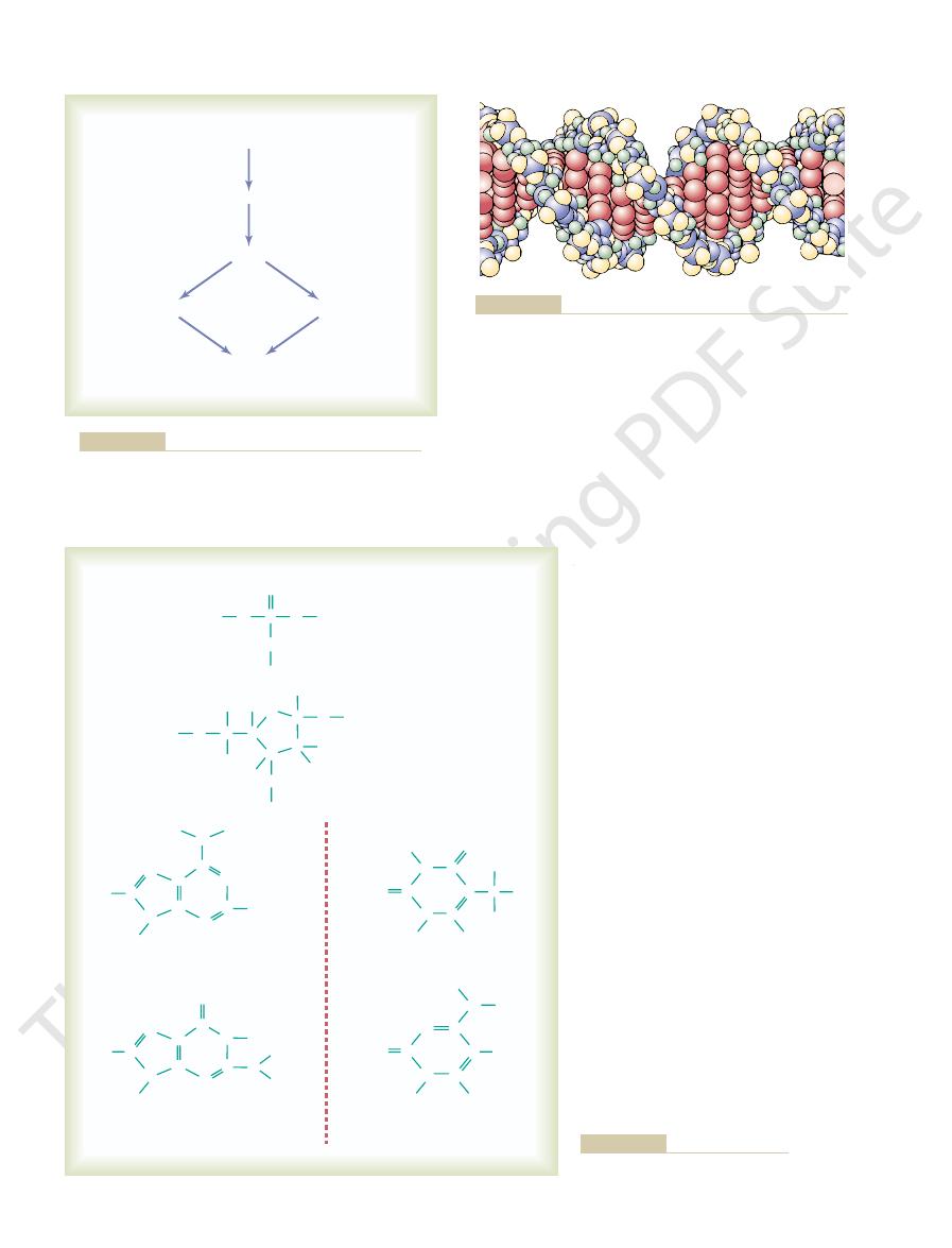
Figure 3–1 shows the general schema of genetic control. Each gene, which is
tures, which enzymes, which chemicals.
day function of all the body’s cells. The genes
from parents to children, but most people do not
the nuclei of all cells of the body, control heredity
Virtually everyone knows that the genes, located in
Synthesis, Cell Function,
C
H
A
P
T
E
R
3
27
Genetic Control of Protein
and Cell Reproduction
realize that these same genes also control day-to-
control cell function by determining which sub-
stances are synthesized within the cell—which struc-
a nucleic acid called
automatically controls the
formation of another nucleic acid,
spreads throughout the cell to control the formation of a specific protein.
Some of the cellular proteins are
teins are enzymes
Genes in the Cell Nucleus
Basic Building Blocks of DNA.
phosphoric acid, (2) a
sugar called
and (3) four nitrogenous bases
adenine
and
thymine and cytosine
Nucleotides.
each of the four bases:
and
Introduction to Physiology: The Cell and General Physiology
28
Unit I
Cell structure
Cell enzymes
Gene (DNA)
RNA formation
Protein formation
Cell function
General schema by which the genes control cell function.
Figure 3–1
helix are purine and pyrimidine bases; these determine the “code”
bose. The internal molecules connecting the two strands of the
strands are composed of phosphoric acid and the sugar deoxyri-
The helical, double-stranded structure of the gene. The outside
Figure 3–2
of the gene.
C
C
H
Guanine
Cytosine
Purines
Pyrimidines
Phosphoric acid
Deoxyribose
Bases
Thymine
C
C
O
C
C
C
H
H
H
H
N
N
N
N
N
H
Adenine
C
C
N
C
C
C
H
C
C
C
C
H
H
O
H
H
H
O
O
H
O
H
P
O
O
H
O
H
H
N
H
H
N
N
N
O
C
H
C
H
C
O
N
H
N
N
H
H
H
H
C
C
O
O
N
C
N
H
H
H
H
H
H
C
H
C
H
Figure 3–3
The basic building blocks of DNA.
Genetic Control of Protein Synthesis, Cell Function, and Cell Reproduction
Chapter 3
29
C
C
N
C
C
C
H
H
N
H
H
N
N
N
P
O
O
O
H
C
C
C
C
H
H
O
H
H
O
O
C
H
H
H
H
H
Adenine
Phosphate
Deoxyribose
Figure 3–4
Deoxyadenylic acid, one of the nucleotides that make up DNA.
D
A
P
D
G
P
D
T
P
D
C
P
Deoxyadenylic acid
Deoxyguanylic acid
Deoxythymidylic acid
Deoxycytidylic acid
and one of the four nucleotide bases: A, adenine; T, thymine;
Symbols for the four nucleotides that combine to form DNA.
Figure 3–5
Each nucleotide contains phosphoric acid (P), deoxyribose (D),
G, guanine; or C, cytosine.
D
P
P
D
P
P
D
P
D
P
D
P
D
P
D
P
D
P
D
P
D
P
D
P
D
P
D
P
D
P
D
P
D
P
D
D
P
C
G
C
G
G
C
T
A
C
G
T
A
G
C
A
T
A
T
form the genetic code.
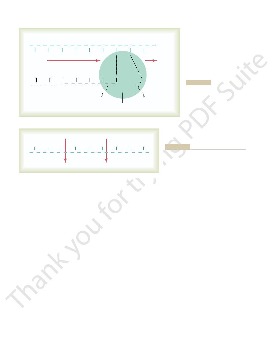
strand in Figure 3–7. It is these projecting bases that
the side of each DNA strand, as shown by the top
two strands of a DNA molecule are split apart, this
That is, when the
genetic code.
the formation of proteins in the cell. It does this by
The importance of DNA lies in its ability to control
molecule, as shown in Figure 3–2.
are present in each full turn of the helix in the DNA
and twist them into a helix. Ten pairs of nucleotides
ical perspective, one could merely pick up the two ends
To put the DNA of Figure 3–6 into its proper phys-
the two strands can pull apart with ease, and they do
AT. Because of the looseness of the hydrogen bonds,
pairs of bases is CG, CG, GC, TA, CG, TA, GC, AT, and
Thus, in Figure 3–6, the sequence of complementary
cytosine.
2. Each purine base
strand, and
1. Each purine base
are held together. But note the following:
and pyrimidine bases, the two respective DNA strands
deoxyribose molecules. Then, by means of loose
acid and deoxyribose molecules. In turn, purine and
DNA strand is comprised of alternating phosphoric
central dashed lines. Note that the backbone of each
weak cross-linkages, illustrated in Figure 3–6 by the
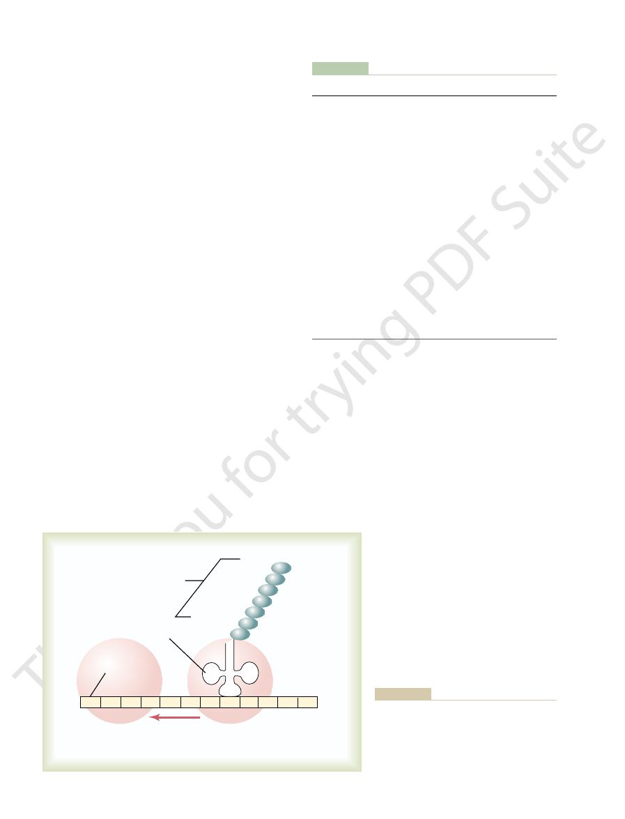
strands are, in turn, loosely bonded with each other by
bound together to form two strands of DNA. The two
Figure 3–6
Arrangement of deoxyribose nucleotides
in a double strand of DNA.
pyrimidine bases are attached to the sides of the
hydrogen bonds (dashed lines) between the purine
adenine of one strand always
bonds with a pyrimidine base thymine of the other
guanine always bonds with a
pyrimidine base
so many times during the course of their function in
the cell.
Genetic Code
means of the so-called
exposes the purine and pyrimidine bases projecting to
cytosine,
adenine, guanine,
These nucleotides contain the bases
rate nucleotides are used in the formation of RNA.
described for DNA synthesis. Here again, four sepa-
RNA nucleotides,
of RNA form
The basic building blocks
dine,
ture. Second, thymine is replaced by another pyrimi-
ribose,
in the formation of RNA. In its place is another sugar
two differences. First, the sugar deoxyribose is not used
RNA are almost the same as those of DNA, except for
The basic building blocks of
cytoplasm.
these codons, in turn, will control the sequence of
) in the RNA;
The code triplets in the DNA cause formation of
used as a template for synthesis of an RNA molecule.
molecule separate temporarily; one of these strands is
During synthesis of RNA, the two strands of the DNA
where it controls protein synthesis.
nuclear pores into the cytoplasmic compartment,
The RNA, in turn, diffuses from the nucleus through
ferred to the RNA; this process is called
nucleus. Thus, as shown in Figure 3–7, the code is trans-
mation of which is controlled by the DNA of the
mediary of another type of nucleic acid, RNA, the for-
of the cytoplasm. This is achieved through the inter-
the cytoplasm, there must be some means for the DNA
Because the DNA is located in the nucleus of the cell,
of Transcription
Cytoplasm—The Process
an RNA Code in the Cell
Nucleus Is Transferred to
proline, serine,
acids,
follow this genetic code through Figures 3–7 and 3–8,
separated from one another by the arrows. As we
the genetic code GGC, AGA, CTT, the triplets being
the top strand of DNA, reading from left to right, has
to be synthesized in the cell. Note in Figure 3–6 that
The successive triplets eventually control the
word.
bases—that is, each three successive bases is a
The genetic code consists of successive “triplets” of
Introduction to Physiology: The Cell and General Physiology
30
Unit I
code
sequence of amino acids in a protein molecule that is
we see that these three respective triplets are respon-
sible for successive placement of the three amino
and glutamic acid, in a newly
formed molecule of protein.
The DNA Code in the Cell
yet most of the functions of the cell are carried out in
genes of the nucleus to control the chemical reactions
transcription.
Synthesis of RNA
com-
plementary code triplets (called codons
amino acids in a protein to be synthesized in the cell
Basic Building Blocks of RNA.
of slightly different composition,
containing an
extra hydroxyl ion appended to the ribose ring struc-
uracil.
Formation of RNA Nucleotides.
exactly as previously
and uracil. Note that these are the same bases
P
P
R
P
P
R
D
G
P
D
G
P
D
C
P
D
A
P
D
G
P
D
A
P
D
P
D
P
D
T
P
C
T
DNA strand
RNA molecule
RNA polymerase
Triphosphate
R
C
C
P
R
G
R
U
C
P
R
U
P
P
R
G
P
P
P
R
A
P
genetic code from the gene to the
with a strand of DNA to form a
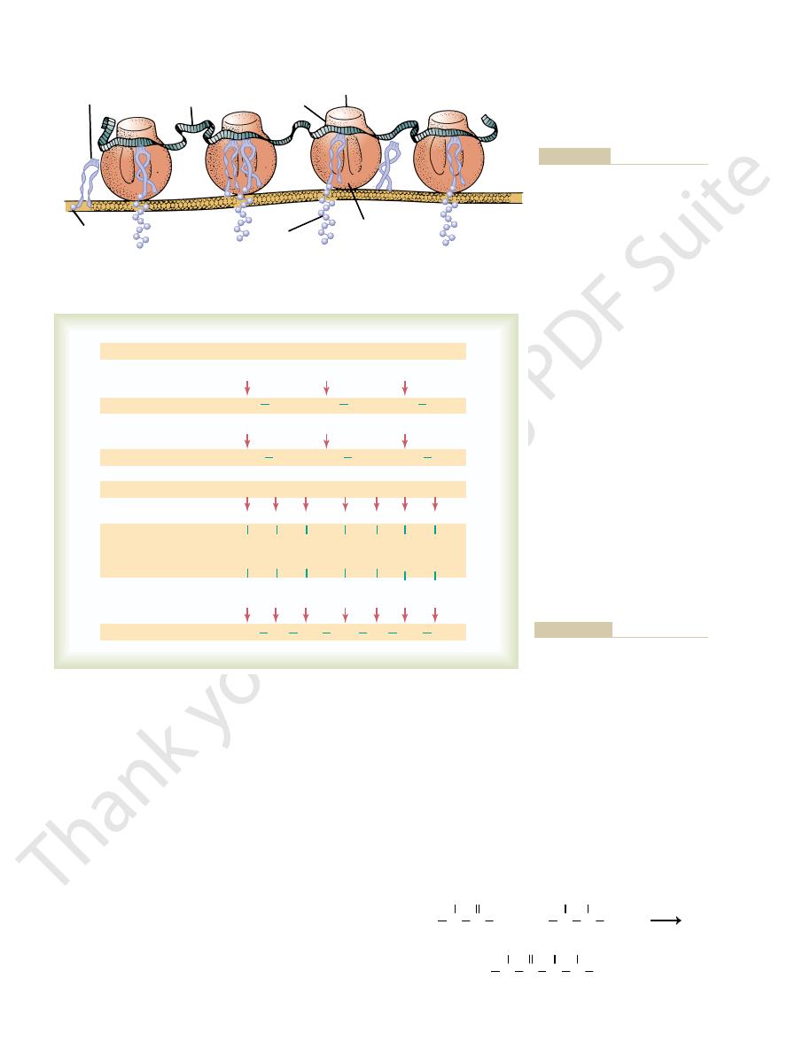
Figure 3–7
Combination of ribose nucleotides
molecule of RNA that carries the
cytoplasm. The RNA polymerase
enzyme moves along the DNA
strand and builds the RNA molecule.
P
P
R
Proline
Serine
Glutamic acid
R
C
C
P
R
G
P
R
U
P
R
C
P
R
U
P
R
G
P
R
A
P
R
A
growing RNA chain.
respectively, to the
proline,
attachment of the three amino acids
“codons”—CCG, UCU, and GAA—which control
Portion of an RNA molecule, showing three RNA
Figure 3–8
serine, and glutamic acid,

RNA chain. The ribose nucleotide bases always
form to the
Thus, the code that is present in the DNA strand is
from the DNA and is released into the
strand. Thus, the RNA chain is forced away
rebonding with its own complementary DNA
away, because the DNA has a high affinity for
hydrogen bonds with the DNA template break
d. As the new RNA strand is formed, its weak
form still more new RNA chains.
to break away from the DNA strand. Then the
polymerase and the newly formed RNA chain
chain-terminating sequence;
sequence of DNA nucleotides called the
end of the DNA gene, it encounters a new
c. When the RNA polymerase reaches the
the ribose on the end of the growing RNA
broken high-energy phosphate bonds; this
away from each of these RNA nucleotides,
b. Then, one at a time, the RNA polymerase
the base of an RNA nucleotide in the
between the end base of the DNA strand and
a. First, it causes a hydrogen bond to form
forming RNA chain by the following steps:
activated RNA nucleotide to the end of the newly
As it moves along, it adds at each stage a new
two DNA strands at each stage of its movement.
strand, temporarily unwinding and separating the
3. Then the polymerase moves along the DNA
of the unwound portions of the two strands.
about two turns of the DNA helix and separation
promoter, the polymerase causes unwinding of
2. After the RNA polymerase attaches to the
of the RNA molecule.
it. This is the essential step for initiating formation
The RNA polymerase has an
promoter.
1. In the DNA strand immediately ahead of the
for formation of the RNA molecule. They are as
This is a large protein
RNA polymerase.
of an enzyme,
the manner shown in Figure 3–7 under the influence
Assembly of the RNA molecule is accomplished in
of “Transcription”
Strand as a Template—The Process
Assembly of the RNA Chain from
RNA nucleotide at the end of the developing RNA
each of the nucleotides, and this energy is used to
large quantities of ATP energy are made available to
The result of this activation process is that
derived from ATP in the cell.
RNA chain formation). These last two phosphates are
3–7 by the two RNA nucleotides to the far right during
phate radicals to form triphosphates (shown in Figure
This
RNA polymerase.
nucleotides by an enzyme,
the synthesis of RNA is “activation” of the RNA
The next step in
thymine in DNA.
as in DNA, except that uracil in RNA replaces
Genetic Control of Protein Synthesis, Cell Function, and Cell Reproduction
Chapter 3
31
“Activation” of the RNA Nucleotides.
occurs by adding to each nucleotide two extra phos-
combined with the nucleotide by high-energy phos-
phate bonds
promote the chemical reactions that add each new
chain.
Activated Nucleotides Using the DNA
enzyme that has many functional properties necessary
follows:
initial gene is a sequence of nucleotides called
the
appropriate complementary structure that
recognizes this promoter and becomes attached to
nucleoplasm.
breaks two of the three phosphate radicals
liberating large amounts of energy from the
energy is used to cause covalent linkage of the
remaining phosphate on the nucleotide with
chain.
this causes the
polymerase can be used again and again to
nucleoplasm.
eventually transmitted in complementary
combine with the deoxyribose bases in the following
combinations:
the RNA codons for the 20 common amino acids
Table 3–1 gives
Figure 3–7.
DNA molecule to the RNA molecule is shown in
tamic acid. The transcription of these codons from the
the codons for the amino acids proline, serine, and glu-
RNA. Its codons are CCG, UCU, and GAA. These are
to the code triplets of the DNA genes. Figure 3–8
thousand RNA nucleotides in unpaired strands, and
strands that are suspended in the cytoplasm. These
molecules are long, single RNA
Messenger RNA
ribosomes,
different proteins, forms
which, along with about 75
Ribosomal RNA,
the protein molecule.
Transfer RNA,
the cytoplasm for controlling the type of protein
Messenger RNA,
types of RNA, each of which plays an independent and
There are three different
Three Different Types of RNA.
thymine . . . . . . . . . . . . . . . . . . . . . . . . . . . . . . . . . .
adenine
adenine . . . . . . . . . . . . . . . . . . . . . . . . . . . . . . . . . .
uracil
cytosine . . . . . . . . . . . . . . . . . . . . . . . . . . . . . . . . . .
guanine
guanine
. . . . . . . . . . . . . . . . . . . . . . . . . . . . . . . . . .
cytosine
DNA Base
RNA Base
entirely different role in protein formation:
1.
which carries the genetic code to
formed.
2.
which transports activated amino
acids to the ribosomes to be used in assembling
3.
the physical
and chemical structures on which protein
molecules are actually assembled.
Messenger RNA—The Codons
molecules are composed of several hundred to several
they contain codons that are exactly complementary
shows a small segment of a molecule of messenger
RNA Codons for the Different Amino Acids.
formation of the protein molecule, the anticodon bases
cloverleaf configuration shown in Figure 3–9). During
of the transfer RNA molecule (at the bottom of the
This is located approximately in the middle
RNA that allows it to recognize a specific codon is
messenger RNA. The specific code in the transfer
RNA also have specificity for a particular codon in the
protein chain, it is essential that each type of transfer
Because the function of transfer RNA is to cause
is always an adenylic acid; it is to this that the trans-
that shown in Figure 3–9. At one end of the molecule
parison with messenger RNA. It is a folded chain of
nucleotides, is a relatively small molecule in com-
Transfer RNA, which contains only about 80
protein molecule.
codon on the messenger RNA (described later) and
specific type of transfer RNA recognizes a particular
protein molecules are forming. In the ribosomes, each
specific type of amino acid to the ribosomes, where
transfer RNA then acts as a
acids that are to be incorporated into proteins. The
RNA combines specifically with 1 of the 20 amino
the protein is being synthesized. Each type of transfer
transfer RNA,
Another type of RNA that plays an essential role in
Transfer RNA—The Anticodons
for “chain-initiating” and CT for “chain-terminating.”
Table 3–1, these two types of codons are designated CI
sent “stop manufacturing the protein molecule.” In
turing the protein molecule,” and three codons repre-
also, one codon represents the signal “start manufac-
found in protein molecules. Note that most of the
Introduction to Physiology: The Cell and General Physiology
32
Unit I
amino acids are represented by more than one codon;
protein synthesis is called
because it
transfers amino acid molecules to protein molecules as
carrier to transport its
thereby delivers the appropriate amino acid to the
appropriate place in the chain of the newly forming
nucleotides with a cloverleaf appearance similar to
ported amino acid attaches at a hydroxyl group of the
ribose in the adenylic acid.
attachment of a specific amino acid to a forming
again a triplet of nucleotide bases and is called an anti-
codon.
combine loosely by hydrogen bonding with the codon
AUG GCC UGU CAU GCC UUU UCC CCC AAA CAG GAC UAU
RNA movement
Ribosome
Ribosome
Transfer RNA
Forming protein
Alanine
Cysteine
Histidine
Alanine
Phenylalanine
Serine
Proline
Messenger
Start codon
GGG
protein.
each specific amino acid to the newly forming
some. The transfer RNA molecule transports
an amino acid is added to the growing protein
ribosomes. As each “codon” passes through,
A messenger RNA strand is moving through two
Figure 3–9
chain, which is shown in the right-hand ribo-
Table 3–1
Stop (CT)
UAA
UAG
UGA
Start (CI)
AUG
Valine
GUU
GUC
GUA
GUG
Tyrosine
UAU
UAC
Tryptophan
UGG
Threonine
ACU
ACC
ACA
ACG
Serine
UCU
UCC
UCA
UCG
AGC
AGU
Proline
CCU
CCC
CCA
CCG
Phenylalanine
UUU
UUC
Methionine
AUG
Lysine
AAA
AAG
Leucine
CUU
CUC
CUA
CUG
UUA
UUG
Isoleucine
AUU
AUC
AUA
Histidine
CAU
CAC
Glycine
GGU
GGC
GGA
GGG
Glutamine
CAA
CAG
Glutamic acid
GAA
GAG
Cysteine
UGU
UGC
Aspartic acid
GAU
GAC
Asparagine
AAU
AAC
Arginine
CGU
CGC
CGA
CGG
AGA
AGG
Alanine
GCU
GCC
GCA
GCG
Amino Acid
RNA
Codons
RNA Codons for Amino Acids and for Start and Stop
CI, chain-initiating; CT, chain-terminating.

ing: (1) Each amino acid is
. The stages of the reactions are the follow-
, AA
reactions for three separate amino acids, AA
shown in Figure 3–11. This figure shows representative
instead of into the endoplasmic reticulum. These pro-
the ribosomes are released directly into the cytosol
tory vesicles are formed, most proteins synthesized by
Yet it should be noted that except in glandular cells
response to the same strand of messenger RNA. Note
endoplasmic reticulum. Note the process of translation
senger RNA to the ribosomes and the manner in
Figure 3–10 shows the functional relation of mes-
matrix. This gives a granular appearance to those por-
reticulum; this causes these molecules to penetrate the
attached to the endoplasmic reticulum. This occurs
Chapter 2, it was noted that many ribosomes become
chemical reactions take place.
somes for given types of protein. The ribosome is
in any ribosome; that is, there is no specificity of ribo-
RNA can cause the formation of a protein molecule
polyribosomes.
These clusters are called
attached to a single messenger RNA at the same time.
ribosomes frequently occur, 3 to 10 ribosomes being
development in each ribosome. As a result, clusters of
3–10. The protein molecules are in different stages of
as shown at the bottom left in Figure 3–9 and in Figure
same time because the initial end of the RNA strand
A single messenger RNA molecule can
protein molecule is freed into the cytoplasm.
“chain-terminating”) codon slips past the ribosome,
back head of a tape recorder. Then, when a “stop” (or
way that a tape is “read” as it passes through the play-
the codons of the messenger RNA in much the same
Thus, the ribosome reads
through the ribosome, a protein molecule is formed—
shown in Figure 3–9, while the messenger RNA travels
bases called the “chain-initiating” codon. Then, as
cule specified by an appropriate sequence of RNA
beginning at a predetermined end of the RNA mole-
with a ribosome, it travels through the ribosome,
When a molecule of messenger RNA comes in contact
of “Translation”
Formation of Proteins on
does not contain mature ribosomes.
cell, but not in the cell nucleus, because the nucleus
Therefore, proteins are formed in the cytoplasm of the
are assembled to form mature, functional ribosomes.
plasm. After the subunits enter the cytoplasm, they
the nuclear envelope to almost all parts of the cyto-
somes. These subunits are then released from the
with “ribosomal proteins” to form granular condensa-
is specially processed in the nucleolus, where it binds
the nucleolus may not even be seen. Ribosomal RNA
structure, whereas in cells that synthesize little protein,
ture large amounts of protein, the nucleolus is a large
are being synthesized, as occurs in cells that manufac-
chromosomes. When large amounts of ribosomal RNA
nucleolus,
As the ribosomal RNA forms, it collects in the
mal RNA required for cellular function.
pairs of chromosomes in the nucleus, and each of these
for formation of ribosomal RNA are located in five
The DNA genes
Thus, the ribosome acts as a manufacturing plant in
messenger RNA
tion into the developing protein molecule, whereas
transfer RNA
with the other two types of RNA as well:
thesized. However, it always functions in association
The ribosome is the physical structure in the cyto-
molecules.
remainder of the ribosome is protein, containing about
The
ribosome.
The third type of RNA in the cell is ribosomal RNA;
forming protein molecule.
the messenger RNA chain, thus establishing the
bases of the messenger RNA. In this way, the respec-
Genetic Control of Protein Synthesis, Cell Function, and Cell Reproduction
Chapter 3
33
tive amino acids are lined up one after another along
appropriate sequence of amino acids in the newly
Ribosomal RNA
it constitutes about 60 per cent of the
75 types of proteins that are both structural proteins
and enzymes needed in the manufacture of protein
plasm on which protein molecules are actually syn-
transports amino acids to the ribosome for incorpora-
provides the information necessary
for sequencing the amino acids in proper order for
each specific type of protein to be manufactured.
which the protein molecules are formed.
Formation of Ribosomes in the Nucleolus.
chromosomes contains many duplicates of these par-
ticular genes because of the large amounts of riboso-
a specialized structure lying adjacent to the
tion products that are primordial subunits of ribo-
nucleolus and transported through the large pores of
the Ribosomes—The Process
a process called translation.
the end of a protein molecule is signaled and the
Polyribosomes.
form protein molecules in several ribosomes at the
can pass to a successive ribosome as it leaves the first,
It is especially important to note that a messenger
simply the physical manufacturing plant in which the
Many Ribosomes Attach to the Endoplasmic Reticulum.
In
because the initial ends of many forming protein mol-
ecules have amino acid sequences that immediately
attach to specific receptor sites on the endoplasmic
reticulum wall and enter the endoplasmic reticulum
tions of the reticulum where proteins are being formed
and entering the matrix of the reticulum.
which the ribosomes attach to the membrane of the
occurring in several ribosomes at the same time in
also the newly forming polypeptide (protein) chains
passing through the endoplasmic reticulum membrane
into the endoplasmic matrix.
in which large amounts of protein-containing secre-
teins are enzymes and internal structural proteins of
the cell.
Chemical Steps in Protein Synthesis.
Some of the chemical
events that occur in synthesis of a protein molecule are
1
2
,
and AA
20
activated by a chemical
The successive amino acids in the
for each amino acid added to the protein chain. Thus,
bonds, making a total of four high-energy bonds used
to the protein chain. These chemical events require
the successive amino acids, thus adding progressively
to form a protein molecule. Then, under the influence
porarily to its specific codon of the messenger RNA,
the anticodon of the transfer RNA attaches tem-
messenger RNA molecule in the ribosome, where
monophosphate. (3) The transfer RNA carrying the
and, at the same time, releases the adenosine
specific transfer RNA to form an amino acid–tRNA
having an excess of energy, then
bonds in the process. (2) The activated amino acid,
process in which ATP combines with the amino acid
Introduction to Physiology: The Cell and General Physiology
34
Unit I
to form an adenosine monophosphate complex with the
amino acid, giving up two high-energy phosphate
combines with its
complex
amino acid complex then comes in contact with the
thus lining up the amino acid in appropriate sequence
of the enzyme peptidyl transferase (one of the proteins
in the ribosome), peptide bonds are formed between
energy from two additional high-energy phosphate
the synthesis of proteins is one of the most energy-con-
suming processes of the cell.
Peptide Linkage.
protein chain combine with one another according to
the typical reaction:
Transfer RNA
Messenger
RNA
Ribosome
Amino acid
Polypeptide
chain
Large
subunit
Small
subunit
(Courtesy of Dr. Don W. Fawcett,
the formation of protein molecules.
the endoplasmic reticulum during
as well as their functional relation to
Physical structure of the ribosomes,
Figure 3–10
messenger RNA, transfer RNA, and
Montana.)
Protein chain
+
ATP
tRNA
1
tRNA
1
+
GCC
Messenger RNA
GCC
GTP
+
ATP
AMP
+
tRNA
2
tRNA
2
+
+
ATP
AMP
+
tRNA
20
tRNA
20
+
tRNA
5
UGU
UGU
GTP
tRNA
3
AAU
AAU
GTP
tRNA
9
CAU
CAU
GTP
tRNA
2
CGU
CGU
GTP
tRNA
13
AUG
AUG
GTP
tRNA
20
GUU
GUU
GTP
Activated amino acid
Amino acid
RNA-amino acyl complex
Complex between tRNA,
messenger RNA, and
amino acid
AMP
+
tRNA
1
¸
Ô
Ô
˝
Ô
Ô
˛
AA
1
AA
1
AA
1
AA
1
AA
1
AA
2
AA
2
AA
2
AA
20
AA
20
AA
20
AA
5
AA
5
AA
3
AA
3
AA
9
AA
9
AA
2
AA
2
AA
13
AA
13
AA
20
AA
20
a protein molecule.
Chemical events in the formation of
Figure 3–11
O
OH
C
C
R
NH
2
R
COOH
C
N
H
H
O
C
C
R
NH
2
H
2
O
R
COOH
C
N
H
+
+
(more than 30,000 genes in all), there is at least one
tions of the cell in step with one another. For each gene
respective genes must be controlled as well; otherwise,
of the cells. However, the degree of activation of
From our discussion thus far, it is clear that the genes
Biochemical Activity in Cells
Control of Gene Function and
Chapters 67 through 69. It is by means of all these
to carbohydrate, lipid, and protein metabolism in
pyrimidines, and hundreds of other substances. We
enzymes promote synthesis of lipids, glycogen, purines,
chemical reactions that take place in cells. These
As each additional amino acid is added, an
linkage.
in a single molecule. This process is called
cessive amino acids bond with each other, resulting
water, and the two reactive sites left on the two suc-
other amino acid is removed. These combine to form
acid, and a hydrogen (H
In this chemical reaction, a hydroxyl radical (OH
moter, thereby blocking transcription of the genes of
prevent attachment of RNA polymerase to the pro-
because a “regulatory” protein can bind here and
promoter. This area is called a
Also note in Figure 3–12 an addi-
“Repressor Operator.”
strand to synthesize RNA. Therefore, the promoter is
moter before it can begin traveling along the DNA
discussed. The polymerase must bind with this pro-
has specific affinity for RNA polymerase, as already
This is a group of nucleotides that
promoter.
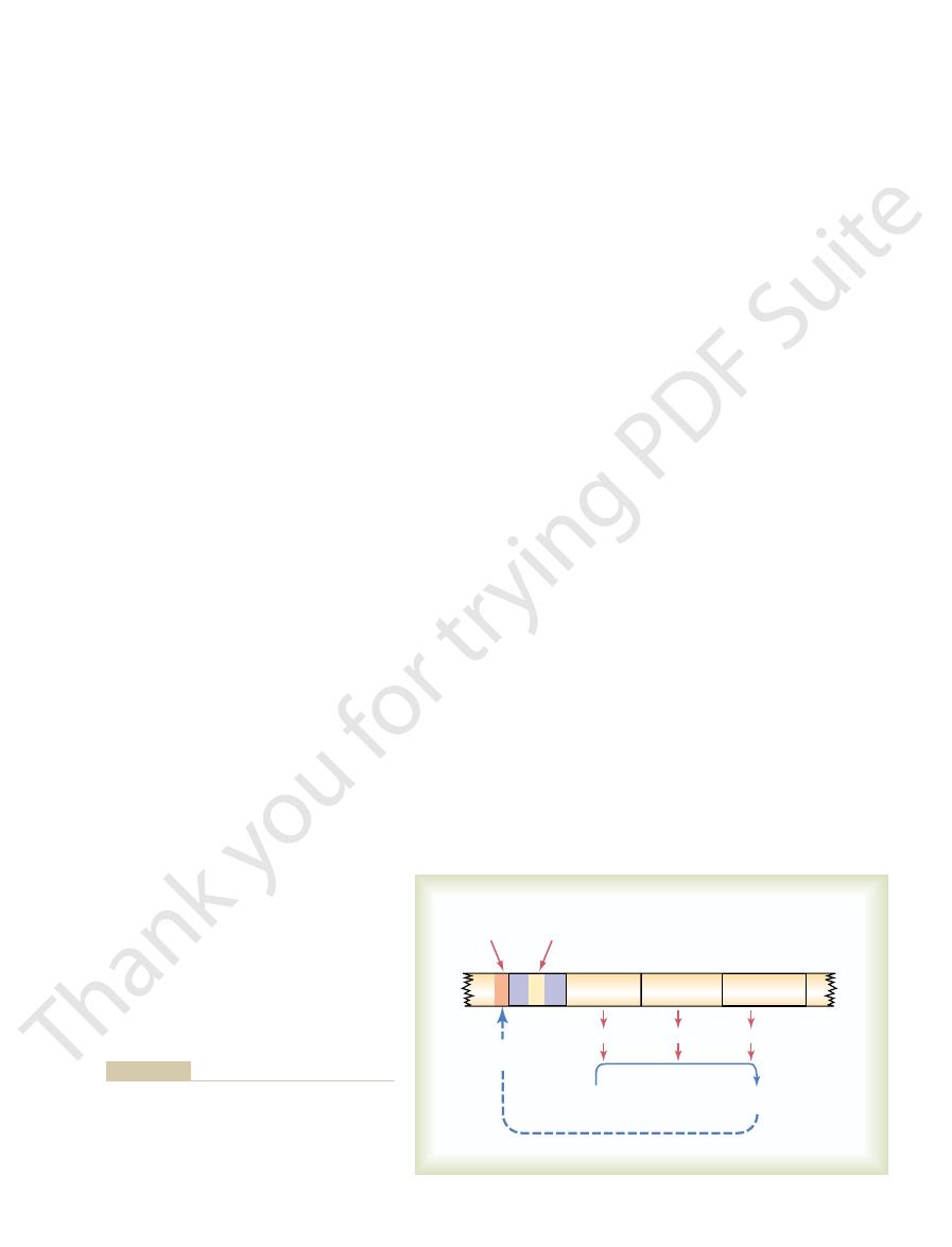
Note in the figure the segment on the DNA strand
structural genes are shown in an operon, and it is
In Figure 3–12, three respective
structural genes.
and the genes respon-
DNA strand is called an
the same chromosomal DNA strand. This area of the
special protein enzyme. Formation of all the enzymes
tions, and each of these reactions is catalyzed by a
thesis—Function of the Promoter.
Genetic Regulation
vation of the genes themselves is controlled, and the
chemical activities in the cell are controlled. One of
There are basically two methods by which the bio-
Genetic Control of Protein Synthesis, Cell Function, and Cell Reproduction
Chapter 3
35
these is genetic regulation, in which the degree of acti-
other is enzyme regulation, in which the activity levels
of already formed enzymes in the cell are controlled.
The “Operon” of the Cell and Its Control of Biochemical Syn-
Synthesis of a cellular
biochemical product usually requires a series of reac-
needed for the synthetic process often is controlled by
a sequence of genes located one after the other on
operon,
sible for forming the respective enzymes are called
demonstrated that they control the formation of three
respective enzymes that in turn cause synthesis of a
specific intracellular product.
called the
an essential element for activating the operon.
Control of the Operon by a “Repressor Protein”—The
tional band of nucleotides lying in the middle of the
repressor operator
–
)
is removed from the COOH portion of the first amino
+
) of the NH
2
portion of the
peptide
additional peptide linkage is formed.
Synthesis of Other
Substances in the Cell
Many thousand protein enzymes formed in the
manner just described control essentially all the other
discuss many of these synthetic processes in relation
substances that the many functions of the cells are
performed.
control both the physical and the chemical functions
some parts of the cell might overgrow or some chem-
ical reactions might overact until they kill the cell.
Each cell has powerful internal feedback control
mechanisms that keep the various functional opera-
such feedback mechanism.
Enzyme B
Enzyme C
¸
Ô
Ô
Ô
˝
Ô
Ô
Ô
˛
Structural
Gene A
Promoter
Inhibition of
the operator
Enzyme A
Synthesized
product
Substrates
(Negative feedback)
Repressor
operator
Activator
operator
Operon
Structural
Gene B
Structural
Gene C
controlling the concentration of the product itself.
the function of the operon, in this way automatically
sized product exerts negative feedback to inhibit
protein intracellular product, such as an intra-
to control synthesis of a non
operon
Figure 3–12
Function of an
cellular metabolic chemical. Note that the synthe-

Thus, cAMP acts as an enzyme activator for the
their energy used for replenishment of the ATP stores.
glycogen-splitting enzyme phosphorylase, liberating
ence of this cAMP, in turn, immediately activates the
formed as a breakdown product of the ATP; the pres-
in a cell. In this case, a considerable amount of cyclic
this occurs when most of the ATP has been depleted
often can be activated when needed. An example of
purines, pyrimidines, vitamins, and other substances.
intracellular concentrations of multiple amino acids,
feedback control; it is responsible for controlling
inactivating the first enzyme: this prevents buildup of
tivates it. One can readily recognize the importance of
enzymes, usually binding directly with the enzyme and
enzyme in a sequence, rather than on the subsequent
specific enzyme systems that synthesize them. Almost
cific intracellular enzymes. Thus, enzyme regulation
regulation, some cell activities are controlled by intra-
Control of Intracellular Function
and products of carbohydrate, lipid, and protein
amino acid derivatives, and intermediate substrates
trolling intracellular concentrations of amino acids,
genetic activity can be controlled is not surprising. The
in each human cell, the large number of ways in which
factors, thus controlling the chemical machinery
some of the body’s hormones, can activate specific
addition, signals from outside the cell, such as
are used for establishing proper cell function. In
chromosome. Thus, still higher orders of control
“transcriptor factor” controls the actual rate of
transcription can occur. Even then, some specific
one part at a time so that partial RNA
RNA. However, multiple control mechanisms are
this compacted state, it cannot function to form
by still other proteins. As long as the DNA is in
histones,
Within each chromosome, the DNA is wound
chromosomes.
in specific structural units, the
4. In nucleated cells, the nuclear DNA is packaged
translation by the ribosomes.
formation in the cytoplasm during RNA
rarely, control might occur at the level of protein
before they are released into the cytoplasm;
processing of the RNA molecules in the nucleus
not even at the DNA strand itself but during the
farther along the strand. Sometimes the control is
point of transcription on the DNA strand but
3. Some operons are controlled not at the starting
simultaneously in this manner, all the operons
operon. When multiple operons are controlled
regulatory protein. In some instances, the same
2. Occasionally, many different operons are
complex of the nucleus. That is, the regulatory
1. An operon frequently is controlled by a
2 decades. Without giving details, let us list some of
Variations in the basic mechanism for control of the
Other Mechanisms for Control of Transcription by the Operon.
product is controlled automatically.
active. In this way, the desired concentration of the
tration decreases, the operon once again becomes
becomes dormant. Conversely, when the synthesized
dant enough for proper cell function, the operon
case, the operon becomes inhibited. Therefore, once
to break its bond with the activator operator. In either
for its synthesis. It can do this either by causing a
Figure 3–12 that the presence of a critical amount
Finally, note in
operon. Therefore, a regulatory protein of this type is
lymerase to the promoter, in this way activating the
binds to this operator, it helps attract the RNA po-
ahead of the promoter. When a regulatory protein
activator operator,
Note in Figure 3–12 another operator,
tor Operator.”
this operon. Such a negative regulatory protein is
Introduction to Physiology: The Cell and General Physiology
36
Unit I
called a repressor protein.
Control of the Operon by an “Activator Protein”—The “Activa-
called the
that lies adjacent to but
called an activator protein.
Negative Feedback Control of the Operon.
of a synthesized product in the cell can cause negative
feedback inhibition of the operon that is responsible
regulatory repressor protein to bind at the repressor
operator or by causing a regulatory activator protein
the required synthesized product has become abun-
product becomes degraded in the cell and its concen-
operon have been discovered with rapidity in the past
them:
regulatory gene located elsewhere in the genetic
gene causes the formation of a regulatory protein
that in turn acts either as an activator or as a
repressor substance to control the operon.
controlled at the same time by the same
regulatory protein functions as an activator
for one operon and as a repressor for another
that function together are called a regulon.
around small proteins called
which in
turn are held tightly together in a compacted state
beginning to be discovered that can cause selected
areas of chromosomes to become decompacted
transcription by the separate operon in the
chromosomal areas and specific transcription
for function of the cell.
Because there are more than 30,000 different genes
gene control systems are especially important for con-
metabolism.
by Enzyme Regulation
In addition to control of cell function by genetic
cellular inhibitors or activators that act directly on spe-
represents a second category of mechanisms by which
cellular biochemical functions can be controlled.
Enzyme Inhibition.
Some chemical substances formed in
the cell have direct feedback effects in inhibiting the
always the synthesized product acts on the first
causing an allosteric conformational change that inac-
intermediary products that are not used.
Enzyme inhibition is another example of negative
Enzyme Activation.
Enzymes that are normally inactive
adenosine monophosphate (cAMP) begins to be
glucose molecules that are rapidly metabolized and
These replicas become the DNA in the two new
of all DNA.
hours. The net result is two exact
hours before mitosis, and this is completed in 4 to 8
The DNA begins to be duplicated some 5 to 10
place.
mosomes.
replication (duplication) of all DNA in the chro-
cell, reproduction begins in the nucleus itself. The first
with Replication of DNA
Cell Reproduction Begins
human body for most nerve cells.
ent cells of the body actually have life cycle periods
the uninhibited life cycle of the cell. Therefore, differ-
duction, inhibitory factors almost always slow or stop
interphase.
represented by the interval between mitosis, called
of the life cycle of even rapidly reproducing cells is
only about 30 minutes, so that more than 95 per cent
later. The actual stage of mitosis, however, lasts for
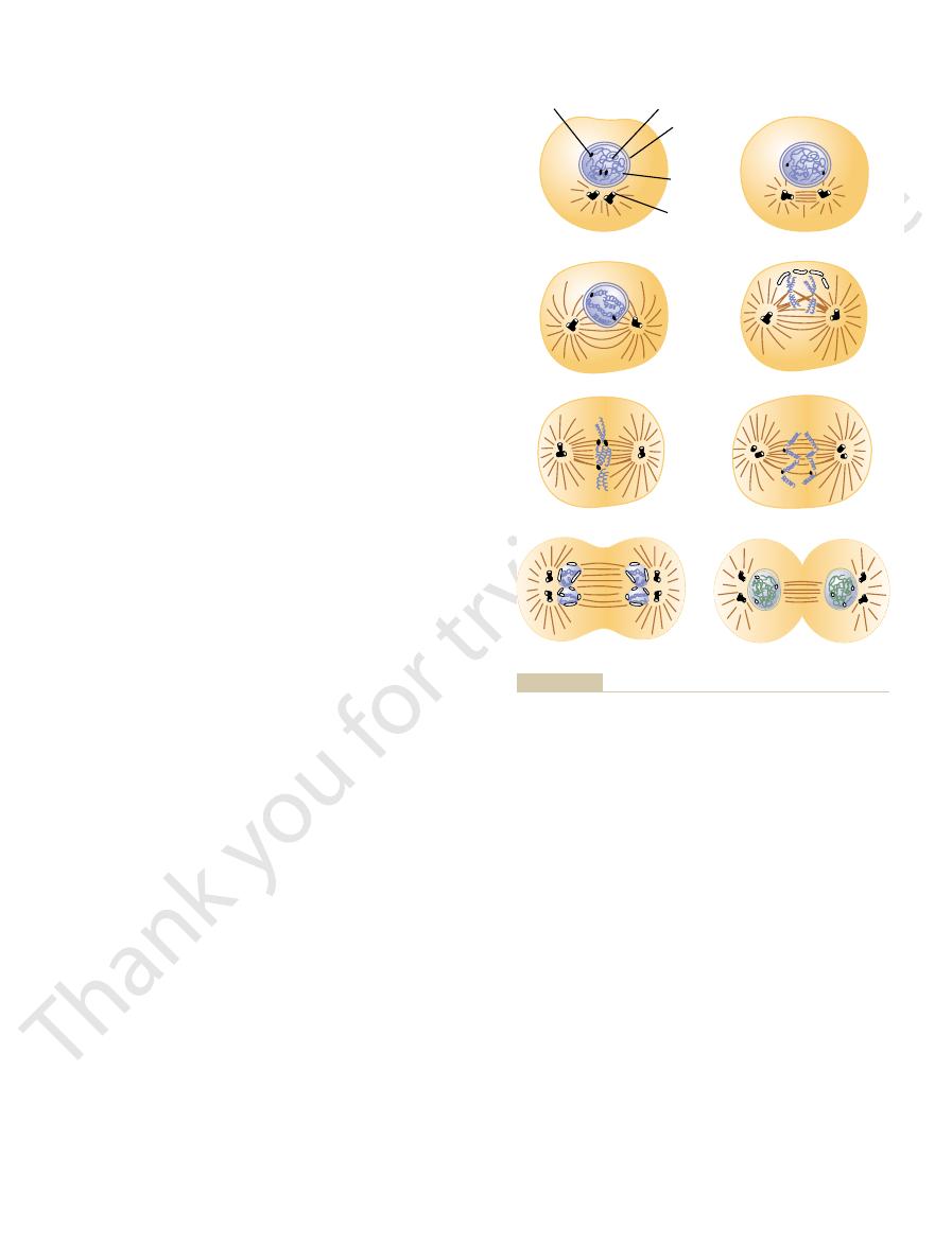
of mitosis are shown in Figure 3–13 and are described
sion of the cell into two new daughter cells. The events
little as 10 to 30 hours. It is terminated by a series of
this life cycle may be as
When mammalian cells
The life cycle of a cell is the period
life, it is the DNA-genetic system.
functioning body. Thus, if there is any central theme to
being, from the single-cell fertilized ovum to the whole
new cells. In this way, the all-important genetic system
processes. The genes and their regulatory mechanisms
tous role that the DNA-genetic system plays in all life
Controls Cell Reproduction
or more of the intracellular control systems.
needed. But on occasion, substances from without the
control systems that continually monitor the cell’s
systems can be either activated or inhibited. These reg-
activated or inhibited, and likewise, the enzyme
nism of enzyme regulation. The genes can be either
quantities of different cellular constituents: (1) the
In summary, there are two principal methods
Summary.
times.
for these two substances, resulting in almost exactly
activate the purine enzymes. In this way, there is con-
versely, the pyrimidines inhibit their own enzymes but
the enzymes for formation of pyrimidines. Con-
formation of additional purines. However, they
formed, they
for formation of DNA and RNA. When purines are
of the purines and pyrimidines. These substances are
intracellular ATP concentration.
Genetic Control of Protein Synthesis, Cell Function, and Cell Reproduction
Chapter 3
37
enzyme phosphorylase and thereby helps control
Another interesting instance of both enzyme inhi-
bition and enzyme activation occurs in the formation
needed by the cell in approximately equal quantities
inhibit the enzymes that are required for
acti-
vate
tinual cross-feed between the synthesizing systems
equal amounts of the two substances in the cells at all
by which cells control proper proportions and proper
mechanism of genetic regulation and (2) the mecha-
ulatory mechanisms most often function as feedback
biochemical composition and make corrections as
cell (especially some of the hormones discussed
throughout this text) also control the intracellular
biochemical reactions by activating or inhibiting one
The DNA-Genetic System Also
Cell reproduction is another example of the ubiqui-
determine the growth characteristics of the cells and
also when or whether these cells will divide to form
controls each stage in the development of the human
Life Cycle of the Cell.
from cell reproduction to the next cell reproduction.
are not inhibited and are repro-
ducing as rapidly as they can,
distinct physical events called mitosis that cause divi-
Except in special conditions of rapid cellular repro-
that vary from as little as 10 hours for highly stimu-
lated bone marrow cells to an entire lifetime of the
As is true of almost all other important events in the
step is
Only after this has occurred can mitosis take
replicas
A
B
C
D
E
F
G
H
Centromere
Nucleolus
Centriole
Aster
Chromosome
Nuclear
membrane
(From Margaret C. Gladbach, Estate of Mary E. and Dan Todd,
Telophase.
Prometaphase.
Prophase.
Stages of cell reproduction.
Figure 3–13
A, B, and C,
D,
E, Metaphase. F, Anaphase. G and H,
Kansas.)

first events of mitosis takes place in the cytoplasm,
cells, mitosis follows automatically within 1 or 2 hours.
been replicated to form the two chromatids, in many
mitosis.
The actual process by which the cell splits into two new
chromatids.
located near their center. These duplicated
helixes collect new protein molecules as needed. The
the DNA helixes has been completed; the new DNA
and enzymes.
genetic regulatory machinery, as activators, inhibitors,
mal structural proteins and, in connection with the
nents of chromosomes, functioning both as chromoso-
RNA.
the DNA and to allow small segments at a time to form
DNA. Further, some of the regulatory proteins have
either the formation of RNA or the replication of new
is packaged tightly, it cannot function as a template for
ulation of DNA activity because as long as the DNA
The histone cores play an important role in the reg-
core after another.
each DNA helix are coiled sequentially around one
numbers of small, bobbin-like cores. Small segments of
The histones are organized into vast
histones.
large amount of protein in the chromosome, composed
In addition to DNA in the chromosome, there is a
not the case.
genes also exist in pairs, although occasionally this is
to each other, so it is usually stated that the different
arranged in 23 pairs. Most of the genes in the two chro-
mosomes. The human cell contains 46 chromosomes
The DNA helixes of the nucleus are packaged in chro-
despite mutations.
almost identical genes. Therefore, one functional gene
a further protection, however, each human genome is
in the passage of the genome from parent to child. As
human generation to another is about 30 years, one
death. Yet, given that there are 30,000 or more genes
cell rather than a needed protein, often leading to
The muta-
mistake is made, this is called a
tion process rarely makes a mistake. But when a
Because of repair and proofreading, the transcrip-
proofreading.
DNA
cation. This repair process is referred to as
polymerases and DNA ligases that are used in repli-
tary nucleotides. This is achieved by the same DNA
plate strand, special enzymes cut out the defective
wherever inappropriate DNA nucleotides have been
repair and “proofreading” of the DNA strands. That is,
beginning of mitosis, there is a period of very active
the hour or so between DNA replication and the
DNA Repair, DNA “Proofreading,” and “Mutation.”
separation, and then resplice the helix. Thus, the
length, rotate each segment enough to cause
special mechanism. This is achieved by enzymes
impossible for the two newly formed DNA helixes
have millions of helix turns, it would be
6. Because the DNA helixes in each chromosome
Therefore, two DNA helixes are coiled together.
original DNA strand that was used as its template.
5. Each newly formed strand of DNA remains
enzyme.
subunits are joined together by the DNA ligase
entire strand is replicated. Then the ends of the
4. Formation of each new DNA strand occurs
phosphate bonds to energize these attachments.
nucleotides to one another, using high-energy
causes bonding of successive DNA
DNA ligase,
DNA template strand while another enzyme,
polymerase. It attaches to and moves along the
which is comparable to RNA
polymerase,
DNA
3. The principal enzymes for replicating DNA
RNA.
portions of them, as occurs in the transcription of
replicated from end to end, rather than small
2. Both entire strands of the DNA helix are
are replicated, not simply one of them.
1. Both strands of the DNA in each chromosome
scribed in response to DNA, except for a few impor-
replicated in much the same way that RNA is tran-
DNA is
place that will lead to the mitotic process.
this period, preliminary changes are beginning to take
2 hours before mitosis begins abruptly. Even during
replication of the DNA, there is another period of 1 to
daughter cells that will be formed at mitosis. After
Introduction to Physiology: The Cell and General Physiology
38
Unit I
Chemical and Physical Events of DNA Replication.
tant differences:
are a complex of multiple enzymes called
simultaneously in hundreds of segments along
each of the two strands of the helix until the
attached by loose hydrogen bonding to the
are approximately 6 centimeters in length and
to uncoil from each other were it not for some
that periodically cut each helix along its entire
two new helixes become uncoiled.
During
matched up with the nucleotides of the original tem-
areas and replace these with appropriate complemen-
mutation.
tion causes formation of some abnormal protein in the
abnormal cellular function and sometimes even cell
in the human genome and that the period from one
would expect as many as 10 or many more mutations
represented by two separate sets of chromosomes with
of each pair is almost always available to the child
Chromosomes and Their Replication
mosomes of each pair are identical or almost identical
mainly of many small molecules of electropositively
charged
been shown to decondense the histone packaging of
Several nonhistone proteins are also major compo-
Replication of the chromosomes in their entirety
occurs during the next few minutes after replication of
two newly formed chromosomes remain attached to
each other (until time for mitosis) at a point called the
centromere
but still attached chromosomes are called
Cell Mitosis
cells is called
Once each chromosome has
Mitotic Apparatus: Function of the Centrioles.
One of the
occurring during the latter part of interphase in or

allowed to collect in the culture medium. This, too,
Third, cells grown in tissue culture often stop growing
they contact a solid object, and then growth stops.
cells are grown in tissue culture; the cells grow until
have run out of space for growth. This occurs when
Second, most normal cells stop growing when they
as the pancreas, fail to grow without a growth factor
For instance, the epithelial cells of some glands, such
in the blood, but others originate in adjacent tissues.
from other parts of the body. Some of these circulate
growth factors
three ways in which growth can be controlled. First,
the body. However, experiments have shown at least
We know little about the mechanisms that maintain
ated cells such as nerve and muscle cells.
marrow, subcutaneous tissue, intestinal epithelium,
liver mass returns almost to normal. The same occurs
the liver can be removed surgically, and the cells of
instance, in some young animals, seven eighths of
appropriate numbers of them are again available. For
In certain tissues, an insufficiency of some types of
fetal life.
life of a person, except during the original period of
ated muscle cells, do not reproduce during the entire
years. A few cells, such as the neurons and most stri-
smooth muscle cells, may not reproduce for many
lium of the gut. Many other cells, however, such as
marrow, the germinal layers of the skin, and the epithe-
time, such as the blood-forming cells of the bone
We know that certain cells grow and reproduce all the
Control of Cell Growth and
each other.
pinches in two, midway between the two nuclei. This is
present in the cytoplasm. Shortly thereafter, the cell
set of chromosomes. This membrane is formed from
pletely apart. Then the mitotic apparatus dissolutes,
), the
In telophase (see Figure 3–13
Telophase.
chromosomes.
separated, forming two separate sets of 46
at the centromere. All 46 pairs of chromatids are
), the
During this phase (see Figure 3–13
of the mitotic spindle.
center of the cell, lining up to form the
each other. Simultaneously, the chromatids are pulled
spines and, using a stepping action as in muscle,
called “motor molecules,” perhaps composed of the
actually push each other away. There is reason to
digitate with each other to form the mitotic spindle,
tubular spines from the two asters, where they inter-
apart. This is believed to occur because the micro-
), the
During metaphase (see Figure 3–13
its partner toward the opposite pole.
still bound to each other; the tubules then pull one
at the centromeres, where the paired chromatids are
the nuclear envelope. At the same time, multiple
During this stage (see Figure 3–13
condensed into well-defined chromosomes.
forming, the chromosomes of the nucleus (which in
While the spindle is
A, B,
shown in Figure 3–13
prophase,
The first stage of mitosis, called
mitotic apparatus.
spindle,
the two sets of chromatids during mitosis. The complex
each end of the cell. Some of the spines of the aster
aster,
triole pairs, forming a spiny star, called the
actually pushing them apart. At the same time, other
of centrioles begin to move apart from each other. This
Shortly before mitosis is to take place, the two pairs
centrosome.
triolar material, is called a
Each pair of centrioles, along with attached pericen-
centrioles of each pair lie at right angles to each other.
structures arranged in the form of a cylinder. The two
in diameter, consisting mainly of nine parallel tubular
the DNA.) Each centriole is a small cylindrical body
during interphase, usually shortly before replication of
like the DNA and chromosomes, were also replicated
other near one pole of the nucleus. (These centrioles,
in Figure 3–13, two pairs of centrioles lie close to each
centrioles.
Genetic Control of Protein Synthesis, Cell Function, and Cell Reproduction
Chapter 3
39
around the small structures called
As shown
about 0.4 micrometer long and about 0.15 micrometer
is caused by polymerization of protein microtubules
growing between the respective centriole pairs and
microtubules grow radially away from each of the cen-
in
penetrate the nuclear membrane and help separate
of microtubules extending between the two new cen-
triole pairs is called the
and the entire set of
microtubules plus the two pairs of centrioles is called
the
Prophase.
is
and C.
interphase consist of loosely coiled strands) become
Prometaphase.
D),
the growing microtubular spines of the aster fragment
microtubules from the aster attach to the chromatids
chromatid of each pair toward one cellular pole and
Metaphase.
E
two asters of the mitotic apparatus are pushed farther
believe that minute contractile protein molecules
muscle protein actin, extend between the respective
actively slide the spines in a reverse direction along
tightly by their attached microtubules to the very
equatorial plate
Anaphase.
F
two chromatids of each chromosome are pulled apart
daughter
One of these sets is pulled toward one
mitotic aster and the other toward the other aster as
the two respective poles of the dividing cell are pushed
still farther apart.
G and H
two sets of daughter chromosomes are pushed com-
and a new nuclear membrane develops around each
portions of the endoplasmic reticulum that are already
caused by formation of a contractile ring of micro-
filaments composed of actin and probably myosin (the
two contractile proteins of muscle) at the juncture of
the newly developing cells that pinches them off from
Cell Reproduction
cells causes these to grow and reproduce rapidly until
the remaining one eighth will grow and divide until the
for many glandular cells and most cells of the bone
and almost any other tissue except highly differenti-
proper numbers of the different types of cells in
growth often is controlled by
that come
from the sublying connective tissue of the gland.
when minute amounts of their own secretions are
could provide a means for negative feedback control
of growth.

genes that control cell growth and cell mitosis. The
tosis in cancer cells.
immune disorders. Some drugs that have been used
Alzheimer’s disease, as well as in cancer and auto-
play a key role in neurodegenerative diseases such as
tissues would shrink or grow excessively. Recent
of new cells in healthy adults. Otherwise, the body’s
however, is precisely balanced with the formation
are replaced by new cells. Programmed cell death,
in adult humans, billions of cells die each hour in
that are being remodeled during development. Even
digested by neighboring phagocytic cells.
thus dismantles itself, and its remains are rapidly
rapidly breaks down proteins within the cell. The cell
activate other procaspases, triggering a cascade that
complex, but once activated, the enzymes cleave and
. The mechanisms of activation of caspases are
. These are enzymes that are
usually remain healthy.
leakage of its contents occurs, and neighboring cells
disassembly and phagocytosis of the cell before any
sis, however, is an orderly cell death that results in
inflammation and injury to neighboring cells. Apopto-
Necrotic cells may spill their contents, causing
necrosis.
loss of cell membrane integrity, a process called cell
In contrast to programmed death, cells that die as a
phagocytic cell, such as a macrophage, can attach to
shrink and condense, to disassemble its cytoskeleton,
. This process involves
threat to the organism, they undergo a suicidal
death. When cells are no longer needed or become a
The 100 trillion cells of the body are members of a
Apoptosis—Programmed
occur.
ation is still hazy, we know many control mechanisms
Thus, although our understanding of cell differenti-
parts.
affecting another part, and this part affecting still other
ops as a result of such inductions, one part of the body
the eye. Therefore, a large share of the embryo devel-
essentially all the organs of the body.
in the surrounding tissues, causes formation of
and, as a result of
a focus around which the rest of the embryo develops.
primary organizer
primordial chorda-mesoderm
For instance, the
in an embryo control differentiation of adjacent cells.
more if all genes were active.
function again. Regardless of the mechanism, mature
group of genes. Therefore, the repressed genes never
ecules. One explanation for this is as follows: It has
densed that they no longer uncoil to form RNA mol-
tron micrographs suggest that some segments of DNA
repression of different genetic operons. In fact, elec-
Therefore, it has become clear that differentiation
structures required in the frog’s body.
which is a well-differentiated cell, carries all the
result is often the formation of a normal frog. This
which the original ovum nucleus was removed, the
When the nucleus from an intestinal mucosal cell of
cesses follows.
structures and organs. The description of an especially
Cell Differentiation
in turn cause the cell to grow larger.
increased production of RNA and cell proteins, which
larger. It is assumed that this results simply from
it normally does, and the cell grows proportionately
nucleus contains far greater quantities of DNA than
replication of the DNA continues. In this event, the
spindle and therefore to prevent mitosis, even though
colchicine,
that size. Conversely, it is possible, by use of the chem-
nucleus. If replication of the DNA does not occur, the
entirely by the amount of functioning DNA in the
Introduction to Physiology: The Cell and General Physiology
40
Unit I
Regulation of Cell Size.
Cell size is determined almost
cell grows to a certain size and thereafter remains at
ical
to prevent formation of the mitotic
A special characteristic of cell growth and cell division
is cell differentiation, which refers to changes in
physical and functional properties of cells as they pro-
liferate in the embryo to form the different bodily
interesting experiment that helps explain these pro-
a frog is surgically implanted into a frog ovum from
demonstrates that even the intestinal mucosal cell,
necessary genetic information for development of all
results not from loss of genes but from selective
helixes wound around histone cores become so con-
been supposed that the cellular genome begins at a
certain stage of cell differentiation to produce a regu-
latory protein that forever after represses a select
human cells produce a maximum of about 8000 to
10,000 proteins rather than the potential 30,000 or
Embryological experiments show that certain cells
is called
the
of the embryo because it forms
It differentiates into a mesodermal axis that contains
segmentally arranged somites
induc-
tions
Another instance of induction occurs when the
developing eye vesicles come in contact with the ecto-
derm of the head and cause the ectoderm to thicken
into a lens plate that folds inward to form the lens of
by which differentiation could
Cell Death
highly organized community in which the total number
of cells is regulated not only by controlling the rate of
cell division but also by controlling the rate of cell
pro-
grammed cell death, or apoptosis
a specific proteolytic cascade that causes the cell to
and to alter its cell surface so that a neighboring
the cell membrane and digest the cell.
result of an acute injury usually swell and burst due to
Apoptosis is initiated by activation of a family of
proteases called caspases
synthesized and stored in the cell as inactive procas-
pases
A tremendous amount of apoptosis occurs in tissues
tissues such as the intestine and bone marrow and
studies suggest that abnormalities of apoptosis may
successfully for chemotherapy appear to induce apop-
Cancer
Cancer is caused in all or almost all instances by muta-
tion or by some other abnormal activation of cellular

by day, cancer cells soon demand essentially all the
proliferate indefinitely, their number multiplying day
tissues for nutrients. Because cancer cells continue to
usually is simple. Cancer tissue competes with normal
The answer to this question
to grow into the cancer, thus supplying the nutrients
new cancerous growths. (3) Some cancers also produce
through the body, where they form nidi for numerous
enter the blood stream, and to be transported all
they have a tendency to wander through the tissues, to
sive to one another than are normal cells. Therefore,
normal cells. (2) Cancer cells often are far less adhe-
usual cellular growth limits; the reason for this is that
are the following: (1) The cancer cell does not respect
The major dif-
cancer.
itself into the animal cell genome, leading to
from the RNA. The transcribed DNA then inserts
that causes DNA to be transcribed
cancer. In the case of RNA viruses, some of these
of DNA viruses, the DNA strand of the virus can
This usually results in one of two ways. In the case
cause some kinds of cancer, including leukemia.
5. In laboratory animals, certain types of viruses can
cancer begins to grow.
genome. Therefore, far fewer additional mutations
cancer, it is presumed that one or more cancerous
more mutations before cancer occurs. In those
to cancer. This results from the fact that
4. In many families, there is a strong
the cells. The more rapid the mitosis, the greater
tract by some types of food. The damage to
also can lead to cancer, such as
deaths.
They cause about one quarter of all cancer
number of deaths are those in cigarette smoke.
The
carcinogens.
predisposition to cancer. Chemical substances that
substances, if unprotected, have a special
derivatives are likely to cause cancer, so that
high propensity for causing mutations. It was
DNA strands, thus causing many mutations.
can predispose individuals to cancer. Ions formed
radioactive substances, and even ultraviolet light
x-rays, gamma rays, and particle radiation from
1. It is well known that
certain chemical, physical, or biological factors, includ-
However, the probability of mutations can be
occurrence.
tions to take place, so we can suppose that a large
Thus, chance alone is all that is required for muta-
characteristics.
cellular precautions, probably one newly formed cell
is allowed to proceed. Yet, despite all these inherited
any abnormal DNA strand before the mitotic process
replicated in each cell before mitosis can take place,
precision with which DNA chromosomal strands are
mutant cancerous cells? The answer is the incredible
each year in humans, a better question might be, Why
form the needed blood vessels.
reproduction of a cell line, but no cancer occurs
For instance, one such gene might promote rapid
genes are required simultaneously to cause a cancer.
Fourth, usually several different activated onco-
as fivefold.
ability of a cancer’s developing is multiplied as much
drugs after kidney or heart transplantation, the prob-
pressed, such as in those taking immunosuppressant
cells, destroying them. In support of this is the fact
sitized lymphocytes that react against the cancerous
immune system, causing it to form antibodies or sen-
altered genes, and these proteins activate the body’s
the following way: Most mutated cells form abnormal
system before they grow into a cancer. This occurs in
often, if not usually, destroyed by the body’s immune
Third, those cells that are potentially cancerous are
cancerous, because even most mutated cells still
bility than normal cells and simply die. Second, only
this. First, most mutated cells have less survival capa-
body ever lead to cancer. There are several reasons for
activation of oncogenes that lead to cancer.
fore, loss of or inactivation of antioncogenes can allow
suppress the activation of specific oncogenes. There-
antioncogenes,
As many as 100
oncogenes.
Genetic Control of Protein Synthesis, Cell Function, and Cell Reproduction
Chapter 3
41
abnormal genes are called
different oncogenes have been discovered.
Also present in all cells are
which
Only a minute fraction of the cells that mutate in the
a few of the mutated cells that do survive become
have normal feedback controls that prevent excessive
growth.
proteins within their cell bodies because of their
that in people whose immune systems have been sup-
because there is not a simultaneous mutant gene to
But what is it that causes the altered genes? Con-
sidering that many trillions of new cells are formed
is it that all of us do not develop millions or billions of
and also the proofreading process that cuts and repairs
in every few million still has significant mutant
number of cancers are merely the result of an unlucky
increased manyfold when a person is exposed to
ing the following:
ionizing radiation, such as
in tissue cells under the influence of such
radiation are highly reactive and can rupture
2. Chemical substances of certain types also have a
discovered long ago that various aniline dye
workers in chemical plants producing such
can cause mutation are called
carcinogens that currently cause the greatest
3. Physical irritants
continued abrasion of the linings of the intestinal
the tissues leads to rapid mitotic replacement of
the chance for mutation.
hereditary
tendency
most cancers require not one mutation but two or
families that are particularly predisposed to
genes are already mutated in the inherited
must take place in such family members before a
insert itself directly into one of the chromosomes
and thereby cause a mutation that leads to
carry with them an enzyme called reverse
transcriptase
Invasive Characteristic of the Cancer Cell.
ferences between the cancer cell and the normal cell
these cells presumably do not require all the same
growth factors that are necessary to cause growth of
angiogenic factors that cause many new blood vessels
required for cancer growth.
Why Do Cancer Cells Kill?

Elsevier Science, 2002.
Pollard TD, Earnshaw WC: Cell Biology. Philadelphia:
disease therapy: past and future. Nat Med 10:135, 2004.
Nabel GJ: Genetic, cellular and immune approaches to
Lewin B: Genes IV. Oxford: Oxford University Press, 2000.
evolution. Science 303:1626, 2004.
Kazazian HH Jr: Mobile elements: drivers of genome
Physiol Sci 18:7, 2003.
toward the cell periphery: a guided tour for mRNAs. News
Jockusch BM, Hüttelmaier S, Illenberger S: From the nucleus
Annu Rev Med 48:35, 1997.
Hall JG: Genomic imprinting: nature and clinical relevance.
II transcription machinery. Nat Struct Mol Biol 11:394,
Hahn S: Structure and mechanism of the RNA polymerase
tivity. Int J Oncol 24:1039, 2004.
implications for DNA damage signaling and drug sensi-
Fedier A, Fink D: Mutations in DNA mismatch repair genes:
Cullen BR: Nuclear RNA export. J Cell Sci 116:587, 2003.
FASEB J 17:1195, 2003.
DNA damage: mechanisms, mutation, and disease.
Cooke MS, Evans MD, Dizdaroglu M, Lunec J: Oxidative
N Y Acad Sci 1002:56, 2003.
mediated RNA interference (RNAi) in human cells. Ann
Caplen NJ, Mousses S: Short interfering RNA (siRNA)-
J Med 349:969, 2003.
Burke W: Genomics as a probe for disease biology. N Engl
Matters of Life and Death. London: Chapman & Hall,
Bowen ID, Bowen SM, Jones AH: Mitosis and Apoptosis:
cancer. Nat Genet 33(Suppl):238, 2003.
Balmain A, Gray J, Ponder B: The genetics and genomics of
expression. Physiol Rev 81:1269, 2001.
Aranda A, Pascal A: Nuclear hormone receptors and gene
the Cell. New York: Garland Science, 2002.
Alberts B, Johnson A, Lewis J, et al: Molecular Biology of
of the body. As a result, normal tissues gradually suffer
Introduction to Physiology: The Cell and General Physiology
42
Unit I
nutrition available to the body or to an essential part
nutritive death.
References
1998.
2004.