2
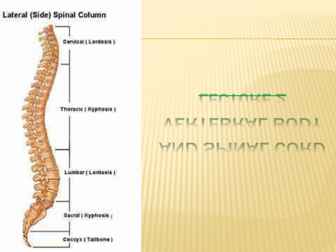
LECTURE
VERTEBRAL BODY
AND SPINAL CORD
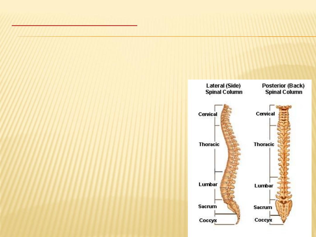
The vertebral column
is the central bony pillar of the body.
It supports the skull, pectoral girdle, upper limbs, and thoracic cage and, by
way of the pelvic girdle, transmits body weight to the lower limbs. Within its
cavity lie the spinal cord, the roots of the spinal nerves, and the covering
meninges, to which the vertebral column gives great protection.
Composition of vertebral
body
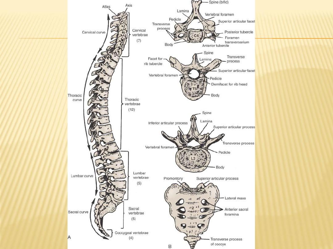
Composed of 33 vertebrae, 7 cervical,
12 thoracic, 5 lumbar, 5 sacrasl
(fused to form the sacrum) and 4
coccygeal (the lower 3 are
commonly fused)
Intervertebral discs pads of
fibrocartilage between each two
adjacent vertebrae.

A typical vertebra composed of:
Body anteriorly and vertibral arch posteriorly
Vertebral arch composed of:
-
Pedicles, lamenae and seven processes
-
- spinous process
-
- articular process 2 sup and 2 inf
-
- two transverse processes
-
- sup. and inf. vertebral notch both form the intervertebral
-
Foramen
-
Joints:
-
- Intervertebral disc between two bodies
-
- Articular Joints between two vertebral arches with a
.
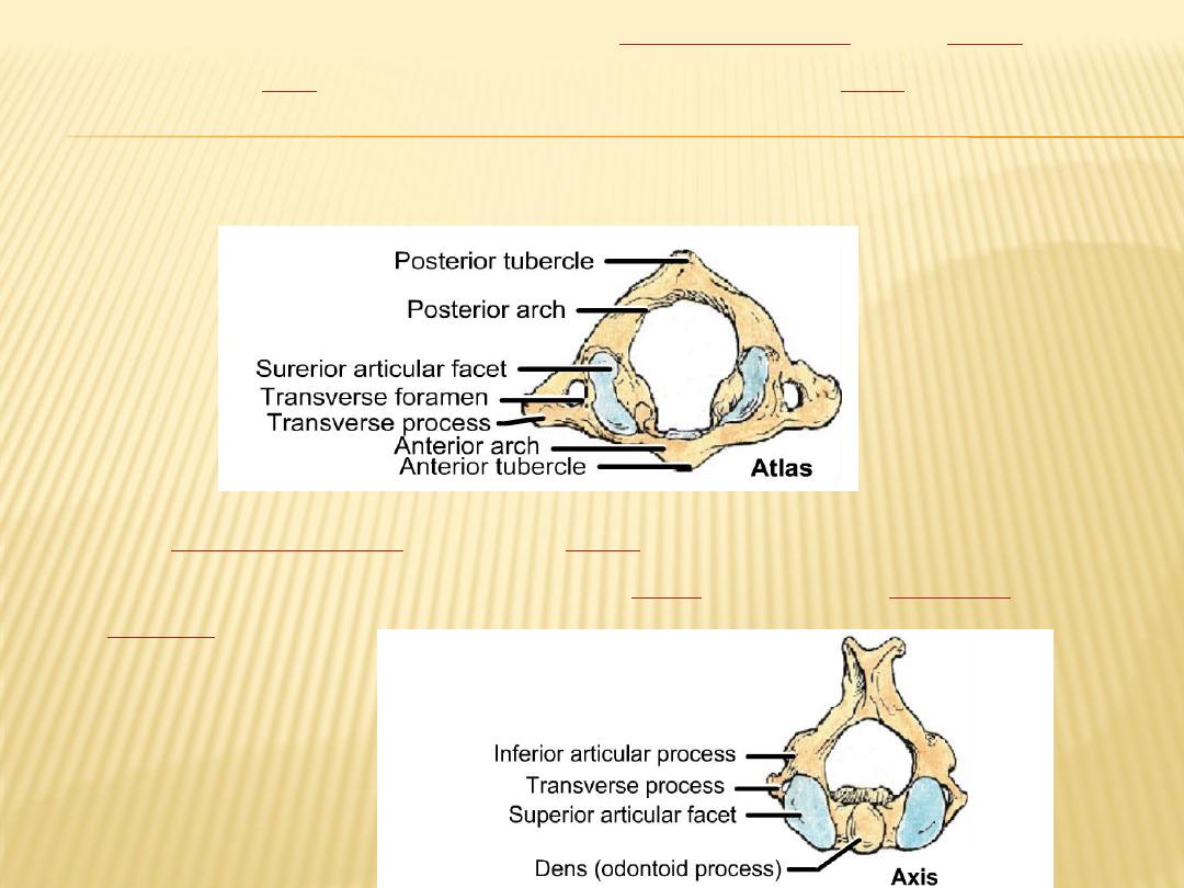
spine
of the
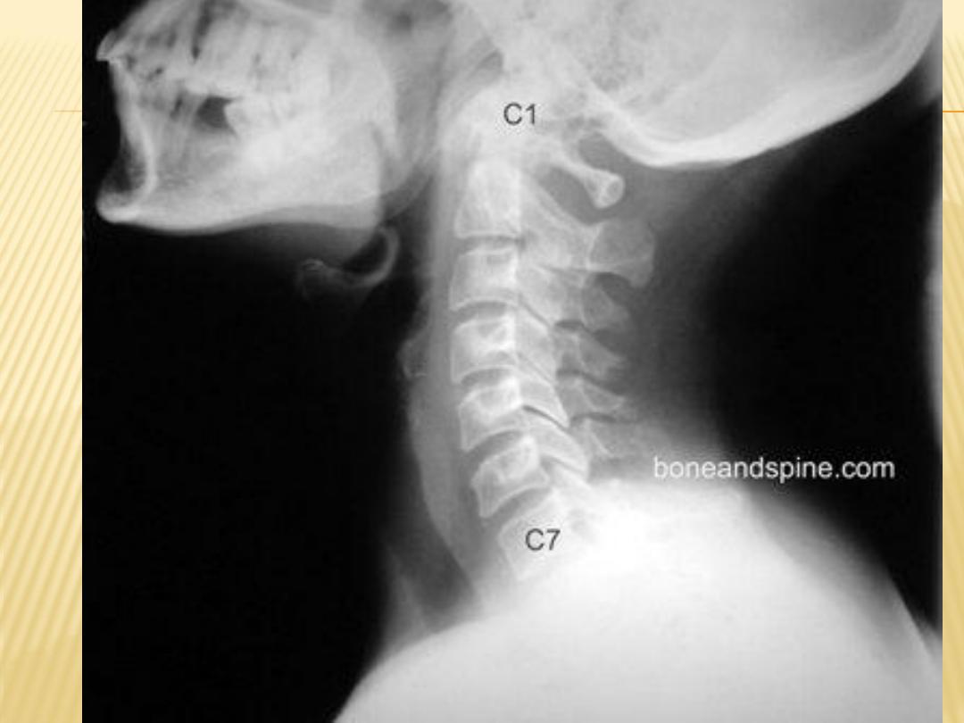
cervical vertebra
) is the most superior (first)
1
(C
atlas
the
along with the
Axis
(C2) – forms the joint connecting the
skull
and spine (the
atlanto-occopital joint). The atlas and axis are specialized to allow a greater
range of motion than normal vertebrae. They are responsible for the
nodding and rotation movements of the head.
second
cervical vertebra
(C2) of the
spine
is named the axis
the most distinctive characteristic of this
bone
is the strong
odontoid
process
("dens")
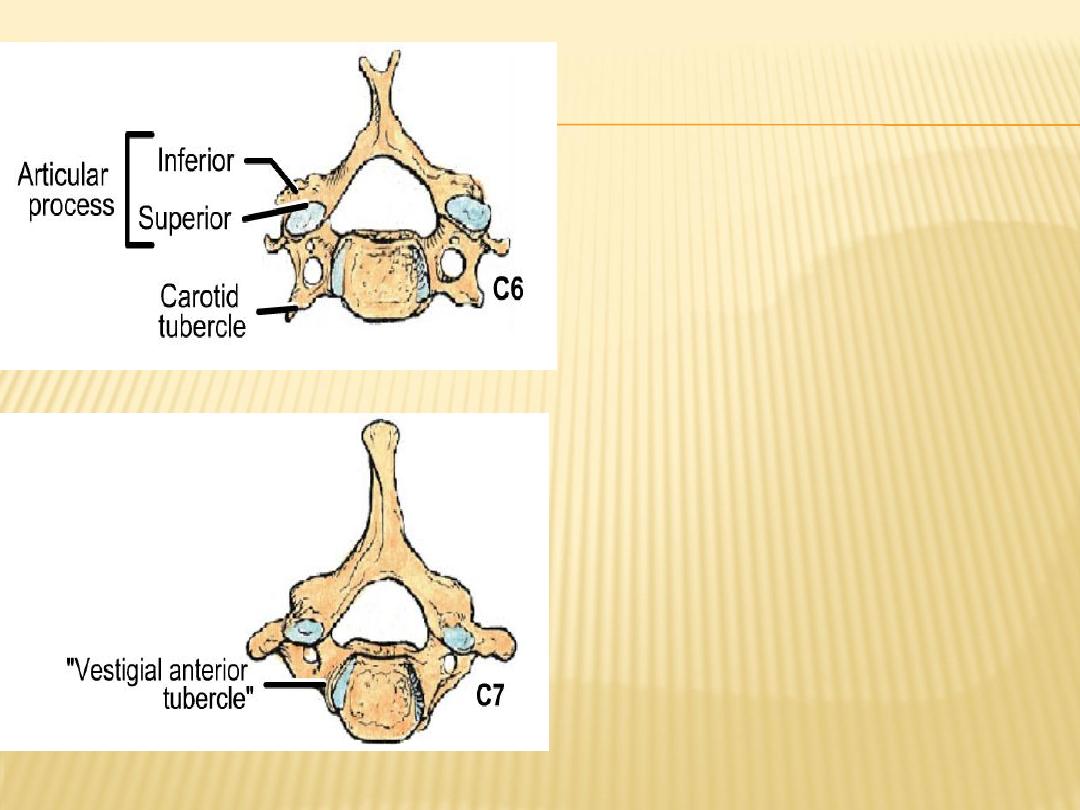
Spinous processes of C2 to C6 are
bifid while that of C7 are not
C7 also called vertebra prominence
(due to palpable spinous
prosess
Vertebral artery pass through
formaina in transverse
prosesses of C2 to C6

Intervertebral Discs
The intervertebral discs are thickest in the cervical and lumbar regions,
where the movements of the vertebral column are greatest. They serve as
shock absorbers when the load on the vertebral column is suddenly
increased.
Unfortunately, their resilience is gradually lost with advancing age.
Each disc consists of a peripheral part, the anulus fibrosus, and a central
part, the nucleus pulposus
The anulus fibrosus is composed of fibrocartilage, which is strongly
attached to the vertebral bodies and the anterior and posterior
longitudinal ligaments of the vertebral column.
The nucleus pulposus in the young is an ovoid mass of gelatinous
material. It is normally under pressure and situated slightly nearer to the
posterior than to the anterior margin of the disc. The upper and lower
surfaces of the bodies of adjacent vertebrae that abut onto the disc are
covered with thin plates of hyaline cartilage.
With advancing age, the nucleus pulposus becomes smaller and is
replaced by fibrocartilage. The collagen fibers of the anulus degenerate
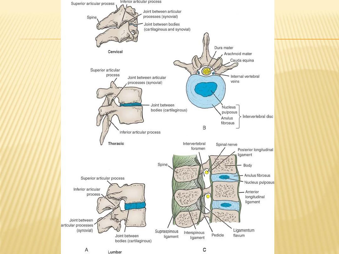

Ligaments
The anterior and posterior longitudinal ligaments run as continuous bands
down the anterior and posterior surfaces of the vertebral column from the
skull to the sacrum
The anterior ligament is wide and is strongly attached to the front and sides of
the vertebral bodies and to the intervertebral discs.
The posterior ligament is weak and narrow and is attached to the posterior
borders of the discs
Other ligaments
Supraspinous ligament:
This runs between the tips of adjacent spines.
Interspinous ligament:
This connects adjacent spines.
Intertransverse ligaments:
These run between adjacent transverse processes
.
Ligamentum flavum:
This connects the laminae of adjacent vertebrae.
Ligamentum nuchae :
the supraspinous and interspinous ligaments are greatly
thickened to form the strong ligamentum nuchae

Nerve Supply of joints
The joints between the vertebral bodies are innervated by the small meningeal
branches of each spinal nerve
The joints between the articular processes are innervated by branches from the
posterior rami of the spinal nerves
the joints of any particular level receive nerve fibers from two adjacent spinal
nerves.
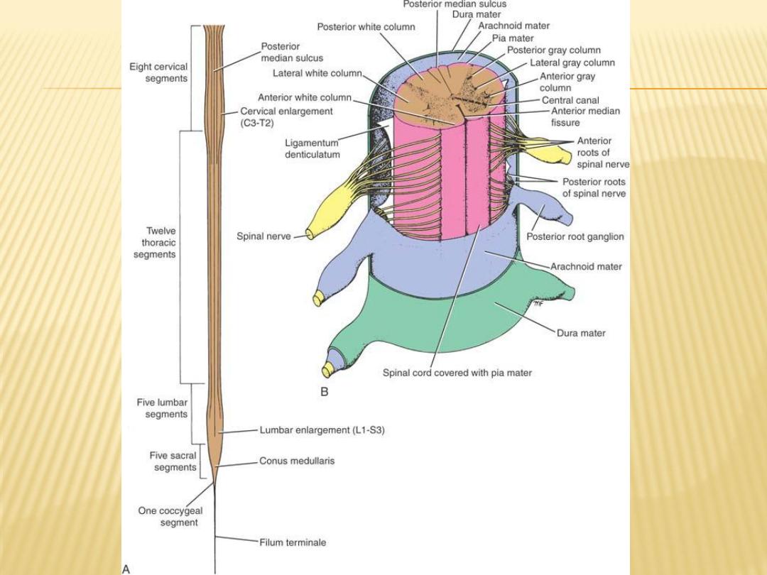
SPINAL CORD
Gross appearance
The spinal cord is roughly cylindrical in shape. It begins superiorly at the
foramen magnum in the skull, where it is continuous with the medulla
oblongata of the brain, and it terminates inferiorly in the adult at the level of
the lower border of the first lumbar vertebra.
In the young child, it is relatively longer and usually ends at the upper border of
the third lumbar vertebra. Thus, it occupies the upper two-thirds of the
vertebral canal of the vertebral column and is surrounded by the three
meninges, the dura mater, the arachnoid mater, and the pia mater. Further
protection is provided by the cerebrospinal fluid, which surrounds the spinal
cord in the subarachnoid space

In the cervical region, where it gives origin to the brachial plexus, and in the
lower thoracic and lumbar regions, where it gives origin to the lumbosacral
plexus, the spinal cord is fusiformly enlarged; the enlargements are referred
Inferiorly, the spinal cord tapers
the cervical and lumbar enlargements
to as
from the apex of which a prolongation of the
conus medullaris,
off into the
, descends to be attached to the posterior
the filum terminale
pia mater,
surface of the coccyx.
anterior median
The cord possesses a deep longitudinal fissure called the
posterior
in the midline anteriorly and a shallow furrow called the
fissure
on the posterior surface
median sulcus
31 pairs of spinal nerves
anterior or motor root and the posterior or sensory root unite to form each
spinal nerve
Each root is attached to the cord by a series of rootlets
Each posterior nerve root possesses a posterior root ganglion

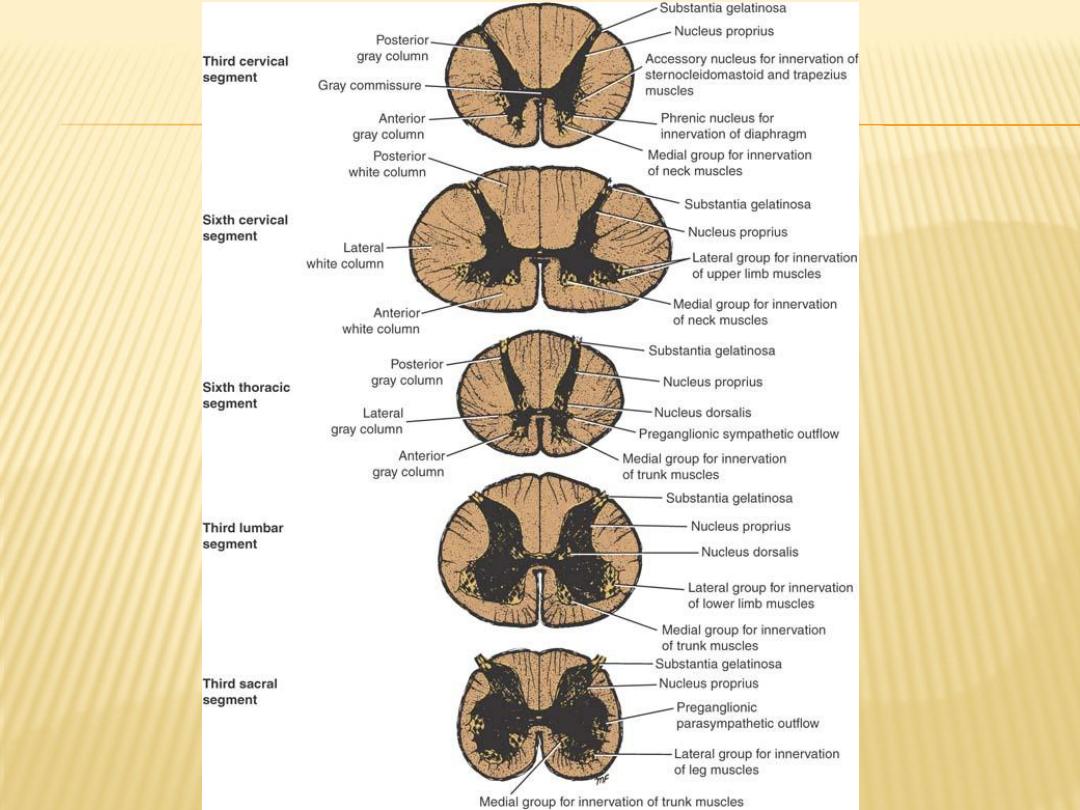
Sturcture of Spinal cord
The spinal cord is composed of an inner core of gray matter, which is
surrounded by an outer covering of white matter
Gray Matter
On cross section, the gray matter is seen as an H-shaped pillar with anterior
and posterior gray columns, or horns, united by a thin gray commissure
containing the small central canal
A small lateral gray column or horn is present in the thoracic and upper lumbar
segments of the cord. The amount of gray matter present at any given level
of the spinal cord is related to the amount of muscle
White matter
The white matter, may be divided into anterior, lateral, and posterior white
columns or funiculi

Blood supply of spinal cord
- Arteries of the spinal cord
Posterior spinal arteries
Anterior spinal artery
Segmental spinal arteries
Feeder arteries (great anterior medullary artery of adamkiewicz)
-
- Veins of the spinal cord
-
Six tortuous longitudinal channels communicate superiorly within the skull
with the veins of the brain and venous sinuses
Meninges of the spinal cord
1- Dura Mater
Dense strong fibrous membrane that encloses the spinal cord
and cauda equina
2- Arachnoid mater
Delicate impermeable membrane separated from the pia
mater by subarachnoid space the contain the Cerebrospinal fluid
3- Pia mater
A vascular membrane closely cover the spinal cord