
1
Lecture
-Introduction to Nervous System
-The Skull
Introduction to Nervous System:
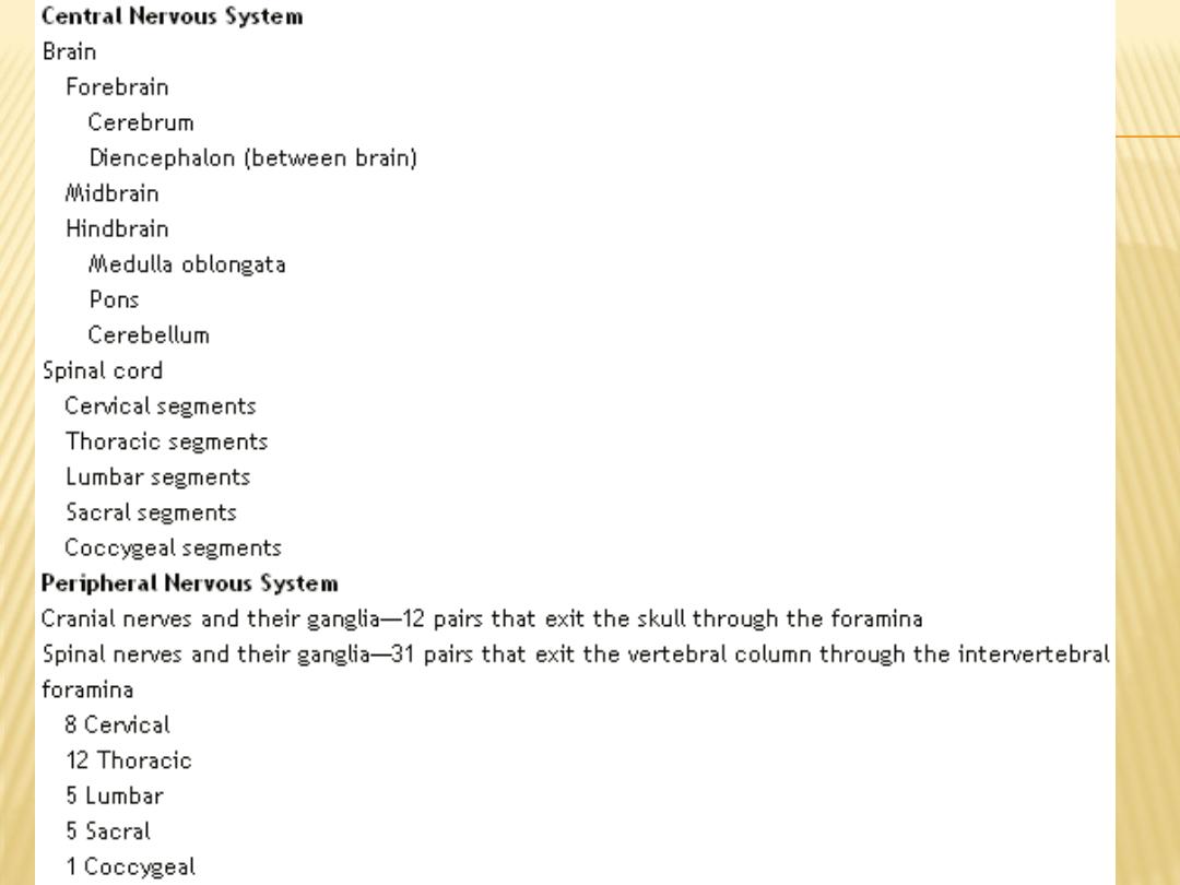
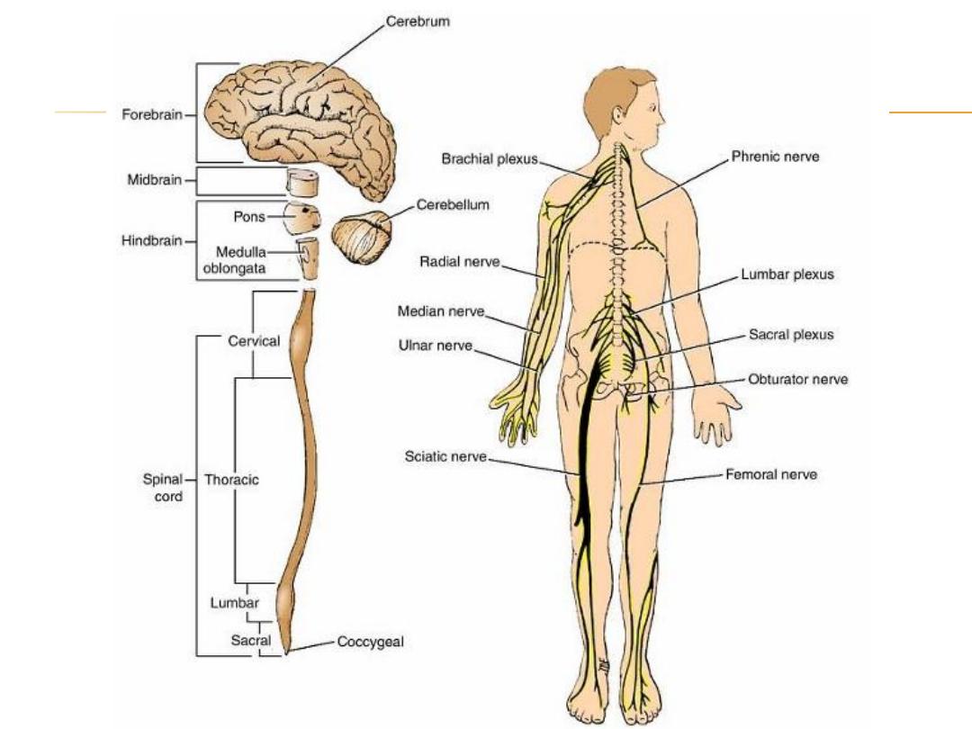
Nervous System
Central NS Peripheral NS
Brain Spinal Cord Spinal ns Cranial ns
Brain:
-
Forebrain: telencephalon (cerebrum) and Diencephalon (Thalamus and
hypothalamus)
-
Midbrain
-
Hindbrain: Cerebellum, pons and medulla oblongata
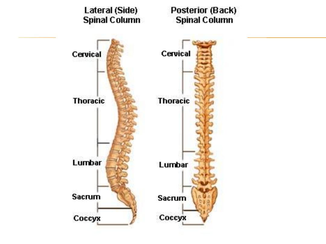

SKULL
Composition
The skull is composed of several separate bones united at immobile joints called
sutures. The connective tissue between the bones is called a sutural ligament.
The mandible is an exception to this rule, for it is united to the skull by the
mobile temporomandibular joint.
The bones of the skull can be divided into those of the cranium and those of the
face. The vault is the upper part of the cranium, and the base of the skull is the
lowest part of the cranium
The skull bones are made up of external and internal tables of compact bone
separated by a layer of spongy bone called the diploë The internal table is
thinner and more brittle than the external table. The bones are covered on the
outer and inner surfaces with periosteum.

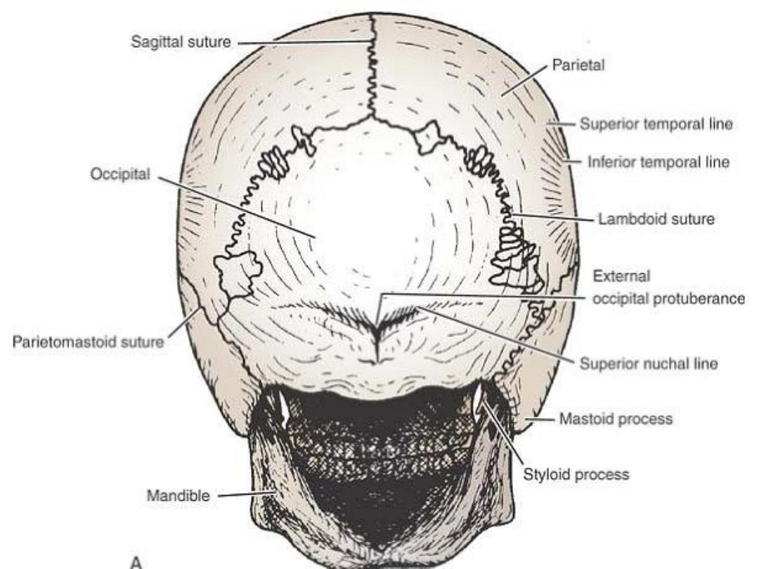
The cranium consists of the following bones, two of which are
paired
• Frontal bone 1
• Parietal bones 2
• Occipital bone 1
• Temporal bones 2
• Sphenoid bone 1
• Ethmoid bone 1

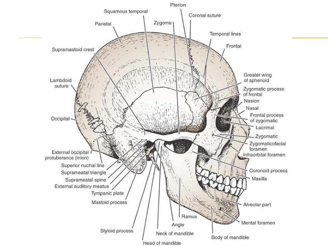
The facial bones consist of the following, two of which are single:
• Zygomatic bones 2
• Maxillae 2
• Nasal bones 2
• Lacrimal bones 2
• Vomer 1
• Palatine bones 2
• Inferior conchae 2
• Mandible 1

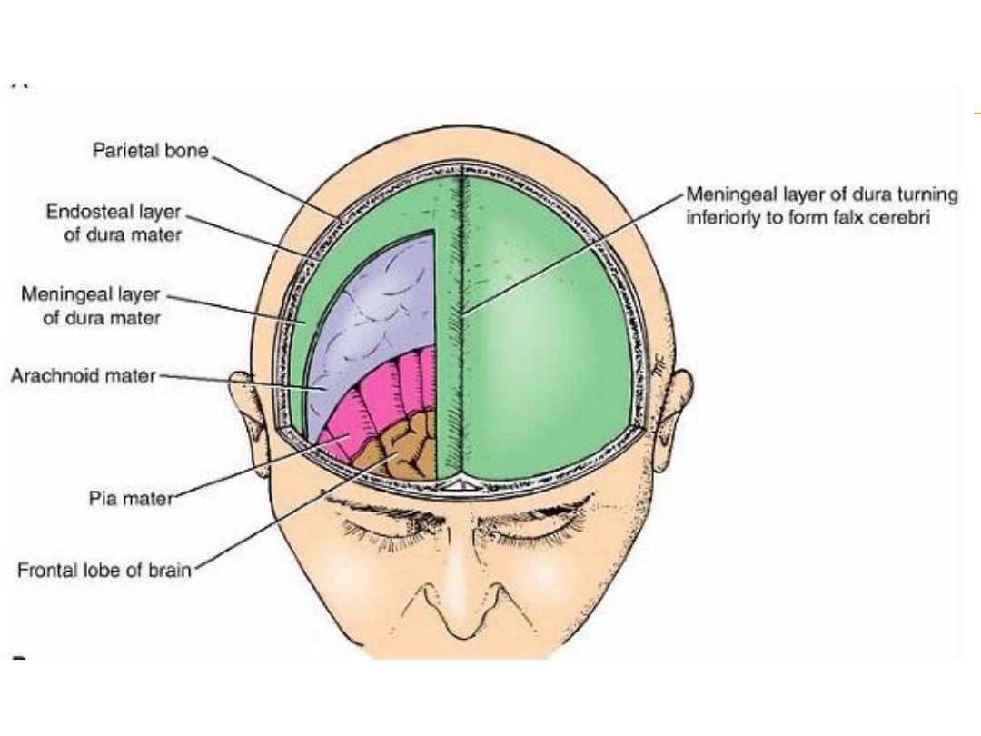
The Cranial Cavity
contains the brain and its surrounding meninges, portions of the cranial nerves,
arteries, veins, and venous sinuses.
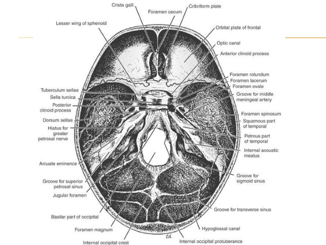
Base of the Skull
is divided into three cranial fossae: anterior, middle, and posterior.
anterior cranial fossa is separated from the middle cranial fossa by the lesser wing
of the sphenoid, and the middle cranial fossa is separated from the posterior
cranial fossa by the petrous part of the temporal bone.

Anterior Cranial Fossa
lodges the frontal lobes of the cerebral hemispheres
bounded anteriorly by the inner surface of the frontal bone, and in the midline is a
crest for the attachment of the falx cerebri. Its posterior boundary is the sharp
lesser wing of the sphenoid
Middle Cranial Fossa
consists of a small median part and expanded lateral parts
The median raised part is formed by the body of the sphenoid, and the expanded
lateral parts form concavities on either side, which lodge the temporal lobes of
the cerebral hemispheres
It is bounded anteriorly by the sharp posterior edges of the lesser wings of the
sphenoid and posteriorly by the superior borders of the petrous parts of the
temporal bones. Laterally lie the squamous parts of the temporal bones, the
greater wings of the sphenoid& the parietal bones
The floor of each lateral part of the middle cranial fossa is formed by the greater
wing of the sphenoid and the squamous and petrous parts of the temporal bone.

Posterior Cranial Fossa
The posterior cranial fossa is deep and lodges the parts of the hindbrain, namely,
the cerebellum, pons, and medulla oblongata. Anteriorly, the fossa is bounded
by the superior border of the petrous part of the temporal bone; posteriorly, it is
bounded by the internal surface of the squamous part of the occipital bone.
The floor of the posterior fossa is formed by the basilar, condylar, and squamous
parts of the occipital bone and the mastoid part of the temporal bone.

Mandible
The mandible, or lower jaw, is the largest and strongest bone of the face, and it
articulates with the skull at the temporamandibular joint
The mandible consists of a horseshoe-shaped body and a pair of rami. The body
of the mandible meets the ramus on each side at the angle of the mandible.
Bones of the Skull
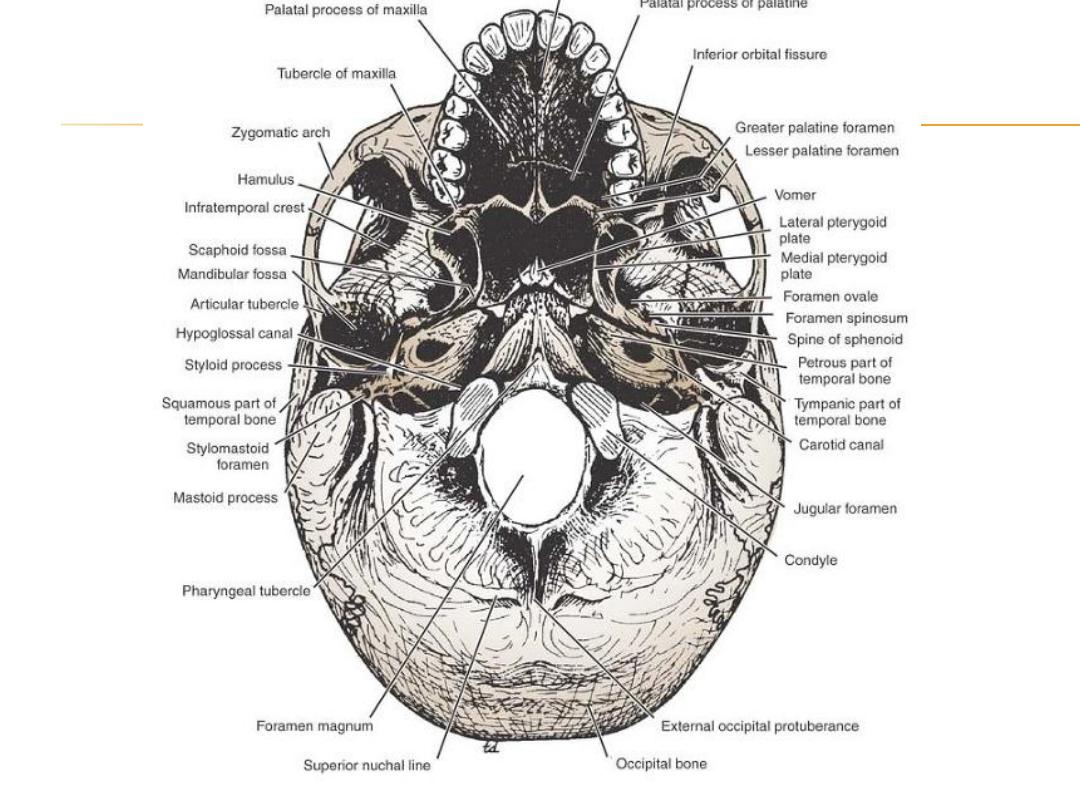
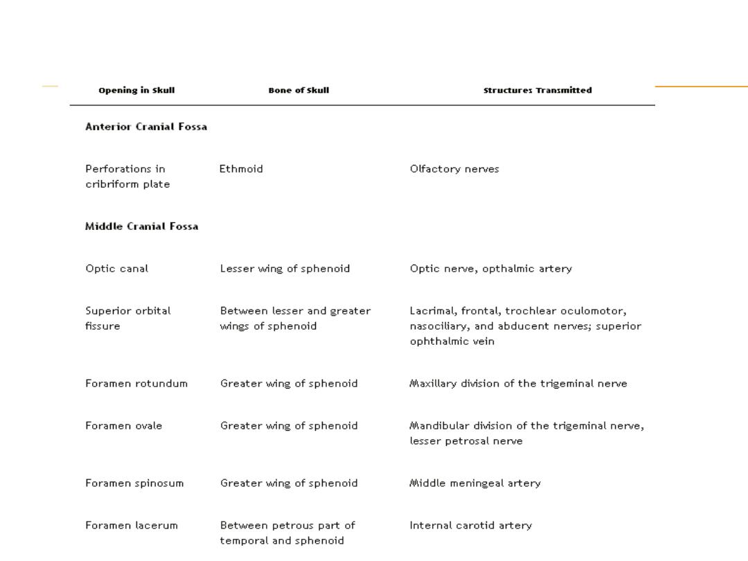
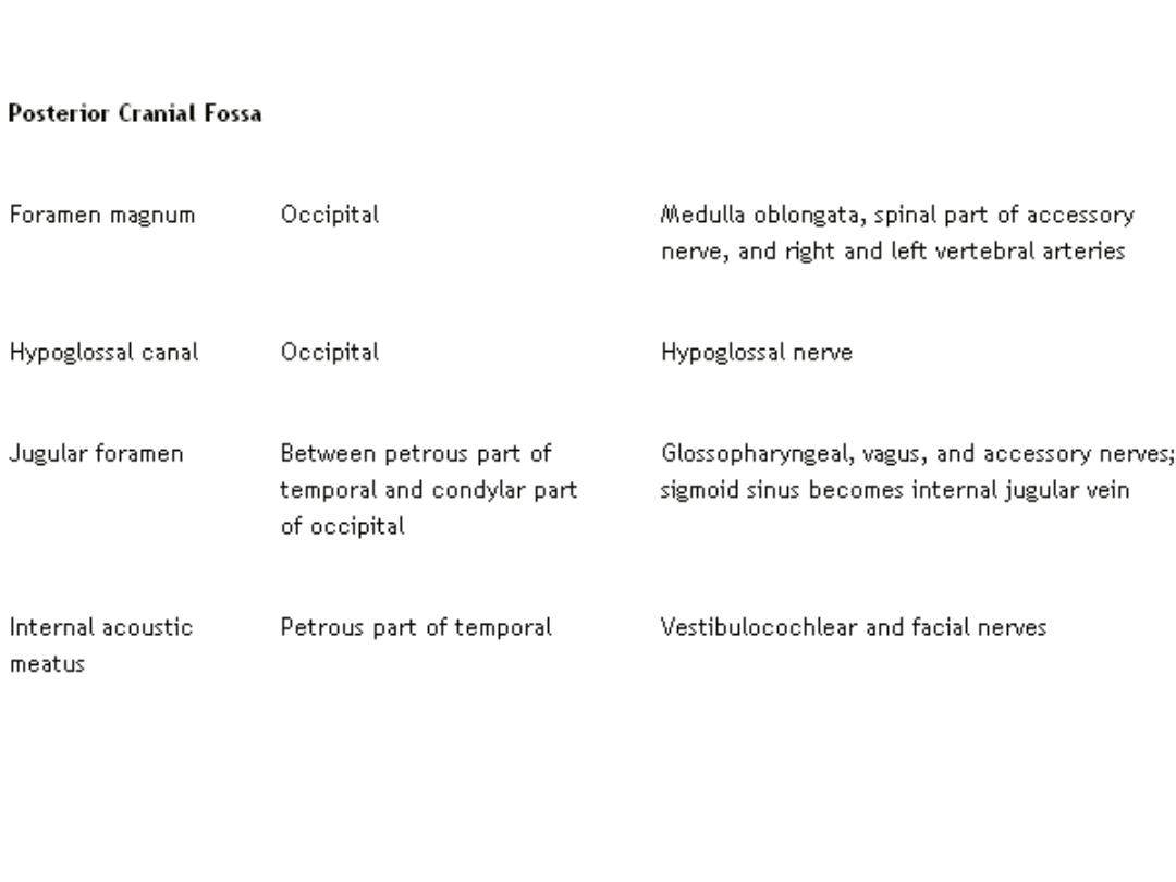
Summary of the More Important Openings in the Base of the
Skull and the Structures That Pass Through Them

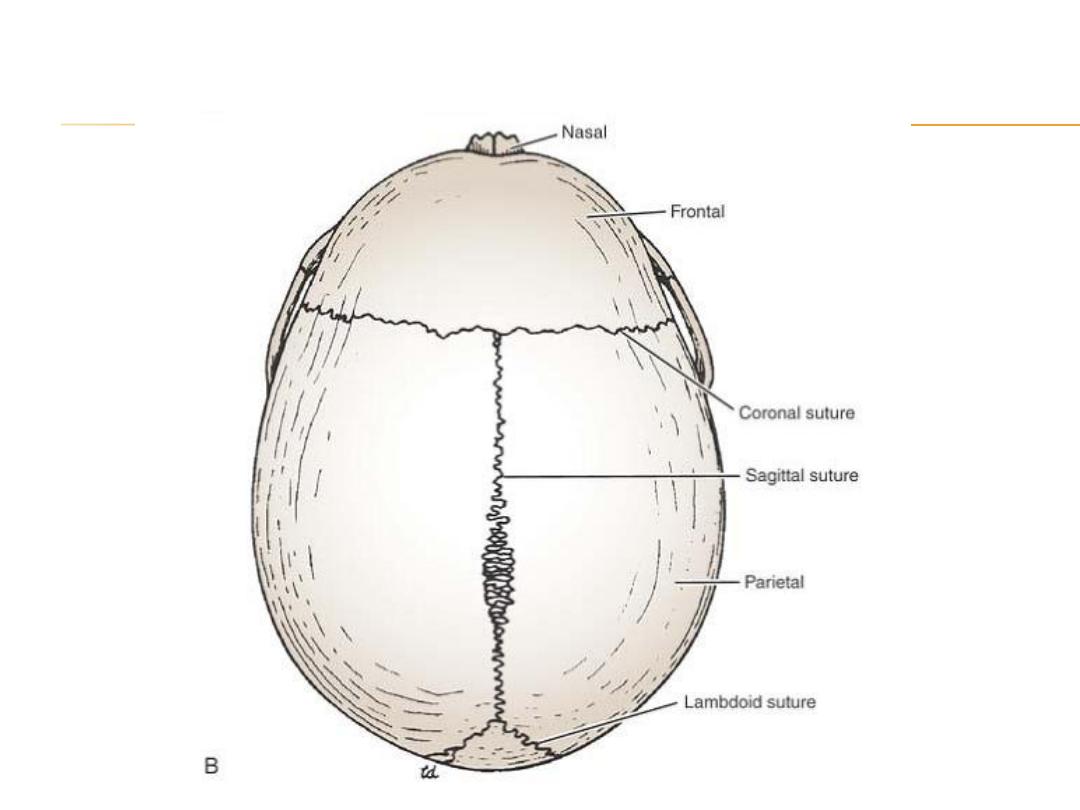
Neonatal Skull
compared with the adult skull, has a disproportionately large cranium relative to the
face. In childhood, the growth of the mandible, the maxillary sinuses, and the
alveolar processes of the maxillae results in a great increase in length of the face.
there being no diploë present
Most of the skull bones are ossified at birth, but the process is incomplete, and the
bones are mobile on each other, being connected by fibrous tissue or cartilage.
fontanelles.
separated by unossified membranous intervals called
Clinically, the anterior and posterior fontanelles are most important and are easily
examined in the midline of the vault.
is diamond shaped and lies between the two halves of the
The anterior fontanelle
frontal bone in front and the two parietal bones behind .The fibrous membrane
forming the floor of the anterior fontanelle is replaced by bone and is closed by 18
months of age.
is triangular and lies between the two parietal bones in front
The posterior fontanelle
and the occipital bone behind. By the end of the first year, the fontanelle is usually
closed and can no longer be palpated.