Surface Anatomy
717
intervertebral
disc
anterior
medulla
oblongata
spinal cord
surrounded
by meninges
posterior
herniated
nucleus
pulposus
C2
3
4
5
6
7
T1
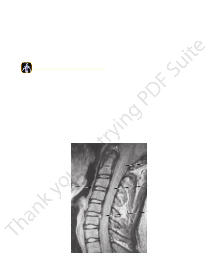
FIGURE 12.34
Sagittal MRI scan of the cervical part of the vertebral column. A herniated disc between the fifth and sixth verte
brae is shown. Note the position of the spinal cord and its meningeal coverings relative to the herniated disc. (Courtesy of Pait.)
-
region.
MRI is now largely replacing myelography or CT in this
to the dural sac can easily be demonstrated. The use of
The herniated fragment of the disc and its relationship
and the posterior longitudinal ligament (Fig. 12.34).
section and shows its relationship to the vertebral body
MRI easily defines the intervertebral disc on sagittal
boundaries of the anulus fibrosus.
Fragments of a herniated disc can be identified beyond the
natomy
face
s
uR
a
The entire posterior aspect of the patient should be exam
runs down the back of the neck connecting the skull to the
a large ligament that
ligamentum nuchae,
covered by the
Cervical spines one to six are
bra (vertebra prominens).
seventh cervical verte
the neck (Fig. 12.35) is that of the
The most prominent spinous process that can be felt in
Cervical Vertebrae
nuchal groove.
the protuberance in the
on the skin in the midline, it can be drawn inferiorly from
the head and neck (Fig. 12.1). If the index finger is placed
The external occipital protuberance lies at the junction of
from above downward.
In the midline, the following structures can be palpated
at the side.
ined from head to foot, and the arms should hang loosely
-
Midline Structures
External Occipital Protuberance
-
spinous processes of the cervical
ebrae.
vert
The crest can
median sacral crest.
midline to form the
are fused with each other in the
spines of the sacrum
The
when the trunk is bent forward.
first thoracic vertebra; the others may be easily recognized
(Fig. 12.35). The most prominent spine is that of the
tebrae
lumbar ver
and the upper four
spines of all the thoracic
that runs down the middle of the back over the tips of the
The nuchal groove is continuous inferiorly with a furrow
Thoracic and Lumbar Vertebrae
compressed.
muscle, and against it the common carotid artery can be
can be palpated medial to the sternocleidomastoid
saignac)
of the sixth cervical transverse process (tubercle of Chas
anterior tubercle
from the lateral side in a thin neck. The
are short but easily palpable
transverse processes
The
-
-
Sacrum
718
CHAPTER 12
The Back
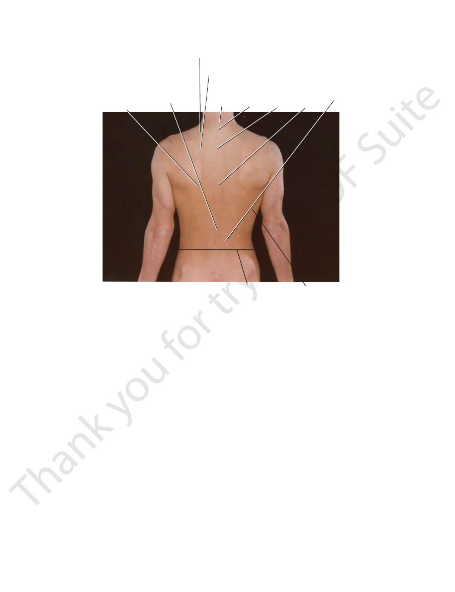
inferior angle
of scapula
superior angle of scapula
base of spine
of scapula
site of lower end
of spinal cord
at lower
border of
first lumbar
vertebra
seventh
cervical
spinous
process
first
thoracic
spinous
process
third
thoracic
spinous
process
seventh
thoracic
spinous
process
erector
spinae
intercristal plane
at level of fourth
lumbar vertebra
latissimus dorsi
FIGURE 12.35
The back of a 27-year-old man.
can be palpated
crest of the spine of the scapula
The
opposite the seventh thoracic spine (Figs. 12.1 and 12.35).
can be palpated
inferior angle
thoracic spine, and the
can be palpated opposite the first
superior angle
The
and traced medially to the medial border of the scapula,
which it joins at the level of the third thoracic spine
(Figs. 12.1 and 12.35).
inferior angle (Fig. 12.35).
which ends above at the superior angle and below at the
of the scapula forms a prominent ridge,
medial border
The
the 1st to the 7th ribs (Figs. 12.1 and 12.35).
ula and its associated muscles. The scapula lies posterior to
The upper lateral part of the thorax is covered by the scap
cyx can be palpated with a gloved finger in the anal canal.
behind the anus (Fig. 12.1). The anterior surface of the coc
in the groove between the buttocks about 1 in. (2.5 cm)
The inferior surface and tip of the coccyx can be palpated
groove between the buttocks.
cm) above the tip of the coccyx and beneath the skin of the
(epidural space) terminates. The hiatus lies about 2 in. (5
the lower end of the sacrum, and here the extradural space
is situated on the posterior aspect of
sacral hiatus
The
between the buttocks.
be felt beneath the skin in the uppermost part of the cleft
the second sacral vertebra and the middle of the sacroiliac
the latter lies beneath a skin dimple at the level of
posterior superior iliac
and behind at the
rior iliac spine
anterior supe
puncture. Each crest ends in front at the
and are used as a landmark when performing a lumbar
(Fig. 12.1). They lie at the level of the fourth lumbar spine
The iliac crests are easily palpable along their entire length
its associated gluteal muscles.
aspect of the upper part of the bony pelvis (false pelvis) and
The lower lateral part of the back is formed by the posterior
located.
ity of the spine of the scapula. It is subcutaneous and easily
forms the lateral extrem
acromion of the scapula
The
-
Lower Lateral Part of the Back
Iliac Crests
-
spine;
Coccyx
-
Upper Lateral Part of the Thorax
-
Scapula

Surface Anatomy
the vertebrae (Figs. 12.1, 12.9, and 12.35). They should be
The muscles are large and lie on either side of the spines of
tains the postural curves of the column, can be palpated.
controls the movements of the vertebral column and main
which mainly
posterior vertebral musculature,
The
buttocks.
the external occipital protuberance to the cleft between the
with reference to an imaginary line passing inferiorly from
Observe the back as a whole and compare the two sides
perior iliac spine.
vertebra (Fig. 12.7), which lies at the level of the posterosu
extends inferiorly to the lower border of the second sacral
cerebrospinal fluid,
with its
subarachnoid space,
The
lumbar spine.
(Fig. 12.7). In young children, it may extend to the third
the lower border of the spine of the first lumbar vertebra
in adults extends inferiorly to the level of
spinal cord
The
of the fifth lumbar spine.
anterosuperior iliac spine. The iliac tubercle lies at the level
surface of the iliac crest about 2 in. (5 cm) posterior to the
joint. The iliac tubercle is a prominence felt on the outer
719
Spinal Cord and Subarachnoid Space
-
Symmetry of the Back
-
examined with the flat of the hand. If they exhibit normal
persons usually exhibit a lateral thoracic curve to the left.
exhibit a lateral thoracic curve to the right; left-handed
involves extreme and prolonged muscular effort, usually
sons. Right-handed persons, especially those whose work
spines, reveals a slight lateral curvature in most normal per
ticular reference to the vertical alignment of the vertebral
Inspection of the posterior surface of the back, with par
lumbosacral angle.
meets the sacrum at a sharp angle, the
together have an anterior concavity. The lumbar region
(Fig. 12.2). The anterior surface of the sacrum and coccyx
in the thoracic region, and concave in the lumbar region
posterior surface is concave in the cervical region, convex
by inspecting the lateral contour of the back. Normally, the
can be examined
curves of the vertebral column
The
the muscular contraction.
produces a concavity of the vertebral column on the side of
harder than normal; it is also shorter than normal, which
tone, they are firm to the touch. A spastic muscle feels
-
-
www.thePoint.lww.com/Snell9e.
Clinical Cases
and
Review Questions
are available online at