
Basic Anatomy
Knowledge of the detailed attachments of the various
supply a band of skin at a lower level than the intervertebral
The posterior rami run downward and laterally and
of the head and supplies the skin of the scalp.
) ascends over the back
greater occipital nerve
nerve (the
ply the skin. The posterior ramus of the second cervical
nerves supply the deep muscles of the back and do not sup
eighth cervical nerves and the fourth and fifth lumbar
nerves. The posterior rami of the first, sixth, seventh, and
tal manner by the posterior rami of the 31 pairs of spinal
The skin and muscles of the back are supplied in a segmen
inguinal nodes (see page 127).
below the level of the iliac crests drain into the superficial
the iliac crests drain into the axillary nodes, and those from
drain into the cervical nodes, those from the trunk above
sacral nodes. The lymph vessels from the skin of the neck
the deep cervical, posterior mediastinal, lateral aortic, and
The deep lymph vessels follow the veins and drain into
Lymph Drainage of the Back
lumbar, and lateral sacral veins.
bral plexus and in turn drain into the vertebral, intercostal,
Here, they are joined by tributaries from the external verte
with the spinal nerves through the intervertebral foramina.
which pass outward
intervertebral veins,
is drained by the
and from the meninges and spinal cord. The internal plexus
(Fig. 12.10)
basivertebral veins
the vertebrae by way of the
The internal vertebral plexus receives tributaries from
of the prostate, page 696).
fact is of considerable clinical significance (see carcinoma
ences that exist at any given time between the regions. This
with the direction of flow depending on the pressure differ
thorax, the abdomen, the pelvis, and the vertebral plexuses,
may therefore take place between the skull, the neck, the
venous sinuses within the skull. Free venous blood flow
communicate through the foramen magnum with the
channels have incompetent valves or are valveless. They
cious venous network whose walls are thin and whose
The external and internal vertebral plexuses form a capa
cord (Fig. 12.10).
vertebral canal but outside the dura mater of the spinal
lies within the
internal vertebral venous plexus
The
surrounds the vertebral column.
lies external and
external vertebral venous plexus
The
coccyx.
extending along the vertebral column from the skull to the
The veins draining the structures of the back form plexuses
Veins
artery.
and lateral sacral arteries, branches of the internal iliac
, branches arise from the iliolumbar
sacral region
In the
and lumbar arteries.
, branches arise from the subcostal
lumbar region
In the
intercostal arteries.
, branches arise from the posterior
thoracic region
In the
695
■
■
■
■
■
■
■
■
■
■
-
-
-
Nerve Supply of the Back
-
-
muscles of the back has no practical value to a clinical profes
deep cervical artery, a branch of the costocervical trunk.
tebral artery, a branch of the subclavian; and from the
artery, a branch of the external carotid; from the ver
branches arise from the occipital
cervical region,
In the
the quadratus lumborum muscle.
verse processes of the lumbar vertebrae; it lies anterior to
medially and is attached to the anterior surface of the trans
the quadratus lumborum. The anterior lamella passes
anterior to the deep muscles of the back and posterior to
of the transverse processes of the lumbar vertebrae; it lies
middle lamella passes medially, to be attached to the tips
cles of the back and is attached to the lumbar spines. The
three lamellae. The posterior lamella covers the deep mus
Medially, the lumbar part of the deep fascia splits into
oblique muscles of the abdominal wall (see page 117).
ers of the transversus and the upper fibers of the internal
aponeurosis and laterally gives origin to the middle fib
between the iliac crest and the 12th rib. It forms a strong
The lumbar part of the deep fascia is situated in the interval
of the abdomen, and the iliac crest.
dorsi, the posterior border of the external oblique muscle
the abdominal wall. The boundaries are the latissimus
The lumbar triangle is the site where pus may emerge from
Lumbar Triangle
the medial border of the scapula.
The boundaries are the latissimus dorsi, the trapezius, and
breath sounds may be most easily heard with a stethoscope.
The auscultatory triangle is the site on the back where
Auscultatory Triangle
Muscular Triangles of the Back
terior rami of the spinal nerves.
All the deep muscles of the back are innervated by the pos
Nerve Supply
into the transverse processes of the upper cervical vertebrae.
has a similar origin but is inserted
splenius cervicis
The
the temporal bone.
nuchal line of the occipital bone and the mastoid process of
upper four thoracic spines and is inserted into the superior
from the lower part of the ligamentum nuchae and the
arises
splenius capitis
back. It consists of two parts. The
The splenius is a detached part of the deep muscles of the
sional, and the attachments are therefore omitted in this text.
-
Splenius
-
Deep Fascia of the Back
(Thoracolumbar Fascia)
-
-
-
Blood Supply of the Back
Arteries
■
■
-
696
CHAPTER 12
The Back
Vertebral Venous Plexus and Carcinoma of the
This is especially likely to occur if the intra-abdominal pressure
tebral venous plexus and by this route may also enter the skull.
venous plexus communicates above with the intracranial venous
Prostate
Because the longitudinal, thin-walled, valveless vertebral
sinuses and segmentally with the veins of the thorax, abdomen,
and pelvis, it is a clinically important structure. Pelvic venous
blood enters not only the inferior vena cava, but also the ver-
is increased. The internal vertebral venous plexus is not subject
to external pressures when the intra-abdominal pressure rises.
A rise in pressure on the abdominal and pelvic veins would tend
to force the blood backward out of the abdominal and pelvic cav-
ities into the veins within the vertebral canal. The existence of
this venous plexus explains how carcinoma of the prostate may
metastasize to the vertebral column and the cranial cavity.
C L I N I C A L N O T E S
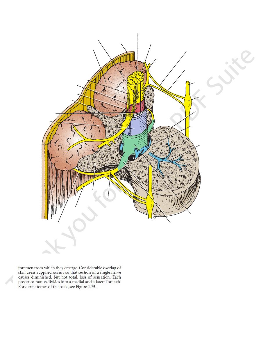
anterior root
posterior vertebral muscles
posterior root
pia mater
ligamentum denticulatum
spine
subarachnoid
space
arachnoid
mater
posterior ramus
anterior ramus
transverse
process
spinal nerve
posterior root ganglion
dura mater
basivertebral
vein
internal vertebral
venous plexus
body of
vertebra
sympathetic
ganglion
sympathetic trunk
gray ramus
white ramus
anterior horn
lateral horn
posterior horn
skin
FIGURE 12.10
Oblique section through the first lumbar vertebra showing the spinal cord and its covering membranes. Note
venous plexus.
the relationship between the spinal nerve and sympathetic trunk on each side. Note also the important internal vertebral