Basic Anatomy
315
fatty layer of superficial fascia
urogenital diaphragm
anal canal
fat of ischiorectal fossa
perineal body
superior fascial layer
of urogenital diaphragm
inferior fascial layer of urogenital
diaphragm (perineal membrane)
scrotum
dartos muscle
Colles' fascia
superficial perineal pouch
deep perineal pouch
membranous layer of superficial fascia (Scarpa's fascia)
FIGURE 8.12
Arrangement of the superficial fascia in the urogenital triangle. Note the superficial and deep perineal pouches.
which covers the distal
glans penis,
expands to form the
and 8.16). At its distal extremity, the corpus spongiosum
spongiosum applied to their ventral surface (see Figs. 8.13
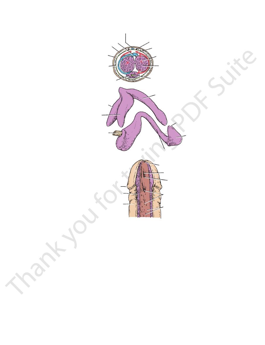
two dorsally placed corpora cavernosa and a single corpus
The erectile tissue is made up of
(Buck’s fascia).
cylinders of erectile tissue enclosed in a tubular sheath of
The body of the penis is essentially composed of three
Body of the Penis
(see Figs. 8.13 and 8.16).
corpora cavernosa
side in the dorsal part of the body of the penis, forming the
The two crura converge anteriorly and come to lie side by
(see Fig. 8.17).
corpus spongiosum
the penis and forms the
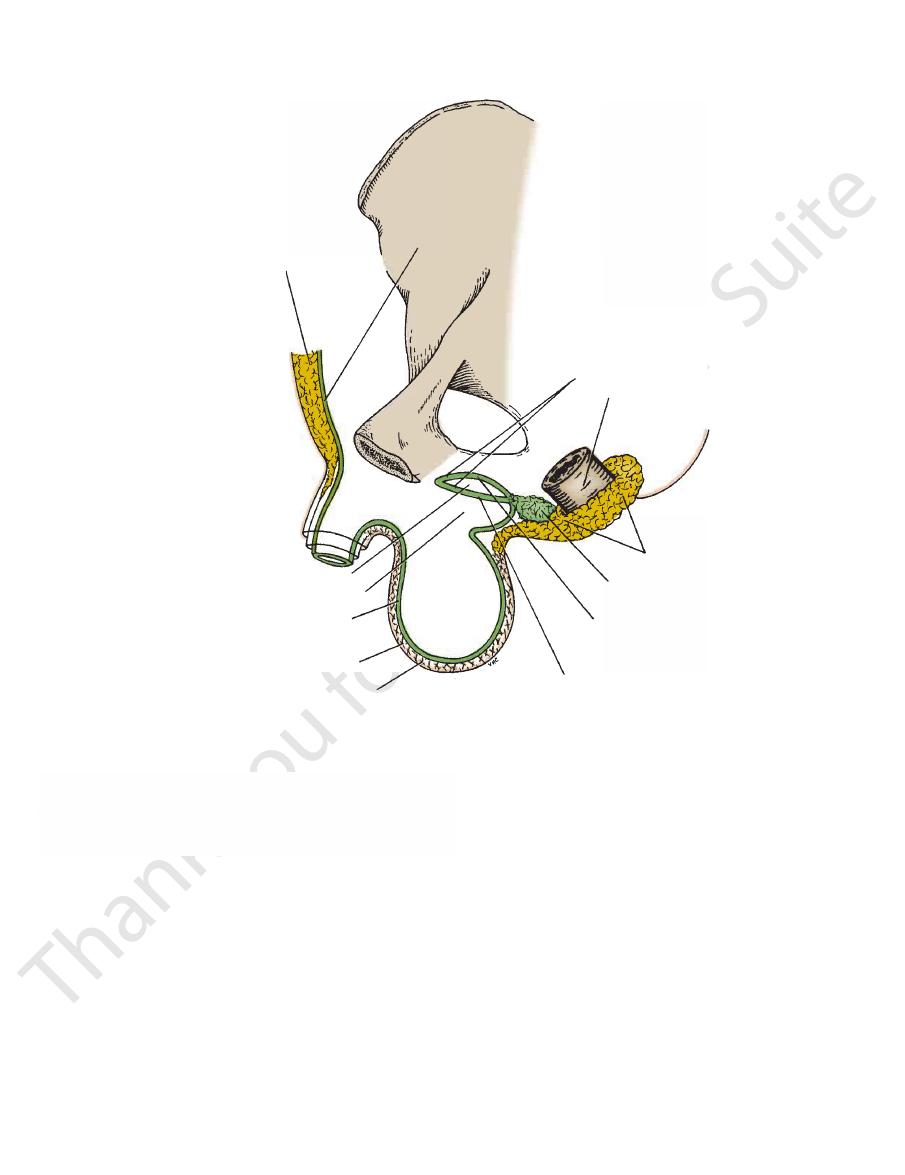
The bulb is continued forward into the body of
muscle.
ischiocavernosus
and is covered on its outer surface by the
Each crus is attached to the side of the pubic arch
muscles.
bulbospongiosus
and is covered on its outer surface by the
of the urogenital diaphragm. It is traversed by the urethra
situated in the midline and is attached to the undersurface
(Figs. 8.13, 8.16, and 8.17). The bulb is
crura of the penis
left
right
bulb of the penis
tissue called the
The root of the penis is made up of three masses of erectile
Root of the Penis
(Figs. 8.4 and 8.16).
The penis has a fixed root and a body that hangs free
Location and Description
In the male, the triangle contains the penis and scrotum.
Triangle
are described in subsequent sections.
The contents of the deep perineal pouch in both sexes
(see Figs. 8.12, 8.14, and 8.15).
perineal pouch
deep
superficial and deep layers of fascia is known as the
Contents of the Male Urogenital
Penis
and the
and
fascia
316
CHAPTER 8
The Perineum
dorsal vein
skin
membranous layer of superficial
deep fascia
(Buck's fascia)
corpus spongiosum
left crus
right crus
membranous part of
urethra
corona
corpus spongiosum
skin
openings of
urethral
glands
lacunae
fold of mucous membrane
fossa terminalis
external urethral
orifice
corpus spongiosum
glans penis
corona
corpus cavernosum
urethra
corpus cavernosum
deep artery
dorsal artery
superficial dorsal artery
superficial dorsal vein
fascia
A
B
C
FIGURE 8.13
The penis.
plexuses.
The nerve supply is from the pudendal nerve and the pelvic
Nerve Supply
are drained into the internal iliac nodes.
superficial inguinal nodes. The deep structures of the penis
The skin of the penis is drained into the medial group of
Lymph Drainage
The veins drain into the internal pudendal veins.
Veins
the internal pudendal artery.
All the above arteries are branches of
artery of the penis.
dorsal
In addition, there is the
artery of the bulb.
by the
(see Fig. 8.13); the corpus spongiosum is supplied
the penis
deep arteries of
The corpora cavernosa are supplied by the
Arteries
symphysis pubis to be attached to the fascia of the penis.
of deep fascia that extend downward from the linea alba and
The body of the penis is supported by two condensations
frenulum.
urethral orifice by a fold called the
covers the glans. It is connected to the glans just below the
is a hoodlike fold of skin that
foreskin
or
prepuce
The
thral meatus.
external ure
is the slitlike orifice of the urethra, called the
ends of the corpora cavernosa. On the tip of the glans penis
The penile urethra slit open to show the folds of mucous membrane and glandular orifices in the roof of
The three bodies of erectile tissue, the two corpora cavernosa, and the corpus spongiosum
A and B.
with the glans. C.
the urethra.
-
Blood Supply
pelvic fascia
levator ani
obturator internus
prostate
sphincter urethrae
muscle
dorsal nerve of penis
membranous part of urethra
artery of bulb
artery of crus
(deep artery of penis)
ischiocavernosus
urethra in bulb
bulbospongiosus
membranous layer of
superficial fascia
deep fascia of thigh
skin of medial side of thigh
scrotal nerves
bulb of penis
bulbourethral gland
crus of penis
inferior
fascial layer
of urogenital
diaphragm
superior fascial
layer of
urogenital
diaphragm
prostatic
urethra
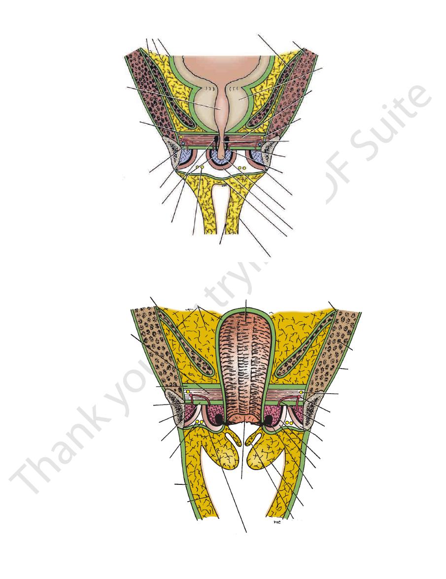
FIGURE 8.14
Coronal section of the male pelvis showing the prostate, the urogenital diaphragm, and the contents of the
superficial perineal pouch.
levator ani
pelvic fascia
vagina
fascia of obturator internus
obturator
internus
obturator
membrane
dorsal nerve
of clitoris
internal pudendal
artery
artery of bulb
ischiocavernosus
bulb of vestibule
bulbospongiosus
greater vestibular gland
labium majus
labium minus
hymen
membranous layer of superficial fascia
sphincter
urethrae
superior
fascial layer of
urogenital diaphragm
artery of crus
(deep artery of clitoris)
crus of clitoris
inferior fascial layer
of urogenital diaphragm
deep fascia of thigh
skin of medial side of thigh
labial nerves
FIGURE 8.15
Coronal section of the female pelvis showing the vagina, the urogenital diaphragm, and the contents of the
317
superficial perineal pouch.
318
CHAPTER 8
The Perineum
deep dorsal vein
dorsal nerve
dorsal artery
crus of penis
corpora cavernosa
corpus spongiosum
glans penis
corona
deep dorsal vein
posterior scrotal nerves
perineal membrane
external urethral meatus
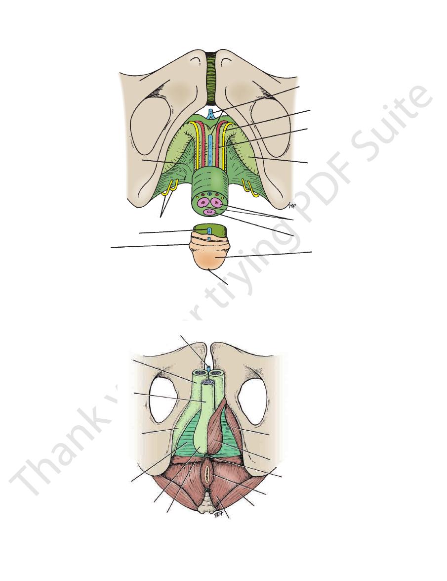
FIGURE 8.16
Root and body of the penis.
deep dorsal vein of penis
urogenital diaphragm
bulb of penis
superficial transverse
perineal muscle
levator ani
gluteus maximus
external anal sphincter
bulbospongiosus
ischiocavernosus
perineal body
corpus cavernosum
corpus spongiosum
crus of penis
FIGURE 8.17
Root of penis and perineal muscles.

Basic Anatomy
two sides and passes medially to encircle the urethra (see
deep perineal pouch. It arises from the pubic arch on the
The sphincter urethrae muscle surrounds the urethra in the
Sphincter Urethrae Muscle
the urethra (see Fig. 8.14).
penile urethra. It is the shortest and least dilatable part of
ous above with the prostatic urethra and below with the
surrounded by the sphincter urethrae muscle; it is continu
(1.3 cm) long and lies within the urogenital diaphragm,
The membranous part of the urethra is about 0.5 in.
Membranous Part of the Urethra
of the penis.
pudendal vessels and their branches, and the dorsal nerves
glands, the deep transverse perineal muscles, the internal
of the urethra, the sphincter urethrae, the bulbourethral
The deep perineal pouch contains the membranous part
the muscles and skin (see Fig. 8.8).
terminates in the superficial perineal pouch by supplying
The perineal branch of the pudendal nerve on each side
Perineal Branch of the Pudendal Nerve
muscle, and superficial transverse perineal muscles.
lowing muscles: external anal sphincter, bulbospongiosus
8.12 and 8.17). It serves as a point of attachment for the fol
the posterior margin of the urogenital diaphragm (see Figs.
This small mass of fibrous tissue is attached to the center of
Perineal Body
plied by the perineal branch of the pudendal nerve.
All the muscles of the superficial perineal pouch are sup
fix the perineal body in the center of the perineum.
into the perineal body. The function of these muscles is to
Each muscle arises from the ischial ramus and is inserted
terior part of the superficial perineal pouch (see Fig. 8.17).
The superficial transverse perineal muscles lie in the pos
Superficial Transverse Perineal Muscles
the penis.
press the crus penis and assist in the process of erection of
side (see Fig. 8.17). The action of each muscle is to com
The ischiocavernosus muscles cover the crus penis on each
Ischiocavernosus Muscles
process of erection of the penis.
drainage of the erectile tissue and thereby assisting in the
the deep dorsal vein of the penis, thus impeding the venous
residual urine or semen. The anterior fibers also compress
to compress the penile part of the urethra and empty it of
rior portion of the corpus spongiosum. Their function is
(see Fig. 8.17), cover the bulb of the penis and the poste
situated one on each side of the midline
giosus muscles,
bulbospon
ischiocavernosus muscles (see Fig. 8.17). The
cover them—namely, the bulbospongiosus muscles and the
ing the root of the penis, together with the muscles that
The superficial perineal pouch contains structures form
the thigh.
the perineal nerves and the posterior cutaneous nerves of
nerves, and the posterior surface is supplied by branches of
guinal nerves and the genital branch of the genitofemoral
The anterior surface of the scrotum is supplied by the ilioin
Nerve Supply
and lymph vessels after it.
nal canal, and into the scrotum, dragging its blood supply
on the posterior abdominal wall, down through the ingui
the testis during development has migrated from high up
of the first lumbar vertebra. This is to be expected because
ends in the lumbar (para-aortic) lymph nodes at the level
the testis and epididymis ascends in the spermatic cord and
superficial inguinal lymph nodes. The lymph drainage of
The wall of the scrotum is drained into the medial group of
Lymph Drainage
The veins accompany the corresponding arteries.
Veins
branches of the internal pudendal arteries supply the scrotum.
The external pudendal branches of the femoral and scrotal
Arteries
trol of the temperature of the testes.
promote heat loss and thus assist in the environmental con
Subcutaneous plexuses and arteriovenous anastomoses
they are fully described in Chapter 4.
tes, and the formation of the inguinal canal are interrelated,
Because the structure of the scrotum, the descent of the tes
anterior, medial, and lateral surfaces of each testis
Tunica vaginalis, which is a closed sac that covers the
versalis
Internal spermatic fascia derived from the fascia trans
Cremasteric fascia derived from the internal oblique
oblique
External spermatic fascia derived from the external
called Colles’ fascia.
nal wall, and Scarpa’s fascia (membranous layer), now
muscle, replaces the fatty layer of the anterior abdomi
Superficial fascia; the dartos muscle, which is smooth
Skin
The wall of the scrotum has the following layers:
(see Fig. 4.21).
epididymides, and the lower ends of the spermatic cords
anterior abdominal wall and contains the testes, the
The scrotum is an outpouching of the lower part of the
Location and Description
319
Scrotum
■
■
■
■
-
■
■
■
■
■
■
-
■
■
-
Blood Supply
-
-
-
Contents of the Superficial Perineal
Pouch in the Male
-
-
-
-
-
Nerve Supply
-
-
Contents of the Deep Perineal Pouch
in the Male
-
Fig. 8.14).

320
CHAPTER 8
penis (see Figs. 8.4, 8.14, 8.16, and 8.17). The external mea
is enclosed in the bulb and the corpus spongiosum of the
is about 6 in. (15.75 cm) long and
penile urethra
The
portion of the urethra (see Fig. 8.14).
by the sphincter urethrae muscle. It is the least dilatable
long and lies within the urogenital diaphragm, surrounded
is about 0.5 in. (1.25 cm)
membranous urethra
The
most dilatable portion of the urethra.
from the base to the apex (see Fig. 8.14). It is the widest and
about 1.25 in. (3 cm) long and passes through the prostate
is described on page 278. It is
prostatic urethra
The
static, membranous, and penile.
glans penis (see Fig. 8.4). It is divided into three parts: pro
from the neck of the bladder to the external meatus on the
The male urethra is about 8 in. (20 cm) long and extends
cles, and the prostate via the inferior hypogastric plexuses.
are then distributed to the vas deferens, the seminal vesi
parts of the sympathetic trunks. The postganglionic fibers
ers may synapse in ganglia in the lower lumbar or pelvic
neurons in the first and second lumbar ganglia. Other fib
fibers. Many of these fibers synapse with postganglionic
second lumbar segments in the preganglionic sympathetic
genital organs are thought to leave the cord at the first and
outflow (T1 to L2). The nervous impulses that pass to the
tem. Impulses pass down the spinal cord to the sympathetic
of nervous impulses takes place in the central nervous sys
At the climax of male sexual excitement, a mass discharge
semen.
or
seminal fluid,
accessory glands constitute the
bladder. The spermatozoa and the secretions of the several
contracts and prevents a reflux of the spermatozoa into the
press the urethra. Meanwhile, the sphincter of the bladder
contractions of the bulbospongiosus muscles, which com
ejected from the penile urethra as a result of the rhythmic
bourethral glands and penile urethral glands and is then
tatic urethra. The fluid now joins the secretions of the bul
seminal vesicles and prostate, are discharged into the pros
and the spermatozoa, together with the secretions of the
vesicles, and the prostate. The smooth muscle contracts,
epididymis and the vas deferens on each side, the seminal
thetic nerve fibers to the smooth muscle of the duct of the
nervous impulses, results in a discharge along the sympa
Friction on the glans penis, reinforced by other afferent
thral glands.
becomes moist as a result of the secretions of the bulboure
ing sex play, the external urinary meatus of the glans penis
During the increasing sexual excitement that occurs dur
vasoconstriction. The penis then returns to its flaccid state.
inhibited, the arteries supplying the erectile tissue undergo
ejaculation takes place, or the excitement passes off or is
Once the climax of sexual excitement is reached and
diameter and assumes the erect position.
ated and maintained. The penis thus increases in length and
is retarded so that the internal pressure is further accentu
By this means, the outflow of blood from the erectile tissue
pressing their draining veins against the surrounding fascia.
giosum become engorged with blood and expand, com
erectile tissue. The corpora cavernosa and the corpus spon
great increase in blood flow through the blood spaces of the
penis. Vasodilatation of the arteries now occurs, producing a
branches, which enter the erectile tissue at the root of the
the internal pudendal arteries and are distributed along their
the postganglionic neurons. The postganglionic fibers join
fibers enter the inferior hypogastric plexuses and synapse on
fourth sacral segments. The parasympathetic preganglionic
to the parasympathetic outflow in the second, third, and
stimuli. Efferent nervous impulses pass down the spinal cord
in a bombardment of the central nervous system by afferent
stimuli from the general body skin and genital skin, result
other psychic stimuli, fortified later by direct touch sensory
various sexual stimuli. Pleasurable sight, sound, smell, and
Erection in the male is gradually built up as a consequence of
the penis (see Fig. 8.14).
through the deep perineal pouch and supplies the skin of
The dorsal nerve of the penis on each side passes forward
Dorsal Nerve of the Penis
which supplies the skin and fascia of the penis.
the penis,
dorsal artery of
(deep artery of penis); and the
of the penis
arteries to the crura
artery to the bulb of the penis;
the deep perineal pouch and passes forward, giving rise to
The internal pudenal artery (see Fig. 8.14) on each side enters
Internal Pudendal Artery
perineal body. These muscles are clinically unimportant.
ischial ramus and passes medially to be inserted into the
sphincter urethrae muscle. Each muscle arises from the
The deep transverse perineal muscles lie posterior to the
Deep Transverse Perineal Muscles
result of erotic stimulation.
of the urethra. The secretion is poured into the urethra as a
of the urogenital diaphragm) and enter the penile portion
ducts pierce the perineal membrane (inferior fascial layer
beneath the sphincter urethrae muscle (see Fig. 8.14). Their
The bulbourethral glands are two small glands that lie
which micturition can be voluntarily stopped.
thra and relaxes during micturition. It is the means by
The muscle compresses the membranous part of the ure
sphincter.
The perineal branch of the pudendal nerve supplies the
Nerve Supply
The Perineum
Action
-
Bulbourethral Glands
the
the
Erection of the Penis
-
-
-
Ejaculation
-
-
-
-
-
-
-
-
-
Male Urethra
-
-
and the orifices of the urethra and the vagina.
In the female, the triangle contains the external genitalia
Triangle
the urogenital diaphragm.
bulbourethral glands open into the penile urethra below
(navicular fossa) (see Fig. 8.4). The
fossa terminalis
the urethra that lies within the glans penis is dilated to form
tus is the narrowest part of the entire urethra. The part of
the
Contents of the Female Urogenital