312
CHAPTER 8
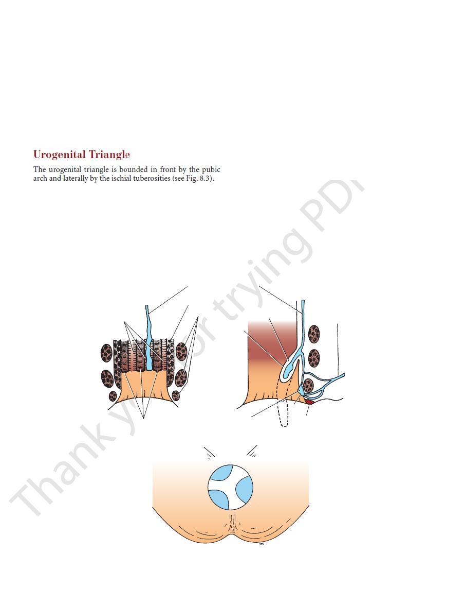
The Perineum
anal columns
anal valves
11 o’clock
3 o’clock
7 o’clock
A
B
C
tributary of superior rectal vein
internal anal sphincter
internal
hemorrhoid
external
hemorrhoid
inferior
rectal vein
perianal hematoma
skin
mucous
membrane
external anal sphincter
FIGURE 8.9
hemorrhoids as seen through a proctoscope with the patient in the lithotomy position.
Positions of three internal
vein forming the internal hemorrhoid. Dotted lines indicate degrees of severity of condition.
Varicosed tributary of the superior rectal
Normal tributary of the superior rectal vein within the anal column.
A.
B.
C.
Development of the Anal Canal
and that of the inferior half of the anal canal is formed from the
The distal end of the hindgut terminates as a blind sac of ento-
derm called the cloaca (see Fig. 7.8). The cloaca lies in contact
with a shallow ectodermal depression called the proctodeum.
The apposed layers of ectoderm and entoderm form the cloacal
membrane, which separates the cavity of the hindgut from the
surface (see Fig. 7.8). The cloaca becomes divided into anterior
and posterior parts by the urorectal septum; the posterior part
of the cloaca is called the anorectal canal. The anorectal canal
forms the rectum and the upper half of the anal canal. The lining
of the superior half of the anal canal is formed from entoderm,
ectoderm of the proctodeum (see Fig. 7.8). The sphincters of the
anal canal are formed from the surrounding mesenchyme. The
posterior part of the cloacal membrane breaks down so that the
gut opens onto the surface of the embryo.
Imperforate Anus
About 1 child in 4000 is born with imperforate anus caused by an
imperfect fusion of the entodermal cloaca with the proctodeum.
E M B R Y O L O G I C N O T E S
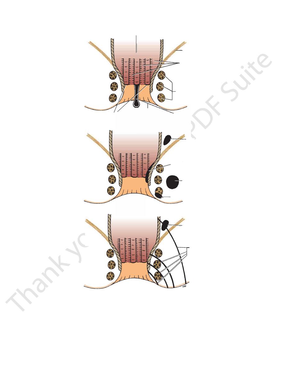
Superficial Fascia
phragm (see Fig. 8.12) and laterally to the margins of the
posteriorly to the posterior border of the urogenital dia
(Colles’ fascia) is attached
membranous layer
The
page 131).
of the scrotal skin (see testicular temperature and fertility,
contracts in response to cold and reduces the surface area
The dartos muscle
dartos muscle.
by smooth muscle, the
cial fascia of the thighs. In the scrotum, the fat is replaced
the fat of the ischiorectal fossa (Fig. 8.12) and the superfi
(fascia of Camper) is continuous with
fatty layer
The
divided into a fatty layer and a membranous layer.
The superficial fascia of the urogenital triangle can be
-
-
Basic Anatomy
313
rectum
levator ani
anal columns
anal fissure
pelvirectal
abscess
ischiorectal
abscess
subcutaneous
abscess
pelvirectal
abscess
fistulae
C
B
A
anal valves
external
anal
sphincter
submucous
abscess
FIGURE 8.10
Common positions of perianal fistulae.
Tearing downward of the anal valve to form an anal fissure.
A.
B. Common locations of perianal abscesses.
C.
314
CHAPTER 8
pubic arch. The closed space that is contained between the
Fig. 8.12). Laterally, the layers of fascia are attached to the
layer of the superficial fascia and the perineal body (see
of fascia fuse with each other and with the membranous
gap beneath the symphysis pubis. Posteriorly, the two layers
Anteriorly, the two layers of fascia fuse, leaving a small
perineal membrane.
diaphragm. The inferior layer of fascia is often referred to
a superior and an inferior layer of fascia of the urogenital
transverse perineal muscles, which are enclosed between
8.15). It is formed by the sphincter urethrae and the deep
filling in the gap of the pubic arch (see Figs. 8.12, 8.14, and
diaphragm situated in the anterior part of the perineum,
The urogenital diaphragm is a triangular musculofascial
sexes are described on pages 319 and 322.
The contents of the superficial perineal pouch in both
anterior abdominal muscles.
superficial fascia of the anterior abdominal wall and the
municates freely with the potential space lying between the
pubic arch (Figs. 8.14 and 8.15). Anteriorly, the space com
fascia and the urogenital diaphragm to the margins of the
by the attachment of the membranous layer of superficial
the fusion of its upper and lower walls. Laterally, it is closed
urogenital diaphragm (see Fig. 8.12). It is closed behind by
membranous layer of superficial fascia and above by the
The superficial perineal pouch is bounded below by the
Fig. 8.12).
scrotum (or labia majora), it forms a distinct layer (see
penis (or clitoris) as a tubular sheath (Fig. 8.13). In the
wall (Scarpa’s fascia). The fascia is continued over the
nous layer of superficial fascia of the anterior abdominal
pubic arch; anteriorly it is continuous with the membra
The Perineum
-
Superficial Perineal Pouch
-
Urogenital Diaphragm
as the
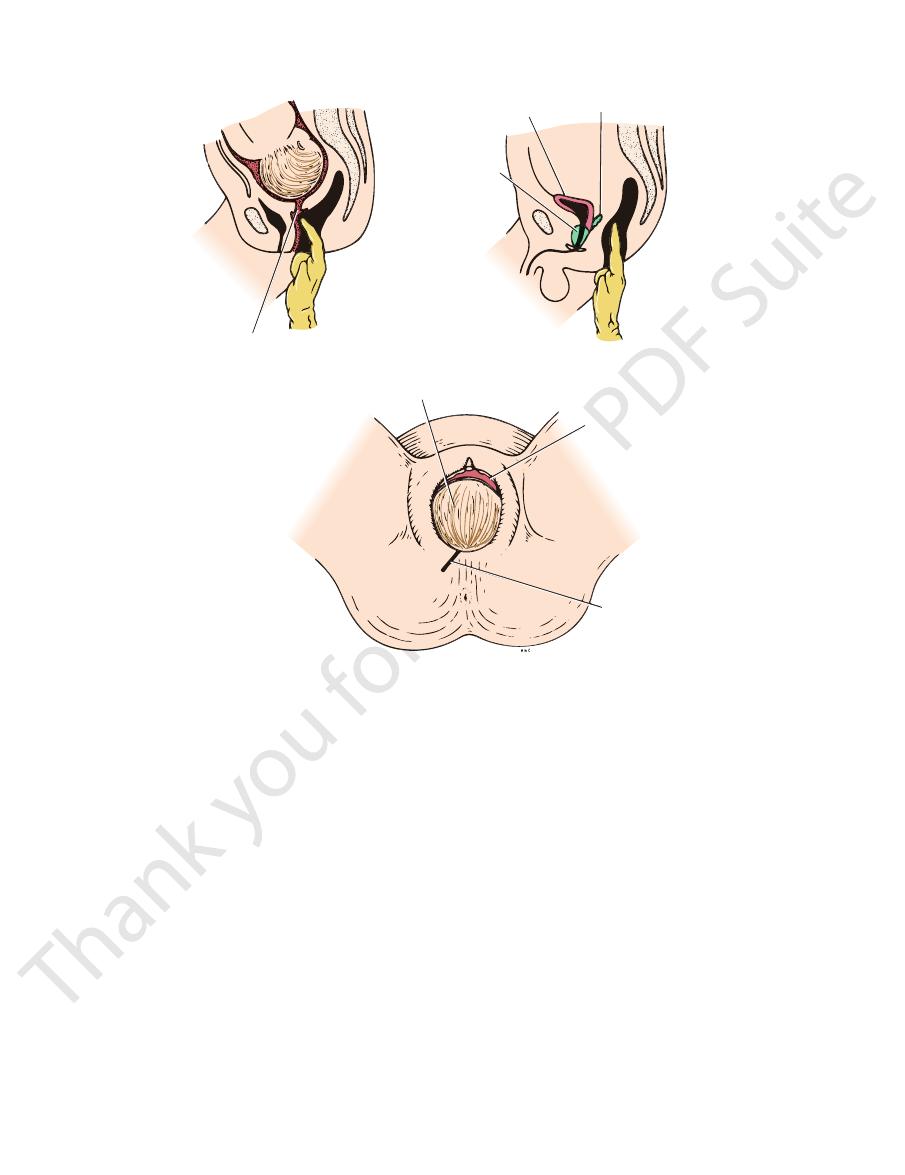
external os of uterus
A
B
C
prostate
bladder
seminal vesicle
head of baby
anterior lip of cervix
site for episiotomy
FIGURE 8.11
baby’s head is presenting at the vaginal orifice.
Position of the episiotomy incision in a woman during the second stage of labor. The
through the anterior rectal wall.
Rectal examination in the male showing how it is possible to palpate the prostate and the seminal vesicles
rior rectal wall.
Rectal examination in a pregnant woman showing how it is possible to palpate the cervix through the ante
A.
-
B.
C.
Basic Anatomy
315
fatty layer of superficial fascia
urogenital diaphragm
anal canal
fat of ischiorectal fossa
perineal body
superior fascial layer
of urogenital diaphragm
inferior fascial layer of urogenital
diaphragm (perineal membrane)
scrotum
dartos muscle
Colles' fascia
superficial perineal pouch
deep perineal pouch
membranous layer of superficial fascia (Scarpa's fascia)
FIGURE 8.12
Arrangement of the superficial fascia in the urogenital triangle. Note the superficial and deep perineal pouches.
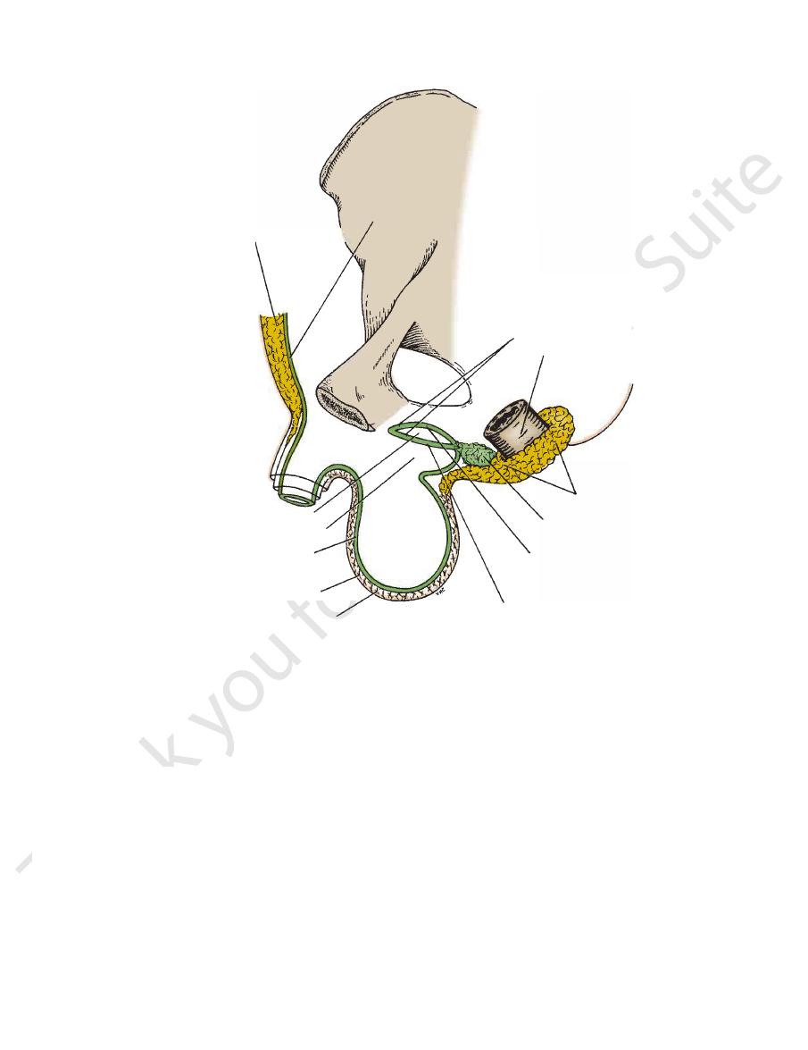
which covers the distal
glans penis,
expands to form the
and 8.16). At its distal extremity, the corpus spongiosum
spongiosum applied to their ventral surface (see Figs. 8.13
two dorsally placed corpora cavernosa and a single corpus
The erectile tissue is made up of
(Buck’s fascia).
cylinders of erectile tissue enclosed in a tubular sheath of
The body of the penis is essentially composed of three
Body of the Penis
(see Figs. 8.13 and 8.16).
corpora cavernosa
side in the dorsal part of the body of the penis, forming the
The two crura converge anteriorly and come to lie side by
(see Fig. 8.17).
corpus spongiosum
the penis and forms the
The bulb is continued forward into the body of
muscle.
ischiocavernosus
and is covered on its outer surface by the
Each crus is attached to the side of the pubic arch
muscles.
bulbospongiosus
and is covered on its outer surface by the
of the urogenital diaphragm. It is traversed by the urethra
situated in the midline and is attached to the undersurface
(Figs. 8.13, 8.16, and 8.17). The bulb is
crura of the penis
left
right
bulb of the penis
tissue called the
The root of the penis is made up of three masses of erectile
Root of the Penis
(Figs. 8.4 and 8.16).
The penis has a fixed root and a body that hangs free
Location and Description
In the male, the triangle contains the penis and scrotum.
Triangle
are described in subsequent sections.
The contents of the deep perineal pouch in both sexes
(see Figs. 8.12, 8.14, and 8.15).
perineal pouch
deep
superficial and deep layers of fascia is known as the
Contents of the Male Urogenital
Penis
and the
and
fascia