Basic Anatomy
157
Stomach 171
Small Intestine 172
Large Intestine 180
Blood Supply of the Gastrointestinal
Tract 184
Differences Between the Small and
Large Intestines 196
Accessory Organs of the Gastrointestinal
Tract 196
Liver 196
Bile Ducts of the Liver 198
Pancreas 201
Spleen 203
Retroperitoneal Space 206
Urinary Tract 206
Kidneys 206
Ureter 209
Suprarenal Glands 211
Location and Description 211
Blood Supply 215
Lymph Drainage 215
Nerve Supply 215
Arteries on the Posterior Abdominal
Wall 215
Aorta 215
Common Iliac Arteries 216
External Iliac Artery 216
Internal Iliac Artery 218
Veins on the Posterior Abdominal
Wall 218
Inferior Vena Cava 218
Inferior Mesenteric Vein 219
Splenic Vein 219
Superior Mesenteric Vein 219
Portal Vein 219
Lymphatics on the Posterior Abdominal
Wall 219
Lymph Nodes 219
Lymph Vessels 220
Nerves on the Posterior Abdominal
Wall 221
Lumbar Plexus 221
Sympathetic Trunk (Abdominal Part) 222
Aortic Plexuses 224
Cross-Sectional Anatomy of the
Abdomen 226
Radiographic Anatomy 226
Radiographic Appearances of the
Abdomen 226
Radiographic Appearances of the
Gastrointestinal Tract 231
Stomach 231
Duodenum 233
Jejunum and Ileum 234
Large Intestine 234
Radiographic Appearances of the Biliary
Ducts 235
Radiographic Appearances of the Urinary
Tract 235
Kidneys 235
Calyces, Renal Pelvis, and Ureter 235
Surface Anatomy of the Abdominal
Viscera 239
C H A P T E R
trauma to the abdomen are just some of the problems facing
mation and bleeding, malignant disease, and penetrating
one can easily involve another. Gastrointestinal tract inflam
packed within the abdominal cavity; therefore, disease of
and parts of the urinary system. These structures are closely
the gastrointestinal tract, liver, biliary ducts, pancreas, spleen,
The abdominal cavity contains many vital organs, including
O B J E C T I V E S
■
■
-
the physician.
■
■
Emergency problems involving the urinary system are common
and may present diverse symptoms ranging from excruciating
pain to failure to void urine.
■
■
Within the abdomen also lie the aorta and its branches, the infe-
rior vena cava and its tributaries, and the important portal vein.
■
■
The purpose of this chapter is to give the student an under-
standing of the significant anatomy relative to clinical problems.
Examiners can ask many good questions regarding this region.
C H A P T E R O U T L I N E
(continued)
natomy
asic
B
a
General Arrangement of the
and 5.2). It occupies the left upper quadrant, epigastric, and
between the esophagus and the small intestine (Figs. 5.1
The stomach is a dilated part of the alimentary canal
(Fig. 5.1).
It is deeply placed, lying behind the left lobe of the liver
about 0.5 in. (1.25 cm) enters the stomach on its right side.
slightly to the left of the midline and after a short course of
ynx to the stomach. The esophagus pierces the diaphragm
The esophagus is a tubular structure that joins the phar
(Figs. 5.1 and 5.2).
or fundus, projects below the inferior border of the liver
undersurface of the right lobe of the liver; its blind end,
The gallbladder is a pear-shaped sac that is adherent to the
across the epigastric region.
under the cover of the ribs and costal cartilages and extends
abdominal cavity (Figs. 5.1 and 5.2). It lies almost entirely
The liver is a large organ that occupies the upper part of the
Abdominal Viscera
Liver
Gallbladder
Esophagus
-
Stomach
158
CHAPTER 5
dix, ascending colon, transverse colon, descending colon,
The large intestine is divided into the cecum, appen
cavity (Fig. 5.3).
the lower right part of the abdominal cavity and the pelvic
of the abdominal cavity, whereas the ileum tends to occupy
(Fig. 5.1). The coils of jejunum occupy the upper left part
nal junction, and the ileum ends at the ileocecal junction
the jejunum. The jejunum begins at the duodenojeju
(6 m) long; the upper two fifths of this length make up
together measure about 20 ft
ileum
jejunum
The
small intestine receives the bile and the pancreatic ducts.
the jejunum (Fig. 5.1). About halfway down its length, the
from the stomach around the head of the pancreas to join
and umbilical regions. It is a C-shaped tube that extends
the posterior abdominal wall. It is situated in the epigastric
of the small intestine, and most of it is deeply placed on
is the first part
duodenum
num, jejunum, and ileum. The
The small intestine is divided into three regions: duode
and then backward and slightly upward.
ribs. Its long axis passes downward and forward to the right
umbilical regions, and much of it lies under cover of the
The Abdomen: Part II—The Abdominal Cavity
Small Intestine
-
and
-
Large Intestine
-
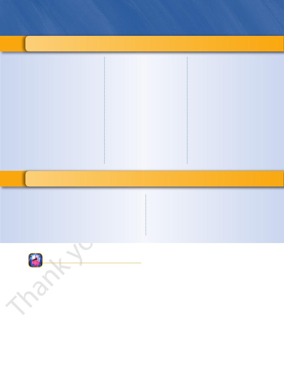
liver
esophagus
diaphragm
stomach
left colic flexure
descending colon
jejunum
sigmoid colon
anus
anal canal
rectum
ileum
appendix
cecum
ileocecal junction
ascending colon
duodenum
transverse colon
right colic flexure
gallbladder
FIGURE 5.1
General arrangement of abdominal viscera.
small intestine.
intestine (Fig. 5.3) and tends to be more fixed than the
intestine arches around and encloses the coils of the small
sigmoid colon, rectum, and anal canal (Fig. 5.1). The large
liver
stomach
gastroepiploic
vessels
greater
omentum
gallbladder
falciform
ligament
FIGURE 5.2
Abdominal organs in situ. Note that the greater
omentum hangs down in front of the small and large
intestines.
Basic Anatomy
the inferior surface of the right lobe of the liver, occupying
extends upward from the cecum to
ascending colon
The
that arises from its medial side (Fig. 5.1).
is a worm-shaped tube
appendix
(Figs. 5.1 and 5.3). The
ward in the right iliac region below the ileocecal junction
is a blind-ended sac that projects down
cecum
The
159
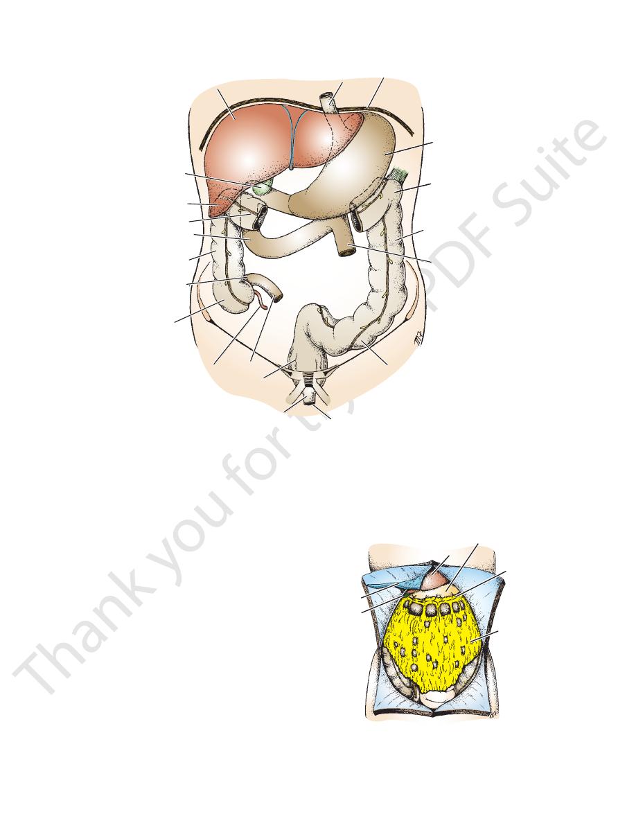
-
greater omentum
coils of
jejunum
descending
colon
coils of ileum
cecum
appendix
ascending
colon
transverse
colon
FIGURE 5.3
Abdominal contents after the greater omentum
the left upper part of the abdomen between the stomach
The spleen is a soft mass of lymphatic tissue that occupies
and extends from the duodenum to the spleen.
gastric region (Fig. 5.4). It is situated behind the stomach
obliquely across the posterior abdominal wall in the epi
The pancreas is a soft, lobulated organ that stretches
with the anal canal in the perineum.
vis by piercing the pelvic floor. Here, it becomes continuous
colon and descends in front of the sacrum to leave the pel
cavity (Fig. 5.1). It is continuous above with the sigmoid
occupies the posterior part of the pelvic
rectum
The
the rectum in front of the sacrum.
down into the pelvic cavity in the form of a loop. It joins
a continuation of the descending colon (Fig. 5.1). It hangs
begins at the pelvic inlet, where it is
sigmoid colon
The
upper and lower quadrants.
to the pelvis below (Figs. 5.1 and 5.3). It occupies the left
extends from the left colic flexure
descending colon
The
ure to become the descending colon.
of the spleen, bends downward, forming the left colic flex
into the pelvis. The transverse colon, on reaching the region
the erect position, the lower part of the U may extend down
ure (Figs. 5.1 and 5.3). It forms a wide U-shaped curve. In
ical region from the right colic flexure to the left colic flex
crosses the abdomen in the umbil
transverse colon
The
flexure.
reaching the liver, it bends to the left, forming the right colic
the right lower and upper quadrants (Figs. 5.1 and 5.3). On
the periphery.
transverse, and descending parts of the colon are located at
the central part of the abdominal cavity, whereas ascending,
has been reflected upward. Coils of small intestine occupy
-
-
-
-
Pancreas
-
Spleen
central tendon of diaphragm
phrenic artery
inferior vena cava
portal vein
right kidney
bile duct
hepatic artery
ascending colon
superior pancreaticoduodenal artery
transverse colon
descending colon
splenic artery
pancreas
phrenicocolic
ligament
left kidney
spleen
left suprarenal gland
gastroduodenal artery
right suprarenal gland
FIGURE 5.4
Structures situated on the posterior abdominal wall behind the stomach.

160
CHAPTER 5
between the viscera.
surfaces of the peritoneum and allows free movement
which lubricates the
peritoneal fluid,
serous fluid, the
and 5.7). The peritoneum secretes a small amount of
(Figs. 5.5
epiploic foramen
or the
ing of the lesser sac,
open
with one another through an oval window called the
ach. The greater and lesser sacs are in free communication
is smaller and lies behind the stom
lesser sac
pelvis. The
partment and extends from the diaphragm down into the
is the main com
greater sac
sac (Figs. 5.5 and 5.6). The
and the lesser
greater sac
and is divided into two parts: the
The peritoneal cavity is the largest cavity in the body
supports the kidneys.
kidneys, this tissue contains a large amount of fat, which
in the area of the
extraperitoneal tissue;
tissue called the
of the abdominal and pelvic walls is a layer of connective
Between the parietal peritoneum and the fascial lining
the uterus, and the vagina.
munication with the exterior through the uterine tubes,
males, this is a closed cavity, but in females, there is com
In
peritoneal cavity.
space of the balloon, is called the
the parietal and visceral layers, which is in effect the inside
covers the organs. The potential space between
toneum
visceral peri
the abdominal and pelvic cavities, and the
lines the walls of
parietal peritoneum
from outside. The
regarded as a balloon against which organs are pressed
the viscera (Figs. 5.5 and 5.6). The peritoneum can be
walls of the abdominal and pelvic cavities and clothes
The peritoneum is a thin serous membrane that lines the
abdominal wall.
the upper poles of the kidneys (Fig. 5.4) on the posterior
The suprarenal glands are two yellowish organs that lie on
muscle.
that runs vertically downward on the psoas
ureter
to a
the liver is smaller than the right). Each kidney gives rise
slightly higher than the right (because the left lobe of
of the vertebral column (Fig. 5.4). The left kidney lies
up on the posterior abdominal wall, one on each side
The kidneys are two reddish brown organs situated high
the 10th left rib.
and the diaphragm (Fig. 5.4). It lies along the long axis of
The Abdomen: Part II—The Abdominal Cavity
Kidneys
Suprarenal Glands
Peritoneum
General Arrangement
-
-
-
-
-
lesser sac
ileum
greater sac
inferior vena
cava
ascending colon
paracolic gutters
descending colon
aorta
mesentery
coils of ileum
greater omentum
hepatic
artery
portal vein
bile duct
free margin of
lesser omentum
inferior vena
cava
right kidney
left kidney
splenicorenal ligament
gastrosplenic
omentum (ligament)
aorta
stomach
lesser sac
greater sac
falciform ligament
liver
A
B
spleen
right
left
T12
L4
median
umbilical
ligament
lateral umbilical ligament
FIGURE 5.5
Transverse sections of the abdomen showing the arrangement of the peritoneum. The
position of the opening of the lesser sac. These sections are viewed from below.
arrow in B indicates the