Unit 7: General Pathology of Infectious Diseases
116
General principles of microbial pathogenesis
Categories of Infectious Agents
Unit 7: General Pathology of Infectious Diseases
117
Unit 7: General Pathology of Infectious Diseases
118

Unit 7: General Pathology of Infectious Diseases
119
Commensals: are microorganisms that live on the expense
of the host without doing harm e.g bacteria live on the skin.
They may become pathogenic in certain conditions.
Opportunistic infections: when infection induce by
commensals microorganisms e.g. E.coli is normal
inhabitant of GIT but when introduce into the urinary tract
it become pathogenic causing UTI. During tooth extraction,
diseased heart valve may become infected by streptococcus.
Viridians which are normal commensal of the mouth such
infection may lead to bacterial endocarditis
Pathogens: microorganisms that injure the host.
Pathogenicity: is the capacity of a particular
microorganism to cause disease.
Virulence: is the degree of pathogenicity.
Transmission & dissemination of
microbes
The outcome of infection depends on the ability of a
microbe to breach host barriers and colonize and damage
host tissues opposed to the ability of host defenses to
eradicate the invader.
Host barriers to infection prevent microbes from entering
the body & consist of innate & adaptive immune defenses.
Innate immunity is typically the first line of defense
against microbes and does not adapt to repeated attacks; it
includes physical barriers to infection, phagocytic and
natural killer (NK) cells, complement, and inflammatory
mediators (e.g., cytokines).
Adaptive immune responses-mediated by T and B
lymphocytes & their products-are stimulated by microbial
exposure and typically improve with successive contacts.
Routes of Entry of Microbes
Microbes can enter the host by inhalation, ingestion,
sexual transmission, insect or animal bites, or injection.
The first barriers to infection are intact host skin and
mucosal surfaces and their secretory products e.g. 10
11
are
required to produce vibrios cholera in human volunteers
with normal gastric pH. Still, some infectious agents
(having greater virulence) are able to overcome these
barriers; thus, 100 Shigella organisms can be sufficient to
cause disease.
Skin
The dense, keratinized outer layer of skin is a natural
barrier to infection; its low pH (about 5.5) and content of
fatty acids inhibit microbial growth other than the normal
bacterial and fungal flora adapted to that environment
(including S. epidermidis &Candida albicans). Moreover,
the keratinized outer layer is constantly shed and renewed
so that colonization is difficult.
Although skin is usually an effective barrier, few
microorganisms are able to traverse the unbroken skin e.g.
Schistosoma larvae penetrate swimmers' skin by releasing
collagenase, elastase that dissolve the extracellular matrix.
Most other microorganisms penetrate through breaks in
the skin, including superficial cuts or abrasions (fungal
infections), wounds (staphylococci), burns (Pseudomonas
aeruginosa), and diabetic (multibacterial infections).
Intravenous catheters in hospitalized patients frequently
cause bacteremia with Staphylococcus species or gram-
negative organisms. Needle sticks expose the recipient to
potentially infected blood and may transmit hepatitis B or
C, or HIV. Bites by fleas, ticks, mosquitoes, mites, and
lice break the skin and transmit diverse infectious
organisms. Animal bites can lead to infections with
bacteria or with rabies virus.
Respiratory tract
A lot of viruses, bacteria, and fungi, are inhaled daily by
human being. Large microbes are trapped in the nose and
the upper respiratory tract in a mucus layer secreted by
goblet cells; from there they are transported by the ciliary
action of the respiratory epithelium to the back of the
throat, where they are swallowed and cleared.
Organisms smaller than 5 μm are inhaled directly into the
alveoli, where they are phagocytosed by alveolar macro-
phages or by neutrophils recruited to the lung by cytokines.
The mucociliary clearance mechanism can be damaged by
smoking (causing metaplasia of the normal bronchial
epithelium with loss of cilia). Intubation or gastric acid
aspiration will also acutely interfere with normal
mucociliary clearance. However, in normal hosts, virulent
respiratory pathogens successfully evade the epithelial
defenses by specifically adhering to respiratory
epithelium. Certain organisms (e.g., Haemophilus
influenzae or Bordetella pertussis) elaborate toxins that
directly paralyze mucosal cilia. Viral damage to epithelial
cells also allows secondary infection by organisms that
normally lack the necessary adherence capability (e.g., S.
pneumoniae or Staphylococcus species).
M. tuberculosis causes respiratory infections because it is
able to escape phagocytotic killing by alveolar macrophages.

Unit 7: General Pathology of Infectious Diseases
120
Intestinal Tract
Normal defenses against ingested pathogens in the GIT
include:
1) Acidic gastric pH,
2) Viscous mucus secretions,
3) Lytic pancreatic enzymes and bile detergents,
4) Antimicrobial peptides called defensins,
5) (IgA) antibodies, secreted by B cells located in mucosa-
associated lymphoid tissues.
6) The normal gut flora. Pathogenic organisms must
compete for nutrients with abundant commensal bacteria
resident in the lower gut.
7) Defecation all gut microbes are intermittently expelled by
defecation.
Pathogenic bacteria in the GI tract cause disease by a
variety of mechanisms:
A. Staphylococcal strains growing in contaminated food
release powerful enterotoxins that cause food poisoning
symptoms without any bacterial multiplication in the gut.
B. Vibrio cholerae and toxigenic E. coli multiply within the
mucous layer, releasing exotoxins that cause the gut
epithelium to secrete large volumes of watery diarrhea.
C. Shigella, Salmonella, and Campylobacter invade and
damage the intestinal mucosa and lamina propria, causing
ulceration, inflammation, and hemorrhage (dysentery).
Salmonella typhi passes from the damaged mucosa
through Peyer's patches &mesenteric lymph nodes and
into the bloodstream, resulting in a systemic infection.
Intestinal protozoan infections particularly rely on cysts
for transmission because they can resist stomach acid.
Cysts eventually convert to motile trophozoites . While G.
lamblia attaches to the epithelial brush border, E.
histolytica invades and ulcerates the colonic mucosa.
Intestinal helminthes such as Ascaris typically cause
disease only when present in large numbers or in ectopic
sites (e.g., by obstructing the gut or invading and
damaging the bile ducts). Hookworms may cause iron
deficiency anemia by chronic loss of blood, which is
sucked from intestinal villi.
Geneto-Urinary tract
The urinary tract is normally sterile because it is flushed
many times per day; successful pathogens (e.g., E. coli,
gonococci) are those that can adhere to the epithelium.
Women have more than 10 times more urinary tract
infections (UTIs) than men because of shorter urethral
length (5 cm in women & 20 cm in men).
Obstruction of urinary flow and/or reflux of urine into the
ureters also increases the risk of UTI. When a UTI
spreads retrograde from the bladder into the kidney, it can
cause acute and chronic pyelonephritis.
From puberty until menopause, the vagina is protected
from pathogens (mostly yeasts) by a low pH resulting
from catabolism of glycogen in the normal epithelium by
commensal lactobacilli. Antibiotics can kill the
lactobacilli and make the vagina susceptible to infection.
To be successful as pathogens, microorganisms have
developed specific mechanisms for attaching to vaginal or
cervical mucosa or for entering via local breaks in the
mucosa during sexual intercourse (genital warts, syphilis).
Spread and Dissemination of Microbes
within the Body
Some microorganisms proliferate only locally at the site
of infection, staying confined to the lumen of hollow
viscera (e.g., cholera) or proliferate exclusively in or on
epithelial cells (e.g., papillomaviruses, dermatophytes);
others breach the epithelial barrier and spread to other
sites via the lymphatics, blood, or nerves. A variety of
pathogenic bacteria, fungi, and helminths are invasive by
virtue of their motility or ability to secrete lytic enzymes
(e.g., staphylococci secrete hyluronidase that degrades the
extracellular matrix, Within the blood, microorganisms
may be transported free or intracellularly, Viruses can
also propagate by cell-cell fusion or by transport within
neurons (e.g., rabies virus).
The major manifestations of infectious disease can arise at
sites distant from those of initial microbial entry. e.g.,
chickenpox viruses enter through the airways but it causes
cutaneous rashes, poliovirus enters through the intestine
but kills motor neurons. The rabies virus travels from the
skin to the brain in a retrograde fashion within nerves,
while the varicella-zoster virus hibernates in dorsal root
ganglia and, on reactivation, travels along nerves to cause
skin shingles.
The placentofetal route is also an important mode of
transmission, Infectious organisms can reach the pregnant
uterus through the cervical orifice or the bloodstream; if
they traverse the placenta, severe fetal damage can result.
Release from the Body & Transmission of
Microbes (Microbial Egress from the Body)
For transmission of disease to occur, exit of
microorganisms from the host is as important as the original
entry. Release may occur through skin shedding, coughing,
sneezing, urination, or defecation, or via insect vectors.

Unit 7: General Pathology of Infectious Diseases
121
Subsequent transmission to the next host depends very
much on the hardiness of the particular microbe. Some
microbes can survive for extended periods in dust, food,
or water. Following defecation many expelled pathogens
will persist in fecally contaminated food or water, with
subsequent transmission by ingestion (fecal-oral route)
e.g. hepatitis A and E viruses, poliovirus. Eggs shed by
some helminths (e.g., hookworms, schistosomes) into
stool gain access to new hosts by larval penetration of the
skin rather than by oral intake. Less hardy
microorganisms must be quickly passed from person to
person, often by direct contact (e.g., respiratory or sexual
routes) or by blood-borne transmission.
Microorganisms can be transmitted from animals to
humans by invertebrate vectors such as insects (i.e.,
mosquitoes), ticks, or mites that either passively spread
infection or occasionally serve as necessary hosts for
microbial replication and development. Transmission can
also occur directly from animals to humans (called
zoonotic infections), either by direct contact or by eating
the infected animal.
Prolonged intimate or mucosal contact during sexual
activity allows the transmission of a variety of agents,
including viruses (e.g., HPV, herpesviruses, HIV),
bacteria (e.g., T. pallidum, N. gonorrhea, Chlamydia
trachomatis), fungi (Candida species), protozoa
(Trichomonas species), and arthropods (crab lice).
Immune evasion by microbes
Microorganisms have developed many strategies to resist
and evade immune system, and such mechanisms are
important determinants of microbial virulence and
pathogenicity. These include:
1)
Remaining inaccessible to the host immune
system:-
this can be achieved by many ways:-
A. Microbes that propagate in the lumen of the intestine
(e.g., toxin-producing Clostridium difficile) or gallbladder
(e.g., Salmonella typhi) are concealed from many host
immune defenses.
B. Viruses that infect keratinized epithelium (poxviruses)
are inaccessible to antibodies and complement.
C. Some organisms rapidly invade host cells before the
humoral response becomes effective (e.g., malaria
sporozoites enter hepatocytes).
D. Viral latency within infected cells: during the latent
state many viral genes are not expressed (e.g., varicella-
zoster virus in dorsal root ganglia).
E. Some organisms can circumvent immune defenses by
covering themselves with host proteins ("the wolf in
sheep's clothing approach").
2)
Varying or shedding antigens
. Neutralizing
antibodies block the ability of microbes to infect cells;
this is the basis for vaccination. However, neutralizing
antibodies cannot effectively protect against microbes
with the capacity to express multiple variants of their
surface antigens. The ability to re-assort viral genomes
(e.g., influenza viruses) leads to viral antigenic variation.
Other example is the S. pneumoniae which has at least 80
different serotypes, distinguished by unique capsular
polysaccharides; the problem is that an antibody produced
in response to one serotype does not usually cross-react
with another, another strategy is used by S. mansoni,
which shed their antigens within minutes of penetrating
the skin, preventing recognition by antibodies.
3)
Resisting innate immune defenses
this may
demonstrate through:-
A. CAMP resistance: Cationic Anti-Microbial Peptides
(CAMPs), including defensins and thrombocidins,
provide important innate defenses against microbes;
CAMP resistance is key to the virulence of many bacterial
pathogens, allowing them to avoid neutrophil and
macrophage killing.
B. The carbohydrate capsules on many bacteria that cause
pneumonia or meningitis shield bacterial antigens from
circulating antibodies and complement proteins and also
prevent neutrophil phagocytosis.
C. Other bacteria make proteins that inhibit phagocytosis,
kill phagocytes, prevent their migration, or diminish their
oxidative burst. Thus, S. aureus expresses protein A
molecules that bind the Fc portion of antibodies and so
inhibit phagocytosis. Neisseria, Haemophilus, and
Streptococcus all secrete proteases that can degrade
antibodies. Several viruses, some intracellular bacteria
e.g. mycobacteria, fungi and protozoa (e.g., leishmania)
all have developed strategies to resist intracellular killing
and can, therefore, multiply within macrophages even
after phagocytosis.
D. Some viruses (e.g., herpes viruses) produce proteins that
block complement activation.
4)
Inhibiting adaptive immunity
. Some microbes use a
strategy of reducing the ability of CD4+ helper T cells
and CD8+ cytotoxic T cells to recognize infected cells.

Unit 7: General Pathology of Infectious Diseases
122
For example, several DNA viruses (e.g., CMV and EBV)
inhibit production of major histocompatibility complex
(MHC) class I proteins impairing peptide presentation to
CD8+ T cells and preventing killing of infected cells.
Although reduced MHC class I expression might be
expected to trigger NK cell killing, herpesviruses can
express MHC class I homologues that inhibit NK activity.
Similarly, herpesviruses can target MHC class II
molecules for early degradation, impairing antigen
presentation to CD4+ T helper cells. Finally, viruses can
directly infect lymphocytes and thereby compromise their
function; HIV infection of CD4+ T cells, macrophages,
and dendritic cells is the best example.
Selected human infectious diseases
Cytomegalovirus
Cytomegalovirus (CMV), a β-group herpesvirus, can
produce a variety of disease manifestations, depending on
the age of the host, and, more important, on the host's
immune status. It infects and remains latent in white
blood cells and can be reactivated when cellular immunity
is depressed. It causes destroying systemic infections in
neonates and in immunocompromised patients. Infected
cells exhibit gigantism of both the entire cell and its
nucleus. Within the nucleus is a large inclusion
surrounded by a clear halo (owl's eye).
Transmission of CMV can occur by several mechanisms,
depending on the age group affected These include the
following:
Transplacental transmission from a newly acquired or
primary infection in a mother who does not have
protective antibodies ("congenital CMV").
Transmission of the virus through cervical or vaginal
secretions at birth or, later, through breast milk from a
mother who has active infection ("perinatal CMV").
Transmission through saliva during preschool years.
Transmission by the venereal route is the dominant mode.
Iatrogenic transmission can occur at any age through
organ transplants or by blood transfusions.
Morphology
Affected cells are strikingly enlarged, often to a diameter of
40 µm, and they show cellular & nuclear polymorphism.
Prominent intranuclear basophilic inclusions spanning half
the nuclear diameter are usually set off from the nuclear
membrane by a clear halo, Within the cytoplasm of these
cells, smaller basophilic inclusions can also be seen.
Viral cellular tropism:
In the glandular organs, the parenchymal epithelial cells
In the brain, the neurons;
In the lungs, the alveolar macrophages and epithelial and
endothelial cells.
In the kidneys, the tubular epithelial and glomerular
endothelial cells.
Hepatitis B Virus
Hepatitis B virus (HBV), the etiologic agent of "serum
hepatitis," is a significant cause of acute and chronic liver
disease worldwide. HBV is a DNA virus that can be
transmitted percutaneously (e.g., intravenous drug use or
blood transfusion), perinatally, and sexually. Viral DNA
can integrate into the host genome. HBV infects
hepatocytes, and cellular injury occurs mainly due to the
immune response to infected liver cells and not to
cytopathic effects of the virus. HBV may evade immune
defenses by inhibiting IFN-β production and by
downregulating viral gene expression. The effectiveness
of the cytotoxic T cell "CTL" response is a major
determinant of whether a person clears the virus or
becomes a chronic carrier. When infected hepatocytes are
destroyed by CTLs, replicating virus is also eliminated
and the infection is cleared. However, if the rate of
infection of hepatocytes outpaces the ability of CTLs to
eliminate infected cells, a chronic infection is established.
This may happen in 5% to 10% of adults and up to 90%
of children infected perinatally. In this setting, the liver
develops a chronic hepatitis, with lymphocytic
inflammation, apoptotic hepatocytes resulting from CTL-
mediated killing, and progressive destruction of the liver
parenchyma. Long-term viral replication can lead to
cirrhosis of the liver and an increased risk for
hepatocellular carcinoma. In some infected individuals,
hepatocytes are infected but the CTL response is dormant,
resulting in the establishment of a "carrier" state, without
progressive liver damage.
Epstein - Barr virus (EBV)
This virus as well as human papilloma virus (HPV), HBV,
and human T-cell leukemia virus-1 (HTLV-1) is called
transforming viruses because they have been implicated in
the causation of human cancer.
EBV causes infectious mononucleosis, a benign, self-
limited lymphoproliferative disorder, and is associated
with the development of a number of neoplasms, most
notably certain lymphomas and nasopharyngeal

Unit 7: General Pathology of Infectious Diseases
123
carcinoma. Infectious mononucleosis is characterized by
fever, generalized lymphadenopathy, splenomegaly, sore
throat, and the appearance in the blood of atypical
activated T lymphocytes (mononucleosis cells). Some
patients develop hepatitis, meningoencephalitis, and
pneumonitis. Infectious mononucleosis occurs principally
in late adolescents or young adults among upper
socioeconomic classes in developed nations. In the rest of
the world, primary infection with EBV occurs in
childhood and is usually asymptomatic.
Pathogenesis
EBV is transmitted by close human contact, frequently
with the saliva during kissing. An EBV envelope
glycoprotein binds to CD21, present on B cells. The viral
infection begins in nasopharyngeal and oropharyngeal
lymphoid tissues, particularly the tonsils. EBV gains
access to sub-mucosal lymphoid tissues. Here, infection
of B cells may take one of two forms. In a minority of B
cells, there is productive infection with lysis of infected
cells and release of virions, which may infect other B
cells. In most B cells, EBV establishes latent infection.
Early in the course of the infection, IgM antibodies are
formed against viral capsid antigens; later, IgG antibodies
are formed that persist for life.
Morphology
The major alterations involve the blood, lymph nodes,
spleen, liver, central nervous system, and, occasionally,
other organs.
1) The peripheral blood shows absolute lymphocytosis with
a total white cell count between 12,000 and 18,000
cells/µl, more than 60% of which are lymphocytes. Many
of these are large, atypical lymphocytes, , characterized
by an abundant cytoplasm containing multiple clear
vacuolations, an oval, indented, or folded nucleus, and
scattered cytoplasmic azurophilic granules . These
atypical lymphocytes, most of which express CD8, are
usually sufficiently distinctive to permit the diagnosis
from examination of a peripheral blood smear.
2) lymph nodes are typically discrete and enlarged
throughout the body, principally in the posterior cervical,
axillary, and groin regions. On histologic examination, the
most striking feature is the expansion of paracortical areas
by activated T cells (immunoblasts).
3) The spleen is enlarged in most cases It is usually soft and
fleshy, with a hyperemic cut surface. The histologic
changes show an expansion of white pulp follicles and red
pulp sinusoids due to the presence of numerous activated
T cells. These spleens are especially vulnerable to rupture,
possibly in part because the rapid increase in size
produces a tense, fragile splenic capsule.
4) Liver function is almost always transiently impaired with
moderate hepatomegaly., The histologic picture may be
difficult to distinguish from that of other forms of viral
hepatitis it show atypical lymphocytes in the portal areas
and sinusoids.
5) The central nervous system may show congestion, edema,
and perivascular mononuclear infiltrates in the
leptomeninges. Myelin degeneration is seen in the
peripheral nerves.
Human Papillomaviruses
Human papillomaviruses (HPVs) are DNA viruses that are
members of the papova virus family .Papillomaviruses are
classified into over 100 types. Some HPVs cause
papillomas (warts), benign tumors of squamous cells on the
skin,. Other HPVs are associated with warts that can
progress to malignancy, particularly squamous cell
carcinoma of the cervix and the anogenital area .
Papillomaviruses are mainly transmitted by skin or genital
contact. HPVs infect squamous epithelial cells, but their life
cycle is not well understood since these viruses cannot be
cultured in vitro. The expression of viral genes depends on
the differentiation state of the epithelial cells. Papilloma
viruses initially infect basal cells in the epithelium, but
there is limited expression of viral genes in these cells.
Mature virions are produced in the cells within the granular
layer and shed from the stratum corneum.
Unit 7: General Pathology of Infectious Diseases
124
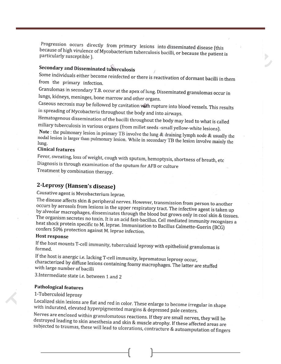
Mycobacterial Infections
Unit 7: General Pathology of Infectious Diseases
125
Unit 7: General Pathology of Infectious Diseases
126
Unit 7: General Pathology of Infectious Diseases
127
