Reticular Activation System
&Limbic System
By
Dr. Mufeed Akram Taha
FIBMS Neurology
Kirkuk College of Medicine

Reticular Activation System
(RAS)
It is composed from collection of an area of highly
organized and differentiated neurons(called
Reticular formation) extending from brain stem
to upper portion of spinal cord.
RF receive collateral nerve ending from the spinal
cord, eye, ears, from cortex (collateral from
pyramidal tract), hypothalamus and cerebellum.

Functions of Reticular Formation
The ascending & descending projections of the
reticular formation are involved in 5 different
types of functions:-
1. Regulation of posture.
2. Control of muscle tone.
3. Modulation of pain sensation.
4. Coordination of autonomic functions.
5. Control of Consciousness

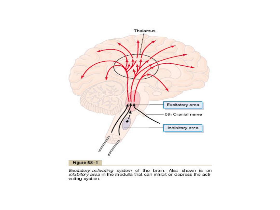
Reticular Excitatory Area
The central driving component of this system is an
excitatory area located in the reticular substance
of the pons and Mesencephalon.
This area is also known by the name bulboreticular
facilitory area.
This area also sends a profusion of signals in the
upward direction; most of these signals go first
to the thalamus where they excite a different set
of neurons that transmit nerve signals to all
regions of the cerebral cortex as well as to
multiple subcortical areas.

Also brain stem reticular area transmits
facilitory signals downward to the spinal
cord to maintain tone in the antigravity
muscles and to control levels of activity of
the spinal cord reflexes.
Also there is positive feedback mechanism
from the cerebral cortex by sending
excitatory signals from the cerebral cortex
back to the brain stem excitatory area.

Reticular Inhibitory Area
This is another area that is important in
controlling brain activity. This is the
reticular inhibitory area, located medially
and ventrally in the medulla.
This area can inhibit the reticular facilitory
area of the upper brain stem and there by
decrease activity in the superior portions of
the brain as well.

The Limbic System
• It is the entire basal system of the brain that
mainly controls the person's emotional behavior.
The hypothalamus is the central elements of the
system surrounded by other cortical structures
of the limbic system (the septal nuclei,
paraolfactory area, epithalamus, anterior nuclei
of the thalamus, and portions of the basal
ganglia, hippocampus, and amygdala).
• The hypothalamus is a major output pathway of
the limbic system and has communicating ways
with all levels of this system.

The functions of the hypothalamus
A. The vegetative control functions:
1. Cardiovascular regulation: Hypothalamus controls
the heart and blood pressure through its effect on
cardiovascular control centers in the reticular
substance of the medulla and pons. This includes the
decrease or increases the heart-rate and the blood
pressure.
2. Regulation of body temperature: Certain areas in
the hypothalamus sensitize the change in the body
temperature and also control the mechanisms for
adjusting the body temperature back to normal
level.
3. Regulation of body water: Hypothalamus regulates
body water by two ways:

(A) - By creating the sensation of thirst through
thirst center. This is achieved by:
(1). Increase in the plasma osmolarity: When the
electrolytes inside the neurons of this center or
in, the allied areas of hypothalamus become too
concentrated, the subject develops an intense
desire to drink water until the electrolyte
concentration of the thirst center neurons
return to normal. Therefore, the neurons of this
center act as osmoreceptors, which are
stimulated by an increased osmotic pressure of
the body fluids to initiate thirst and drinking.

(2). Decrease of the ECF volume: A decrease in
ECF volume also stimulates thirst by a
pathway, which is independent of the
osmolality of the plasma. The effect of ECF
volume depletion on thirst is mediated in part
via renin
—angiotensin system in which
angiotensin II acts on a specialized receptor
area in hypothalamus to stimulate the neural
areas concerned with thirst.

(3) Dryness of mouth and reduced salivary
secretion: These are the most common signals.
For example, eating a very dry food produces the
desire to drink water because salivary secretion
is not adequate to keep the mouth moist.
(B). by controlling the excretion of water in the
urine. When the body fluids become too
concentrated, i.e. the osmotic pressure of the
plasma is increased, the hypothalamus through
osmoreceptor cells, stimulates the secretion of
ADH through the posterior pituitary gland.

This hormone is absorbed into the blood and
acts on the collecting ducts of the kidneys to
cause massive reabsorption of water, thereby
decreasing the loss of water into the urine.
4. Regulation of uterine contractility and milk
ejection by the breast: The neurons of certain
nuclei in hypothalamus secrete oxytocin
through pituitary gland which causes
increased contractility of the uterus and also
contraction of the myoepithelial cells that
surround the alveoli of the breasts causing the
alveoli to empty the milk through the nipples.

5. GIT and feeding regulation: Many GIT
activities and reflexes such as licking the lips
and swallowing are integrated in the
hypothalamus through mammillary bodies.
Feeding regulation is achieved by two
hypothalamic centers and these are hunger
(or feeding) center and satiety center. The
former evokes eating behavior (promotes
appetite) which appears to be chronically
active, while the latter opposes the desire for
food (anorexia) which functions by inhibiting
the feeding center.

6. Regulation of circadian rhythm (biological
clock): Your body has more than 100 circadian
rhythms. Each unique24 hour cycle influences an
aspect of your body's function, including body
temperature, hormone levels, heart rate,blood
pressure, etc, even pain threshold. The
suprachiasmatic nuclei of the hypothalamus and
preoptic nuclei are the dominant pacemakers for
many circadian rhythms in the body to 24-hour
light-dark cycle such as secretion of ACTH and
melatonin, as well as sleep-wake cycles, and the
body temperature rhythm. Circadian periodicities
are changes in biological variables that occur
daily.

It appears that the suprachiasmatic nuclei (the
biological clock) takes the information on day length
from the retina, interprets it, and passes it on to the
pineal gland (a pea-like structure found on the
epithalamus), which then secretes the hormone
melatonin in response. Secretion of melatonin
peaks at night and decrease during the day.
Darkness probably stimulates melatonin secretion
by the pineal gland, which inhibits the secretion of
gonadotropic hormones from the anterior pituitary,
and thus reduces sexual drive. Melatonin secretion
decreases with age. Destruction of the biological
clock disrupts many biological rhythms, such as
oscillations in body temperature, other vegetative
functions and the sleep-wake cycle.

B.Endocrine control functions: Stimulation of
certain areas of the hypothalamus causes the
anterior pituitary gland to secrete its
hormones. As the blood courses through the
hypothalamus before reaching the anterior
pituitary, releasing hormones and inhibitory
hormones are secreted into the blood by
various hypothalamic nuclei. They are then
transported in the blood to the anterior
pituitary where they act, on the glandular cells
to control the release of the anterior pituitary
hormones.

C- Emotional and behavioral control functions:
Reward centers: They are also called pleasant or
satisfaction centers. Stimulation these centers
cause pleasure, satisfaction.These centers are
located mainly in the hypothalamus. Less
potent centers are found in the amygdala, the
hippocampus and other areas of the brain.
Punishment centers: They are also called
unpleasant or aversion centers. Stimulation of
these centers cause terror, pain, fear, defense,
escape reactions, rage, and all the other
elements-of punishment.

The most potent areas for punishment have
been found in the central gray area
surrounding the aqueduct of Sylvius in the
mesencephalon and extending upward into
the hypothalamus and thalamus. Less potent
punishment areas are found in the amygdala
and the hippocampus. In rage (which is an
emotional pattern due to strong stimulation of
the punishment centers of the brain) the
animal takes the position of attack with a
defense posture, lift its tall, develop
piloerection, wide open eyes, and dilated
pupils.

Stimulation of the punishment centers can
frequently inhibit the reward centers
completely indicating that the punishment
centers take precedence over the reward
centers: Almost everything that We do is
related in some way to reward and
punishment. If we are doing something
that is rewarding, we continue to do it, if it
is punishing, we cease to do it. Reward
and punishment are important in learning
and memory

3. Sexual behavior
4. Learning processes: Learning is a change of
behavior caused by neural mechanisms affected by
experience. Memory refers to neural storage
mechanisms for experiences. The hippocampus is
involved in learning and memory.
D. Control of excitement and alertness in association
with other structures of the limbic system:
Stimulation of certain regions of the hypothalamus
greatly excites the RAS and therefore causes
wakefulness, alertness and excitement. In addition,
the sympathetic NS becomes excited in general,
causing increasing the arterial B.P, pupillary
dilatation and enhancing other activities associated
with sympathetic activity.

On the other hand, stimulation of some areas in the
limbic system, hypothalamus, or in the thalamic
portions of the RAS often inhibits the
mesencephalic portion of the RAS, causing
somnolence, and sometimes actual sleep.
The functions of amygdala: Amygdala has extensive
connections with various parts of the brain, play its
important role on the mediation and control of
major affective activities like friendship, love and
affection, on the expression of mood and, mainly on
fear, rage and aggression The amygdala, being the
center for identification of danger, is fundamental
for self preservation.

The amygdala is believed to help in choosing the
pattern of the person's behavioral response so
that it is appropriate for each occasion.
Stimulation of amygdala causes almost all the
same effects as those elicited by stimulation
of hypothalamus plus still other effect.
Bilateral lesions in the amygdala cause
hyperphagia with indiscriminate ingestion of
all kinds of food (omniphagia), loss of fear,
decrease aggressiveness and excessive sex
drive, forgets very rapidly, has a tendency to
place every thing in its mouth.

The functions of hippocampus: Hippocampus
becomes habituated to indifferent signals, but
learns from signals that cause either reward
(pleasure) or punishment. Hippocampus is the
"brain librarian" (helps the cortex to store new
signals into the long lasting long-term
memory). Bilateral removal of the hippocampi
in epileptic patients permanently disrupts the
ability to learn anything new (anterograde
amnesia). Other lesions of the hippocampi
reduce previously learned memory material
(retrograde amnesia).

Thanks