
Special Sensation
Vision & Olfaction
By
Dr. Mufeed Akram Taha
FIBMS Neurology
Kirkuk College of Medicine

Functional Anatomy of the eye.
The principle structures of the eye are the following:
Sclera
… Is the oute p ote tive laye of the eye all.
Cornea
… A te io odifi atio of the s le a. It is
transparent through it the light rays enter the eye.
Choroids
…. The laye i side the s le a it o tai a y
of the blood vessels that nourish the structures in
the eye ball

Retina
…. the eu al tissue o tai i g the e epto ells
lining the posterior 2/3 of the choroids
The Lens
.… is a t a spa e t st u tu es held i pla e y
a circular lens ligament with zonule
Ciliary body
…. thi ke ed a te io pa t of the ho oid
attached to the zonule. It contain circular band
longitudinal muscle fibers.
The Iris
…. The olo ed pa t of the eye lo ated i f o t
of the lens
– it is pigmented and opaque it contain
circular muscle fibers that constrict and radial fibers
that dilate the pupil.

The pupil .... The central opening in the iris. The
diameter of the pupil is changed by the iris
contraction and relaxation.
Vitreous humor
…. lea gelati ous ate ial fills
the space between the lens and the retina.
Aqueous humor
…. A lea li uid that ou ishes
the cornea of the lens.

The retina extends anteriorly almost to the
ciliary body. It is organized in ten layers and
contains the rods and cones, which are the
visual receptors, plus four types of neurons:
bipolar cells, ganglion cells, horizontal cells,
and amacrine cells.
The optic nerve leaves the eye and the retinal
blood vessels enter it at a point 3 mm medial
to and slightly above the posterior pole of the
globe. This region is called optic disk. There
are no visual receptors overlying the disk, and
consequently this spot is blind (the blind
spot).

Near the posterior pole of the eye, there is
a yellowish pigmented spot, the macula
lutea. This marks the location of the
fovea centralis, a thinned-out, rod-free
portion of the retina. In it, the cones are
densely packed, the fovea is the point
where visual acuity is greatest. When
attention is attracted to or fixed on an
object, the eyes are normally moved so
that light rays coming from the object fall
on the fovea.

The potential changes that initiate
action potentials in the retina are
generated by the action of light on
photosensitive compounds in the
rods and cones. When light is
absorbed by these substances, their
structure changes, and this change
triggers a sequence of events that
initiates neural activity.

The cone receptor potential has a sharp
onset and offset, whereas the rod
receptor potential has a sharp onset and
slow offset.
Rods mediate vision under dim light and its
visual pigment is rhodopsin which
contain retinene that formed from vit.A.
so lesion of Rods cause night blindness.
Cones are specialized for colour vision

Neural Pathways :-
The axons of the ganglion cells pass caudally in
the optic nerve and optic tract to end in the
lateral geniculate body, a part of the
thalamus. The fibers from each nasal
hemiretina decussate in the optic chiasm. In
the geniculate body, the fibers from the nasal
half of one retina and the temporal half of the
other synapse on the cells whose axons form
the geniculocalcarine tract. This tract passes
to the occipital lobe of the cerebral cortex.
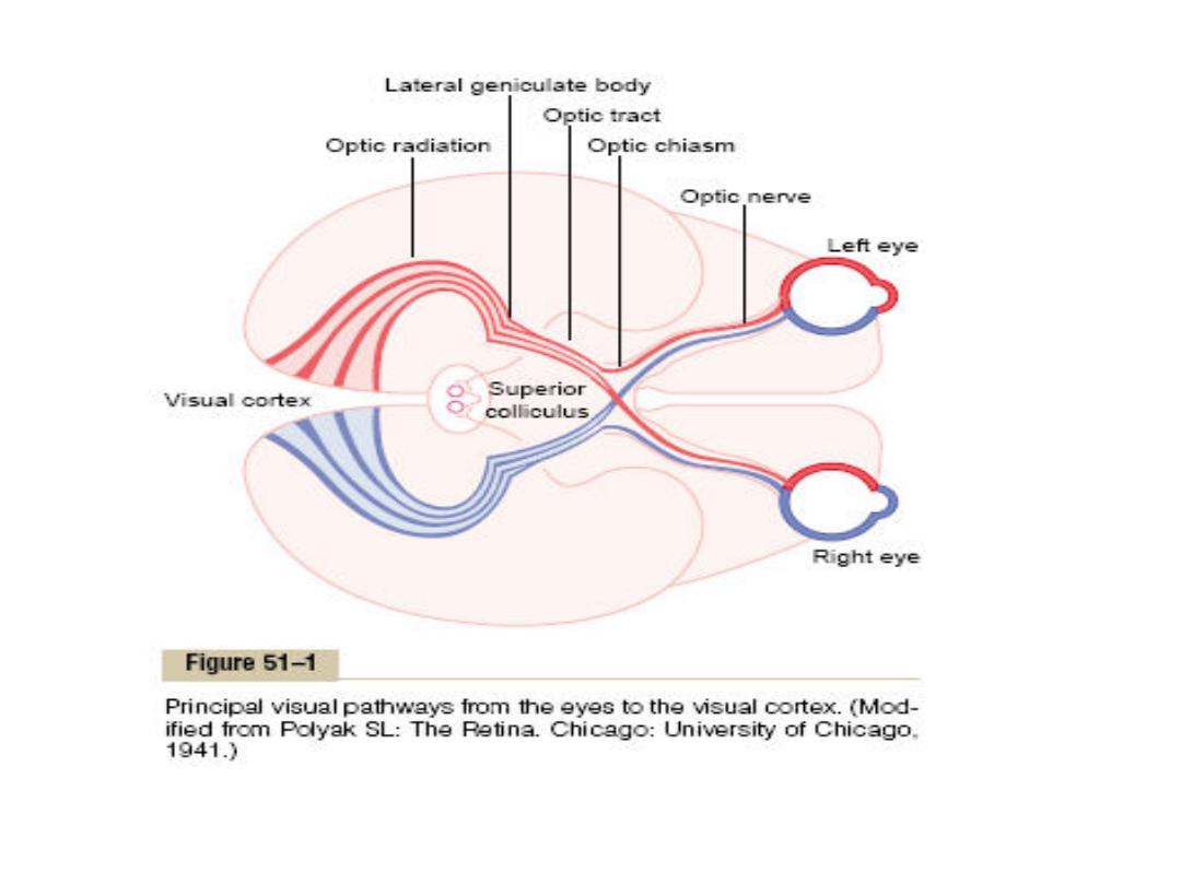

The primary visual receiving area (primary visual
cortex, Brodmann's area 17), is located
principally on the sides of the calcarine fissure.
• Visual fields:-
The visual field of each eye is the visual area seen
by that eye at a given instant.
The area seen at the nasal side is called nasal field,
and the area seen to the lateral side is called
temporal field.
The visual field of each eye is not circular because it
is cut off medially by the nose and superiorly by
the roof of the orbit.

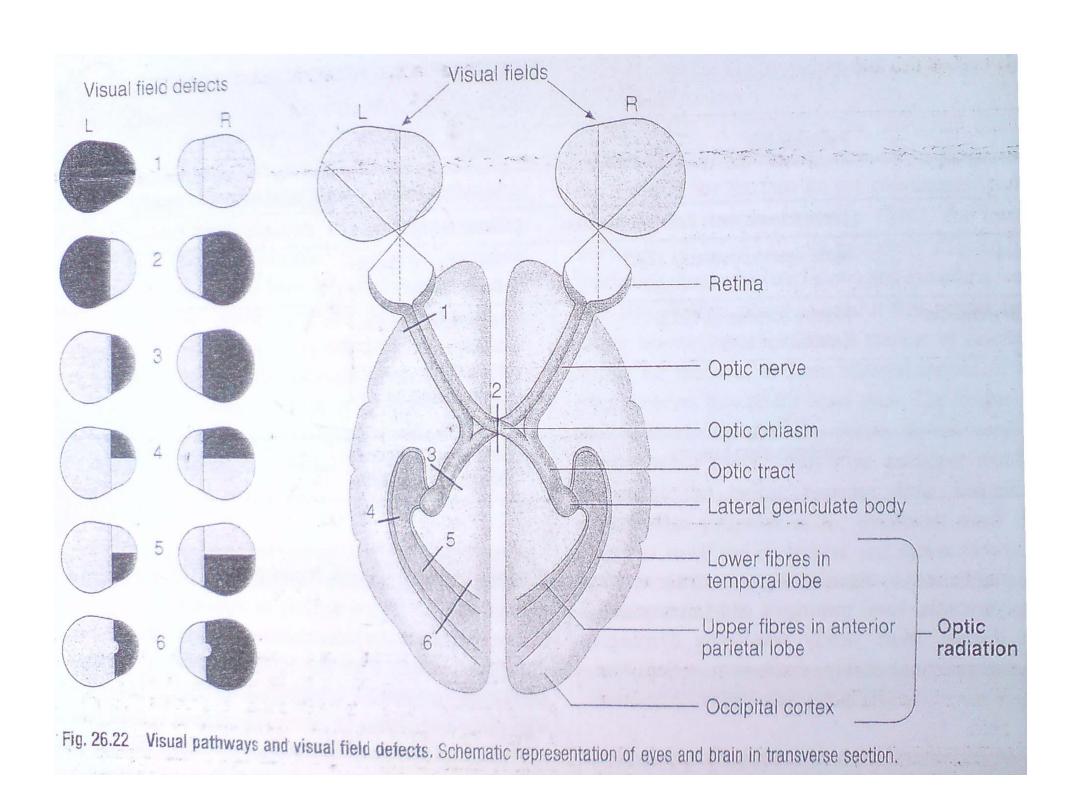
Effect of lesions in the optic pathway
Destruction of an entire optic nerve causes blindness
of the affected eye.
Lesion of the optic chiasm causes loss of visual field
of temporal fields of both eyes and this is called
bitemporal hemianopia.
Lesion in the optic tract causes loss of nasal field of
the same side of the lesion and temporal field of
the opposite side this is called contralateral
homonymous hemianopia.
Lesion in the optic radiation in the temporal lobe
cause contralateral homonymous upper
quadrantanopia and in parietal lobe cause
contralateral homonymous lower quadrantanopia.
Lesion in the occipital lobe also causes homonymous
hemianopia but there may be macular sparing.

Visual acuity :-
-Is the degree to which the details and contours of
the objects are perceived.
-Visual acuity is the function of the central portion
of the retina especially the fovea centralis.
-Clinically visual acuity is often determined by a
special chart called snellen charts viewed at a
distance of 6 meters and the individual being
tested read aloud the smallest line
distinguishable.
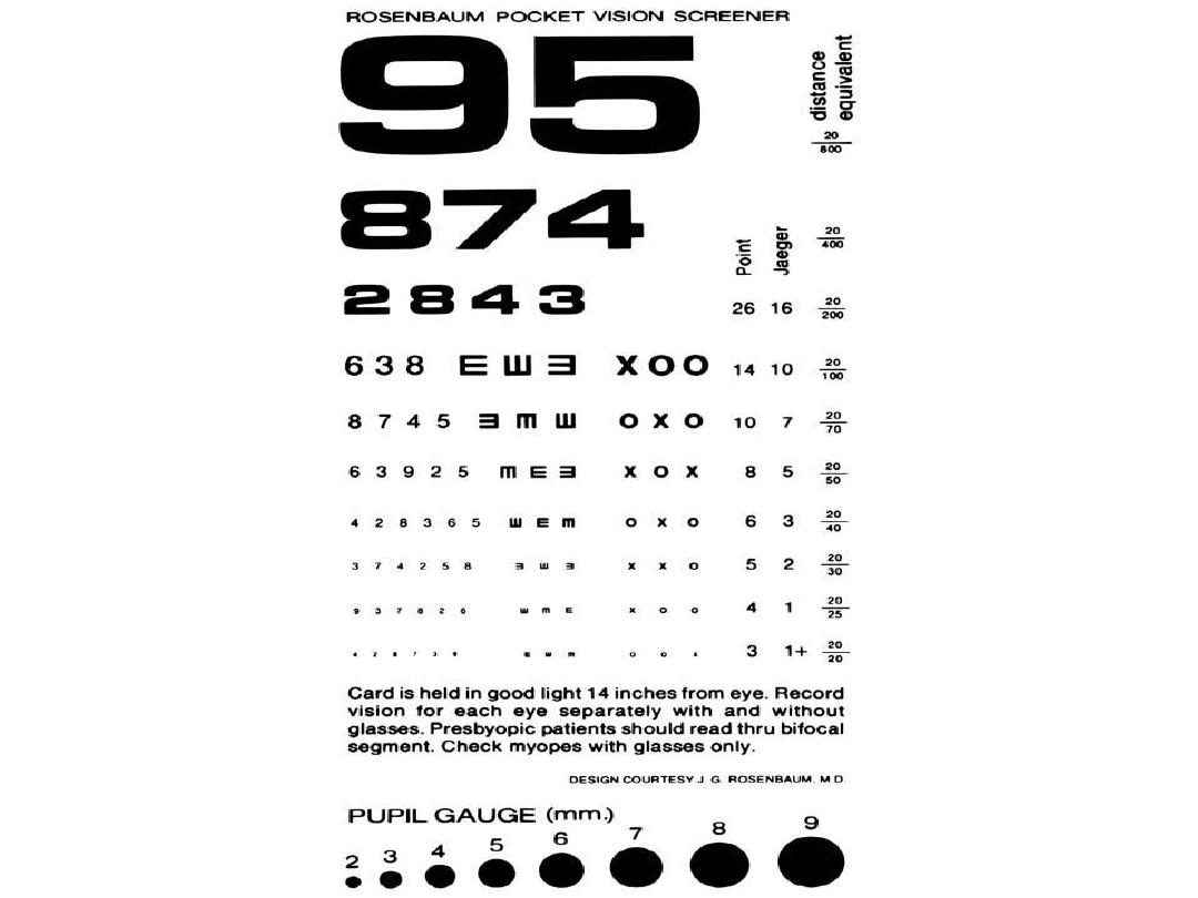

-The result is expressed as a fraction. The
numerator of the fraction 6 meter is the
distance of which the subject reads the chart.
The denominator is the greatest distance from
the chart at which a normal individual can
read the smallest line the subject can read.
For example, patient with V.A of 6/18 mean that
he can only read the line from 6 meters what
a normal person can read at 18 meters
distance.
-Normal V.A is 6/6.

Color vision:-
Sensation of white or any spectral color or even extraspectral
color (Purple) can be produced by mixing various proportions
of red light (wave length 723-647 nm), green light (wave
length 575-492 nm) and blue light (wave length 492-450 nm).
Red, green and blue are therefore called primary colors.
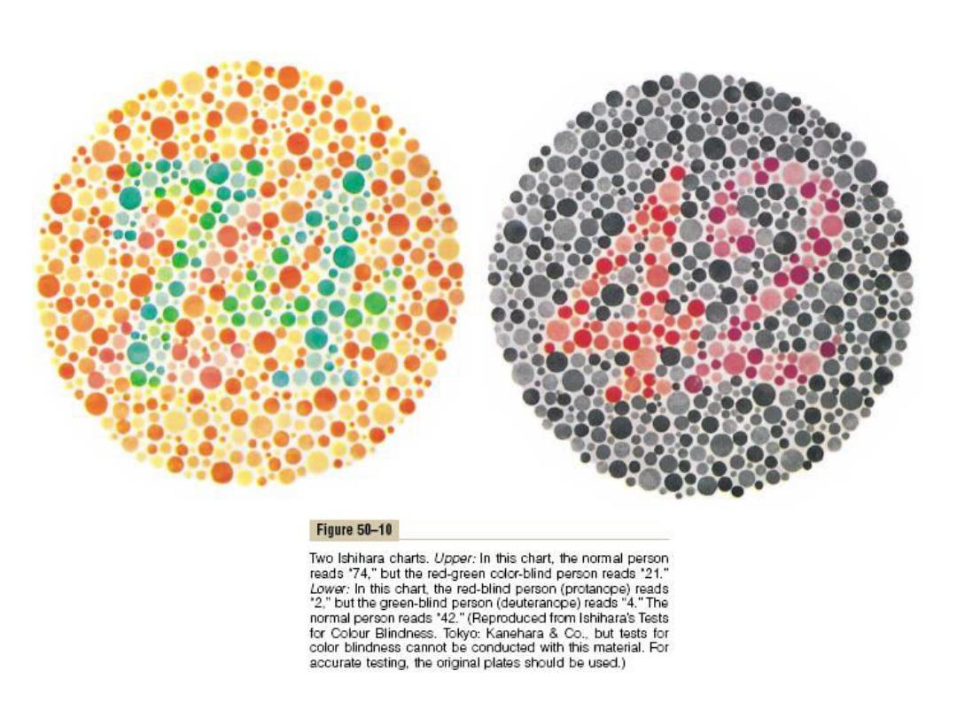
Color Blindness
Red-Green Color Blindness
Normally green, yellow, orange, and red colors, which are the
colors between the wave lengths of 525 and 675 nanometers
(nm), are distinguished from one another by the red and
green cones. If either of these two cones is missing, the
person cannot distinguish red from green and is therefore
said to have red-green color blindness.
Red-green color blindness is X-linked genetic disorder that
occurs almost exclusively in males.

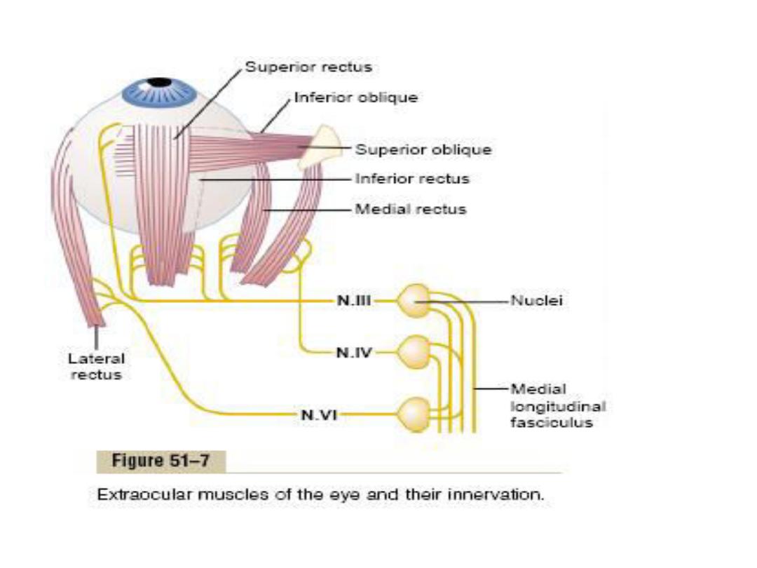
Eye movement:-
-The eye movements are controlled by 3 pairs of
skeletal muscles:-
1- Medial and lateral recti, contraction of which
moves the eye from side to side.
2- Superior and inferior recti, contraction of which
moves the eyes upward and downward.
3-
Supe io a d i fe io o li ue us les΄ fu tio
mainly to rotate the eyeballs to keep visual fields in
upright position.
-The nerve supplies of these muscles are as follows:-
-Oculomotor (3
rd
) cranial nerve supply 4 of these
muscles which are medial rectus, superior and
inferior recti, and inferior oblique.

-Trochlear (4
th
) cranial nerve supply superior
oblique.
-Abducent nerve (6
th
) cranial nerve supply
Lateral rectus.
-The nuclei of 3
rd
,4
th
and 6
th
cranial nerve lies in
the brain stem and these nuclei are
medial
interconnected by a nerve tract called
longitudinal fasciculus.
-Signals from visual areas of occipital cortex pass
to the brain stem nuclei of the three cranial
nerves to direct the stimulation of eye
movements.

Visual Reflexes :-
Pupillary light reflexes
When light is directed into one eye, the pupil constricts
(pupillary light reflex). The pupil of the other eye also
constricts (consensual light reflex). The optic nerve fibers
that carry the impulses initiating these pupillary
responses leave the optic nerves near the lateral
geniculate bodies. On each side, they enter the midbrain
via the brachium of the superior colliculus and terminate
in the pretectal nucleus. From this nucleus, the second-
order neurons project to the ipsilateral Edinger-Westphal
nucleus and the contralateral Edinger-Westphal nucleus..

The third-order neurons pass from this
nucleus to the ciliary ganglion in the
oculomotor nerve, and the fourth-order
neurons pass from this ganglion to the
ciliary body. This pathway is dorsal to the
pathway for the near response.
Consequently, the light response is
sometimes lost while the response to
accommodation remains intact (Argyll
Robertson pupil). One cause of this
abnormality is CNS syphilis.

Accomadation:-
The process by which the curvature of the lens is
increased is called accommodation. At rest, the
lens is held under tension by the lens ligaments.
Because the lens substance is malleable and the
lens capsule has considerable elasticity, the lens is
pulled into a flattened shape. When the gaze is
directed at a near object, the ciliary muscle
contracts, this decreases the distance between
the edges of the ciliary body and relaxes the lens
ligaments, so that the lens springs into a more
convex shape. In young individuals, the change in
shape may add as many as 12 diopters to the
refractive power of the eye.

Function of visual cerebral cortex:-
-The visual cortex located primarily on the medial
aspect of the occipital lobes and it is divided to
primary visual cortex and secondary visual areas.
-The cortical area representing the macula in the
primary visual cortex is very large because this
area is responsible for highest degree of visual
acuity.
-primary visual cortex arranged in 6 layers and the
geniculocalcarine fibers terminate on layer IV.
-the visual cortex is organized structurally into
several millions of vertical columns of neuronal
cells.

-The primary visual cortex detects the existence
of lines and borders in the different areas of
the retinal image and also detects the
direction of orientation of each line and
border that weather vertical or horizontal or
lines at some degree of inclination.
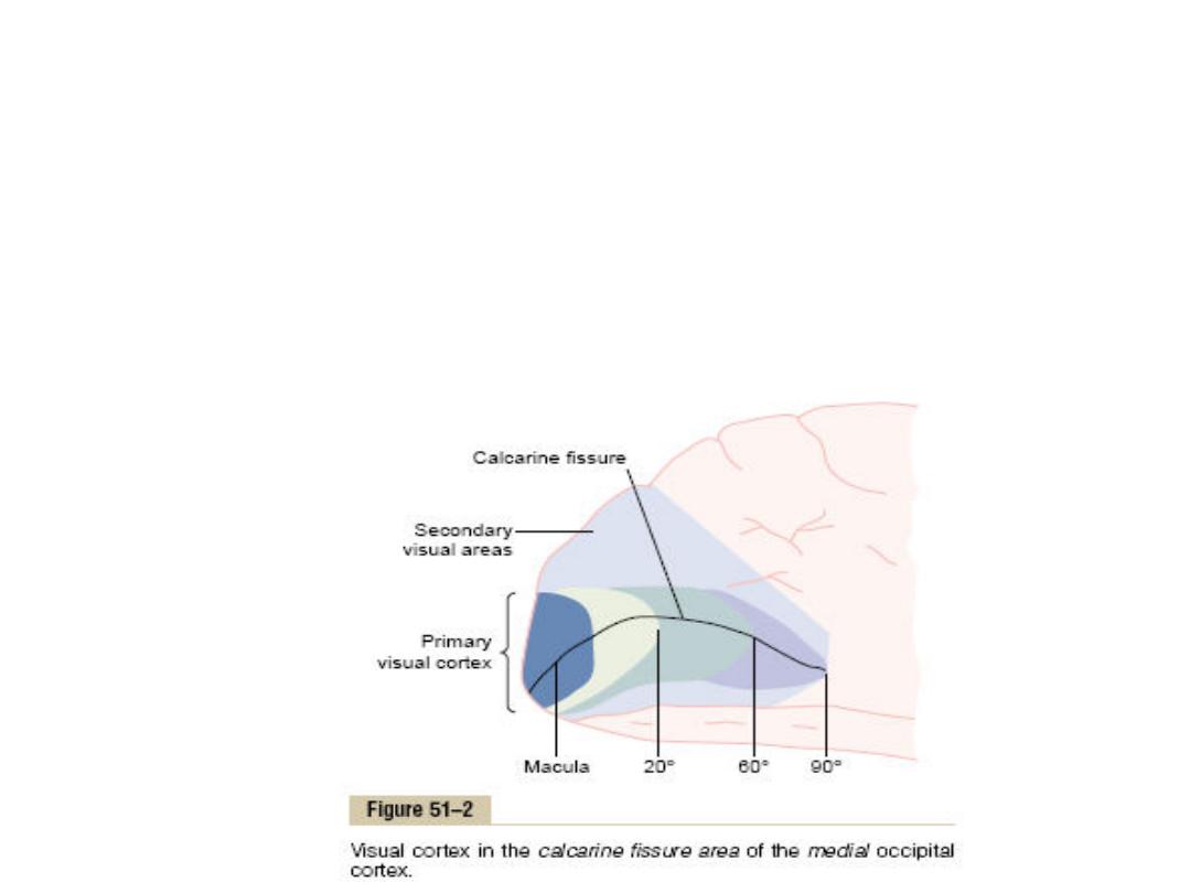
Secondary visual areas
These also called visual association areas, lies
lateral, anterior and inferior to the primary
visual.

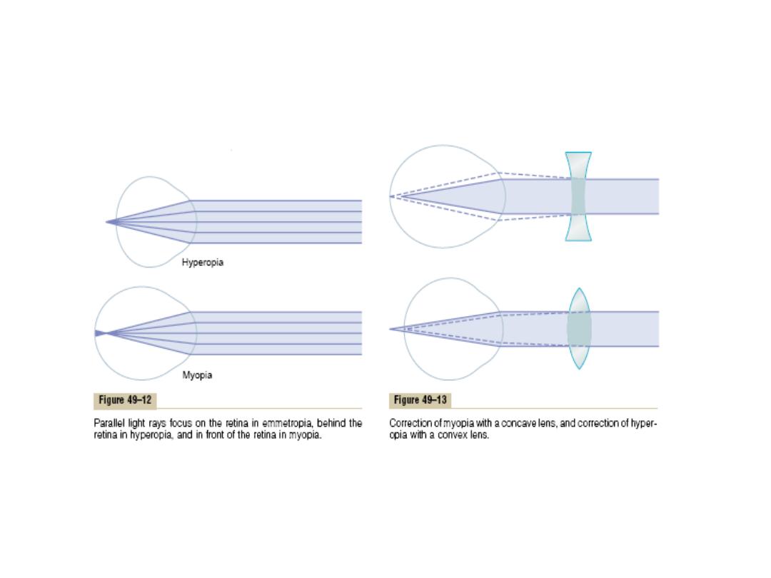
Errors of Refraction
Hyperopia (Farsightedness).
Is usually due to either an eyeball that is too small
or, occasionally, a lens system that is too weak.
Parallel light rays are not bent sufficiently come
to focus by the time they reach the retina. A
farsighted person is capable of focusing distant
objects on the retina by accommodation
mechanism but the closure objects cannot be
focused sharply when the ciliary muscle contract
to its limit. Correction of this defect is by convex
lens in front of the eye (convex lens converges
light rays).

Myopia (Nearsightedness)
is usually due to too long an eyeball, but it can
result from too much refractive power in the
lens system of the eye the light rays coming
from distant objects are focused in front of
the retina
The correction is by placing in front of the eye a
concave spherical lens, which will diverge rays

Astigmatism
Is a common condition in which the curvature of
the eye cornea is not uniform and this causes
the visual image in one plane to focus at a
different distance from that of the plane at
right angles, so that, part of retinal image is
blurred.
Astigmatism can usually be corrected with
cylindrical lenses placed in such away that
they equalize the refraction in all meridians.

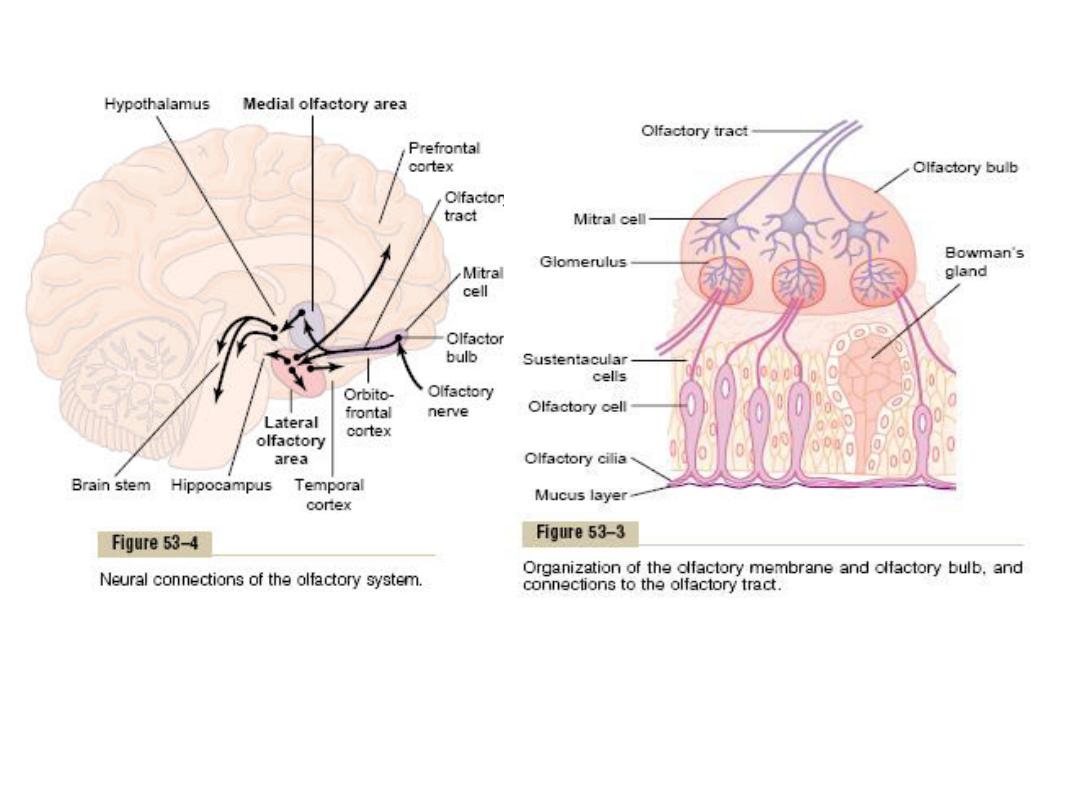
Smell
Olfactory receptors& pathways
The olfactory receptor cells are located in specialized
portion of nasal mucosa.
Olfactory mucus membrane: it covers an area of 5cm²
in the roof of nasal mucosa near the septum.Each
olfactory receptor is a neuron,had short dendrite
with expanded end (Olfactory rods). From the rod
cilia project to the surface of the mucus.
Each receptor neuron has 10-20 cilia. The axon of
olfactory receptor neurons pierces the cribriform
plate of the ethmoid bone &enters the olfactory
bulbs.

-in the olfactory bulbs, the axons of receptor cell
contacts the primary dendrite of Mitral cells
&tufted cells to form the complex globular
synapses (Olfactory glomeruli).
-the axon of mitral &tufted cells pass posteriorly
through the intermediate olfactory Stria &lateral
olfactory stria to olfactory cortex.
-olfactory cortex is the lateral &anterior
orbitofrontal gyri of the frontal lobe.The cortical
representation of olfaction is asymmetric
-Some fibers pass to the amygdala which is
probably involved with the emotional Response
to olfactory stimuli to entorhinal cortex which is
concerned with olfactory memory.

Physiology of smell
The portion of each olfactory cell that responds
to the olfactory chemical stimuli is the
olfactory cilia. The odorant substance first
diffuse into the mucus covers the cilia, then it
binds With receptor protein in the membrane
of cilium &through second messenger
Mechanism opens Na channel and causes
excitation of olfactory neurons and Transmit
the A.P into the C.N.S by the way of olfactory
neuron.

Adaptation
It is common knowledge that when one is continuously
exposed to even the most disagreeable odor, perception of
the odor decreases and eventually ceases. This sometimes
beneficent phenomenon is due to the fairly rapid
adaptation, or desensitization, that occurs in the olfactory
system. It is mediated by Ca
2+
acting via calmodulin on
cyclic nucleotide-gated (CNG) ion channels.
Abnormalities
anosmia (absence of the sense of smell), hyposmia
(diminished olfactory sensitivity), and dysosmia (distorted
sense of smell). Olfactory thresholds increase with
advancing age, and more than 75% of humans over the age
of 80 have an impaired ability to identify smells
-olfactory discrimination is remarkable, humans can recognize
more than 10,000 Different odors

Thank You