Autonomic Nervous System
(ANS)

The Autonomic Nervous System
The portion of the nervous system
that controls most visceral
functions of the body is called the
autonomic nervous system ( ANS)
.

This system helps to control arterial
pressure, gastrointestinal motility &
secretion, urinary bladder emptying,
sweating, body temperature, and
many other activities. some of which
are controlled almost entirely and some
only partially by the autonomic nervous
system.

The autonomic nervous system like the
somatic nervous system is organized
on the base of the reflex arc.
The autonomic nervous system also
often operates by means of visceral
reflexes.

That is, subconscious sensory signals
from a visceral organ can enter the
autonomic ganglia, the brain stem,
or the hypothalamus and then
return subconscious reflex responses
directly back to the visceral organ to
control its activities.

The efferent autonomic signals
are
transmitted to the various organs
of the body through two major
subdivisions:
sympathetic nervous system ,
&
parasympathetic nervous system.

The autonomic nervous system is
activated mainly by centers located
in the spinal cord, brain stem,
hypothalamus and the cerebral
cortex. One of the most striking
characteristics of the autonomic
nervous system is the rapidity and
intensity with which it can change
visceral functions.

For instance, within 3 to 5 seconds
it can increase the heart rate to
twice normal, and within 10 to 15
seconds the arterial pressure can
be doubled; or, at the other
extreme, the arterial pressure can
be decreased low enough within
10 to 15 seconds to cause
fainting.

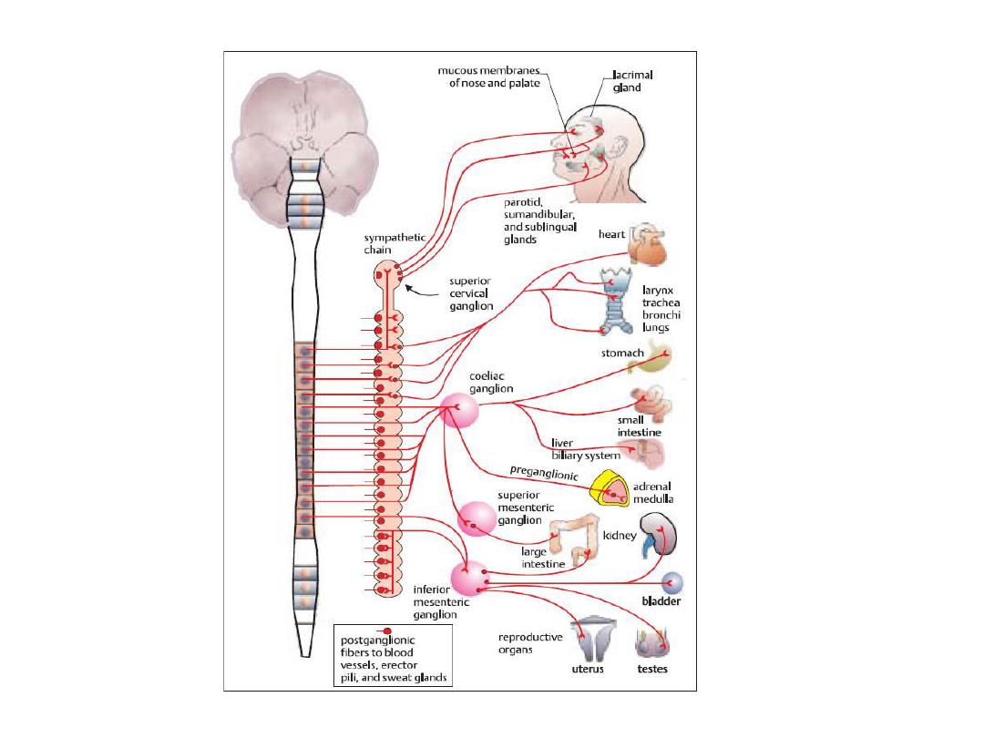
Functional Anatomy of the
Sympathetic Nervous System
The sympathetic nerve fibers
originate in the spinal cord along
with spinal nerves
between
spinal cord segments T-1 and L-2
, so called thoracolumbar ,
then
passes into the sympathetic
chain.

Autonomic Nervous System:
sympathetic Division
The sympathetic preganglionic neuron
cell bodies are situated in the
thoracic and
upper two lumbar segments of
the spinal cord
. The cell bodies lie in the
lateral horn of the spinal gray matter
. The
(usually) short preganglionic fibers leave
the spinal cord in the ventral nerve root,
and join the spinal nerve.

These fibers synapse with the
postganglionic fibers, either in one of
the sympathetic ganglia, which lie in a
bilateral longitudinal, paravertebral
chain ( sympathetic chain ) on either
side of the spinal column, or in one of
the prevertebral ganglia , which
surround the main branches of the
abdominal aorta
.

The prevertebral ganglia include
coeliac, superior mesenteric, and
inferior mesenteric ganglia, and are
unpaired. An exception to this general
arrangement of the sympathetic
division is that of the adrenal medulla.
The adrenal gland lies above the
kidney, and is structurally two separate
organs.

The outer shell of the adrenal gland is
concerned with production of the
steroid hormones, while the inner
core is the adrenal medulla, a
modified sympathetic ganglion. Thus,
preganglionic cholinergic fibers run
to the adrenal medulla, where they
synapse with postganglionic cell
bodies, which are in effect
hormone-secreting cells.

These cells respond to the arrival of
impulses down the preganglionic fibers, by
secreting the catecholamine hormones
epinephrine and norepinephrine into the
bloodstream. The preganglionic fibers, like
those of the parasympathetic division,
release the neurotransmitter, ACh, which
binds to
nicotinic receptors
on ganglionic
postsynaptic cell bodies of postganglionic
fibers.

the postganglionic fibers of the
sympathetic division release their
eurotra s itters “norepinephrine
(noradrenaline) , which binds to or
alpha & beta receptors
on the
postsynaptic membrane of the target
organ.

An exception to this general rule is
the presence in the sympathetic
division of postganglionic fibers,
which innervate the sweat glands.
These are cholinergic, and release
ACh, which acts on muscarinic
receptors on the membranes of the
sweat glands.

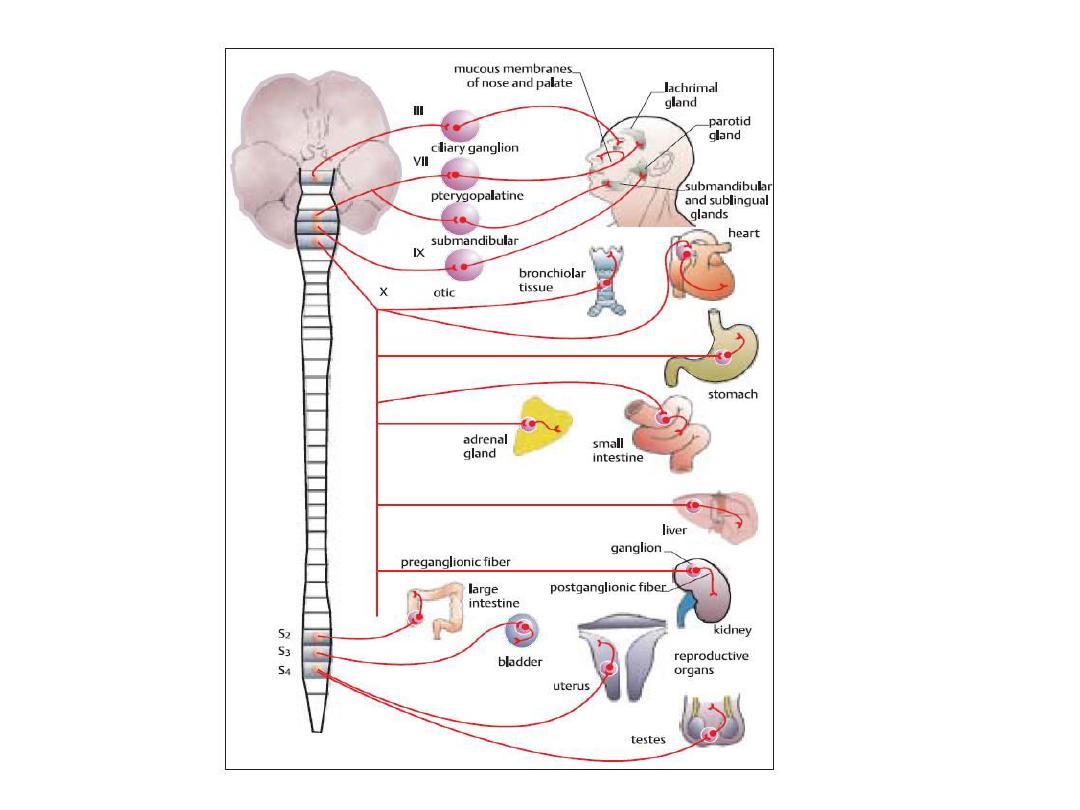
Autonomic Nervous System:
Parasympathetic Division
The parasympathetic division of the
autonomic nervous system (PNS)
consists of preganglionic fibers that
originate in three main areas of the
central nervous system. These are
the midbrain or tectum, the
medulla, and the sacral outflow,so
called :
craniosacral
.

The outflows emerge from two main
regions, (i) the brain stem cranial
outflow, and (ii) the sacral outflow.
Preganglionic fibers are generally
much longer than the postganglionic
fibers and often the ganglia lie on the
organ innervated.
Cranial outflow includes
Χ, іΧ, Ѵіі, ііі
.
Sacral outflow includes S2, S3, S4

The neurotransmitter released by the
postganglionic presynaptic nerve
terminal is ACh, which acts on
postsynaptic muscarinic receptors
on the membrane of the target
organ or tissue.
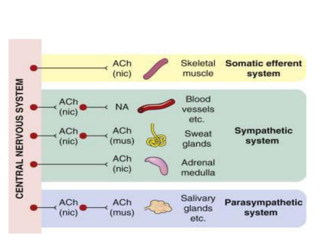
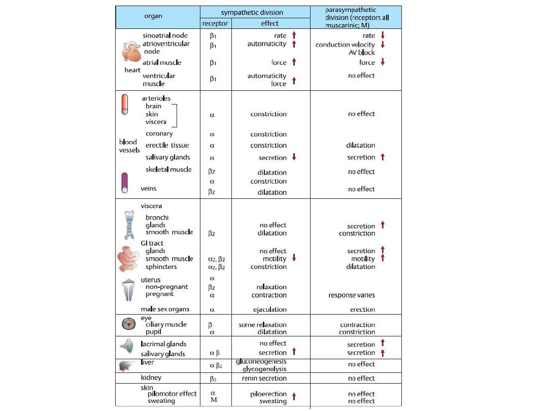
Neurotransmitters and their receptors

Autonomic Nervous System: Effects
Most organs receive dual parasympathetic
and sympathetic innervation.
Generally, these two divisions antagonize
each other, although this is not always so.
In some cases, for example the spleen and
arterioles, the tissues receive only
sympathetic fibers.

Autonomic Reflexes
Many visceral functions of the body are
regulated by autonomic reflexes.
Cardiovascular Autonomic Reflexes.
Several reflexes in the cardiovascular system
help to control especially the arterial blood
pressure and the heart rate. One of these is
the baroreceptor reflex, Briefly, stretch
receptors called baroreceptors are located in
the walls of several major arteries, including
especially the internal carotid arteries and the
arch of the aorta.

When these become stretched by high
pressure, signals are transmitted to
the brain stem, where they inhibit
the sympathetic impulses to the
heart and blood vessels and excite
the parasympathetics; this allows
the arterial pressure to fall back
toward normal.

Gastrointestinal Autonomic Reflexes.
The uppermost part of the gastrointestinal
tract and the rectum are controlled
principally by autonomic reflexes. For
instance, the smell of appetizing food or
the presence of food in the mouth
initiates signals from the nose and
mouth to the vagal, glossopharyngeal,
and salivatory nuclei of the brain stem.

These in turn transmit signals through
the parasympathetic nerves to the
secretory glands of the mouth and
stomach, causing secretion of
digestive juices sometimes even
before food enters the mouth.

Alarm
or Stress Response of the
Sympathetic Nervous System
When large portions of the sympathetic
nervous system discharge at the same
time
—that is, a mass discharge—
this increases in many ways the ability of
the body to perform vigorous muscle
activity, as this response summerized
as follows:-

1. Increased arterial pressure
2. Increased blood flow to active
muscles concurrent with decreased
blood flow to organs such as the
gastrointestinal tract and the kidneys.
3. Increased rates of cellular
metabolism throughout the body

4. Increased blood glucose
concentration
5. Increased glycolysis in the liver and
in muscle
6. Increased muscle strength
7. Increased mental activity
8. Increased rate of blood coagulation

The sum of these effects permits a
person to perform far more
strenuous physical activity than
would otherwise be possible.
Because either mental or
physical stress can excite the
sympathetic system,

it is frequently said that the purpose
of the sympathetic system is to
provide extra activation of the body
in states of stress: this is called the
sympathetic stress response.
When the sympathetic nervous
system is strongly activated in many
emotional states,

For instance, in the state of rage, this
cause massive sympathetic discharge;
most aforementioned sympathetic
events ensue immediately. This is
called the sympathetic alarm reaction.
or also is called the fight or flight
reaction because an animal in this
state decides almost instantly whether
to stand and fight or to run.

Thanks