Physiology of Muscle
Continued....
Lecture 5
By
Dr. Mufeed Akram Taha
FIBMS Neurology
Clinical Attachment Turkey
Excitability of muscle (Electrical
Characteristics of Skeletal Muscle)
The electrical events in skeletal muscle
and the ionic fluxes that underlie
them share distinct similarities to
those in nerve, with quantitative
differences in timing and magnitude.

-The resting membrane potential of
skeletal muscle is about
–90 mV.
- The action potential lasts 2 to 4 ms
and is conducted along the muscle
fiber at about 5 m/s.
-The absolute refractory period is 1 to
3 ms long.

Ion Distribution & Fluxes
The distribution of ions across the muscle
fiber membrane is similar to that across
the nerve cell membrane.
- As in nerves, depolarization is largely a
manifestation of Na
+
influx, and
repolarization is largely a manifestation
of K
+
efflux.

Contractile Responses
Muscle fiber membrane depolarization
normally starts at the motor end
plate, the specialized structure under
the motor nerve ending. The action
potential is transmitted along the
muscle fiber and initiates the
contractile response.

The Muscle Twitch
A single action potential causes a brief
contraction followed by relaxation.
This response is called a muscle
twitch. The twitch starts about 2 ms
after the start of depolarization of
the membrane, before repolarization
is complete.

The duration of the twitch varies with
the type of muscle being tested.
"Fast" muscle fibers, primarily those
concerned with fine, rapid, precise
movement, have twitch durations as
short as 7.5 ms. "Slow" muscle
fibers, principally those involved in
strong, gross, sustained movements,
have twitch durations up to 100 ms.
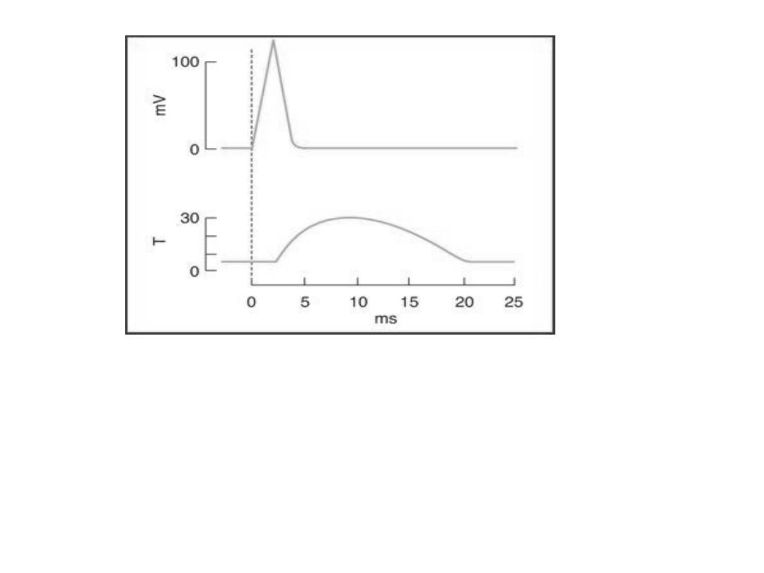
The electrical and mechanical responses of a mammalian skeletal
muscle fiber to a single maximal stimulus. The electrical response (mV
potential change) and the mechanical response (T, tension in arbitrary
units) are plotted on the same abscissa (time). The mechanical response
is relatively long-lived compared to the electrical response that initiates
contraction.

Molecular Basis of Contraction
The process by which the contraction of
muscle is brought about is a sliding of the
thin filaments over the thick filament. The
width of the A bands is constant, where as
the
Z lines move closer together
when
the muscle contracts and further apart
when it relaxes.

-The sliding during muscle
contraction occurs when the
myosin heads bind firmly to actin,
bend at the junction of the head
with the neck, and then detach,
this is called "Power Stroke".

-Each power stroke shortens the
sarcomere about 10 nm.
-The "power stroke" depends on the
simultaneous hydrolysis of ATP.
-Many myosin heads cycle at or near
the same time, and they cycle
repeatedly, producing gross muscle
contraction.

-Each thick filament has about 500
myosin heads, and each head cycles
about five times per second during a
rapid contraction.
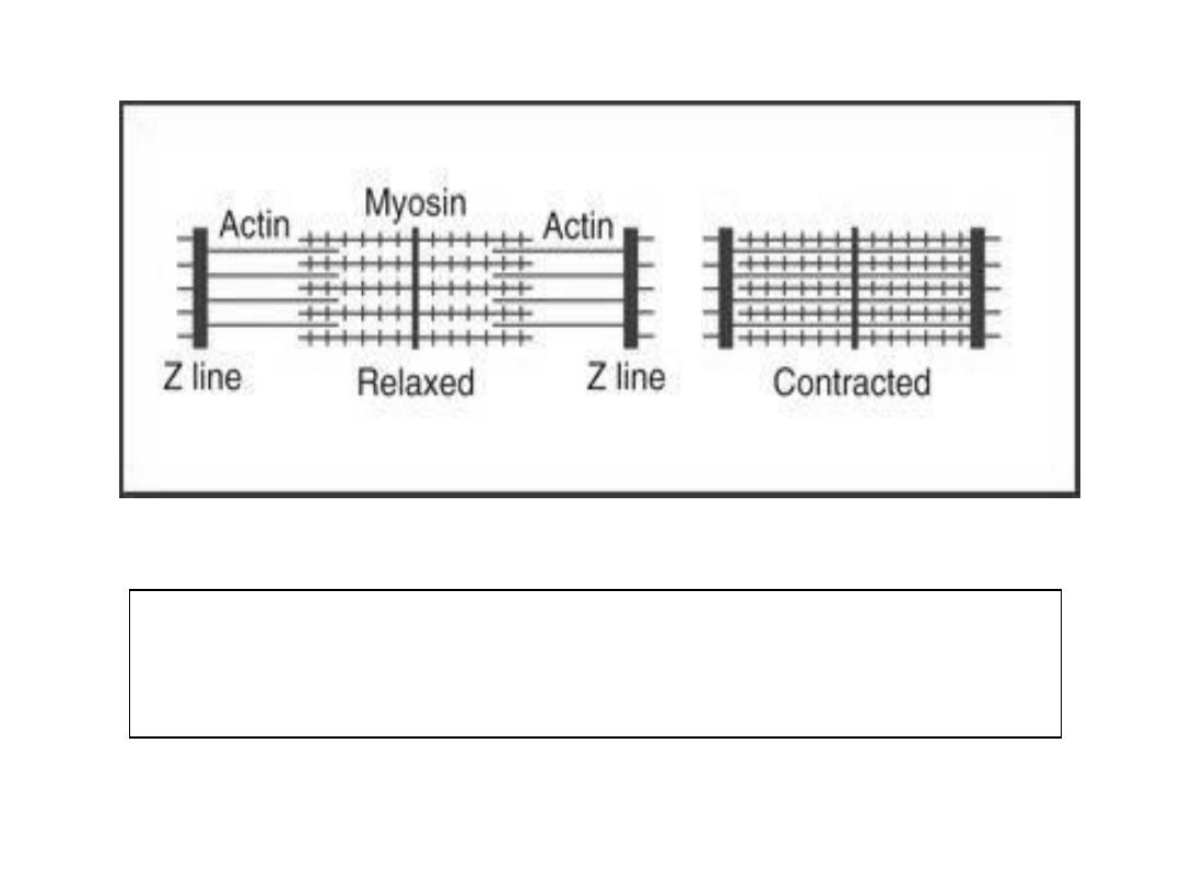
Sliding of actin on myosin during contraction so
that Z lines move closer together.

Excitation
–Contraction coupling
The process by which depolarization of the
muscle fiber initiates contraction is called
excitation
–contraction coupling.
The action potential is transmitted to all
the fibrils in the fiber via the T system. It
triggers the release of Ca
2+
from the
terminal cisterns, Ca
2+
initiate
contraction by binding to troponin C .

-In resting muscle, troponin I is bound to
actin and tropomyosin and covers the
sites where myosin heads interact with
actin.
- Ca
2+
binds to troponin C and this binding
results in a weakening of the troponin I
interaction with actin and this permits
tropomyosin to move laterally .This
movement exposes the actin binding site
for myosin head ,ATP splits to ADP and
contraction occurs .

-Shortly after releasing Ca
2+
The
Sarcoplasmic reticulum pump uses
energy from ATP hydrolysis to remove
Ca
2+
by active transport from the cytosol
back into the terminal cisterns, where it
is stored until released by the next action
potential.
-Once the Ca
2+
concentration outside the
reticulum has been lowered sufficiently,
chemical interaction between myosin
and actin ceases and the muscle relaxes.

-If transport of Ca
2+
into the reticulum
is inhibited, relaxation does not
occur even though there are no
more action potentials; the resulting
sustained contraction is called a
contracture.
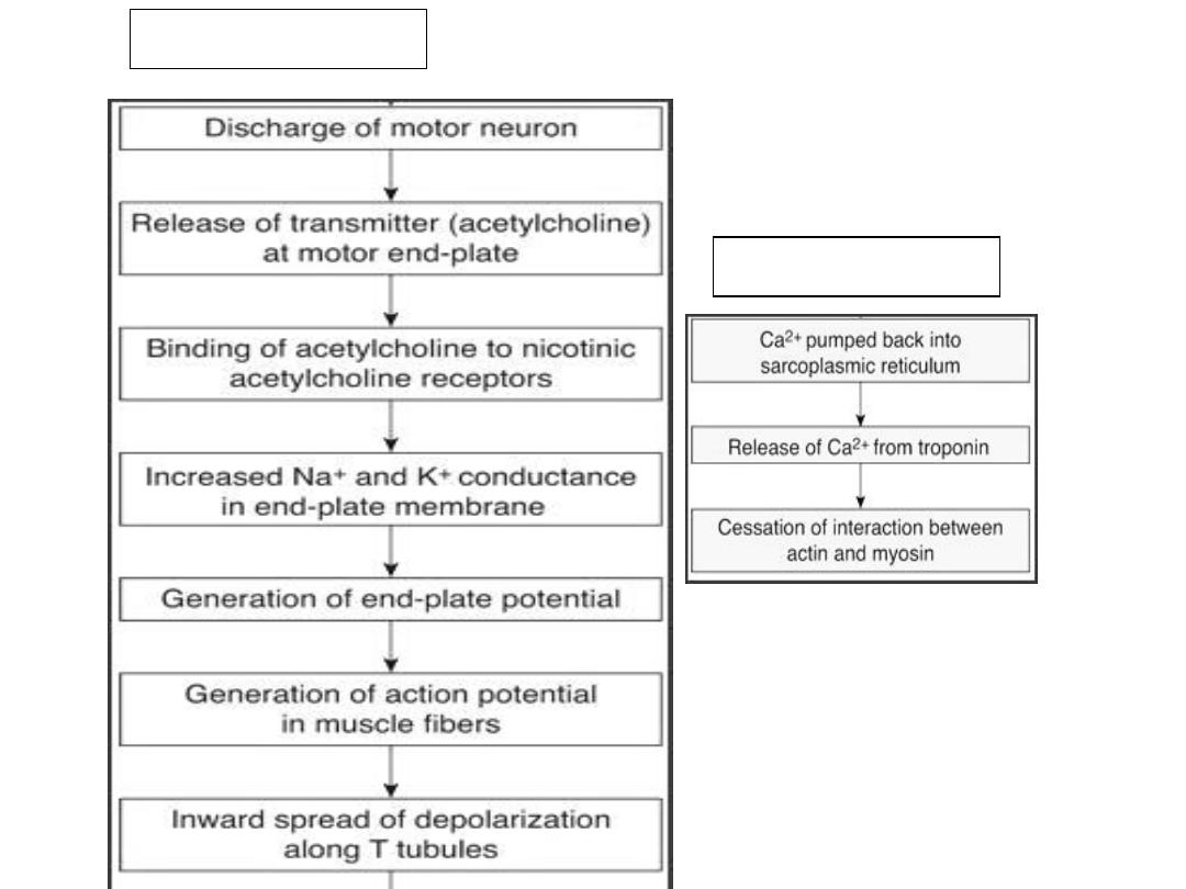
Steps of contraction
Steps of relaxation
Types of Contraction
1) Isometric Contraction
Contraction of muscle without an
appreciable decrease in the length of the
whole muscle
2) Isotonic Contraction
Contraction of muscle with approximation
of the ends of the muscle (shortening of
muscle) .This Contraction is usually
against a constant load.

Summation of Contractions
The electrical response of a muscle fiber
to repeated stimulation is like that of
nerve. The fiber is electrically refractory
only during the rising and part of the
falling phase of the spike potential. At
this time, the contraction initiated by
the first stimulus is just beginning
-But the contractile mechanism does not
have a refractory period

repeated stimulation before relaxation
has occurred produces additional
activation of the contractile elements
and a response that is added to the
contraction already present. This
phenomenon is known as summation
of contractions.
- The tension developed during
summation is considerably greater than
that during the single muscle twitch.

Tetanus
With rapidly repeated stimulation,
activation of the contractile
mechanism occurs repeatedly before
any relaxation has occurred, and the
individual responses fuse into one
continuous contraction. Such a
response is called a tetanus (tetanic
contraction).

Tetanus is of 2 types:
1)) Complete tetanus: when no
relaxation occurs between stimuli
2)) Incomplete tetanus: when periods
of incomplete relaxation take place
between the summated stimuli.

Energy Sources & Metabolism
Muscle contraction requires energy,
and muscle has been called "a
machine for converting chemical
energy into mechanical work." The
immediate source of this energy is
ATP, and this is formed by the
metabolism of carbohydrates and
lipids.

ATP is resynthesized from ADP by the
addition of a phosphate group. Some
of the energy for this endothermic
reaction is supplied by the
breakdown of glucose to CO
2
and
H
2
O.

Phosphorylcreatine
Energy-rich phosphate compound found in
muscle that can supply the energy for
short periods. This compound
(Phosphorylcreatine) which is hydrolyzed
to creatine and phosphate groups with
the release of considerable energy

-At rest, some ATP in the mitochondria
transfers its phosphate to creatine, so that
a phosphorylcreatine store is built up.
-At rest and during light exercise, muscles
utilize lipids in the form of free fatty acids
as their energy source.
-Thus, during exercise, much of the energy
for phosphorylcreatine and ATP
resynthesis comes from the breakdown of
glucose to CO
2
and H
2
O.

-Glucose which is either come from the
bloodstream or form breakdown of glycogen in
the cell will be metabolized by series of
chemical reaction into pyruvate.
-When adequate O
2
is present, pyruvate enters
the citric acid cycle and is metabolized
—
through this cycle and the so-called respiratory
enzyme pathway
—to CO
2
and H
2
O. This
process is called aerobic glycolysis. The
metabolism of glucose or glycogen to CO
2
and
H
2
O liberates sufficient energy to form large
quantities of ATP from ADP.

- If O
2
supplies are insufficient, the
pyruvate formed from glucose does not
enter the tricarboxylic acid cycle but is
reduced to lactate. This process of
anaerobic glycolysis is associated with
the net production of much smaller
quantities of energy-rich phosphate
bonds, but it does not require the
presence of O
2

Thanks