Resistance of
The Body to
Infection
Immunity and Allergy

1. Innate Immunity
•
The human body has the ability to
resist
almost all types of organisms or toxins that tend to
damage the tissues and organs. This capability is
called
immunity
.
Much of immunity is
acquired
immunity
that does not develop until after the body is first attacked by a bacterium,
virus, or toxin, often
requiring
weeks or months to develop the immunity. An additional
portion of immunity results from
general
processes, rather than from processes directed at
specific disease organisms. This is called
innate
immunity
.
It includes the following:
1.
Phagocytosis
of bacteria and other invaders by white blood cells and cells of the tissue
macrophage system.
natural killer lymphocytes
that can recognize and destroy
foreign
cells,
tumor
cells, and even some infected
cells
2. Destruction of swallowed organisms by the
acid
secretions
of the stomach and the digestive
enzymes.
3. Resistance of the
skin
to invasion by organisms.
4. Presence in the blood of certain
chemical
compounds
that attach to foreign organisms or toxins
and destroy them. Some of these compounds are
(a)
lysozyme
, a
mucolytic
polysaccharide that attacks bacteria and causes them to dissolute;
(b)
basic
polypeptides
, which react with and inactivate certain types of
gram-positive
bacteria
;
(c) the
complement
complex
a system of proteins that can be activated in various ways to destroy
bacteria;
2. Acquired (Adaptive) Immunity
•
The human body has the ability to develop extremely powerful
specific immunity
against
individual
invading agents such as lethal
bacteria
,
viruses
,
toxins
, and even
foreign
tissues
from other animals.
•
Basic Types of Acquired Immunity
•
the body develops circulating
antibodies
,
which are globulin molecules in the blood plasma
that are capable of attacking the invading agent. This type of immunity is called
humoral
immunity
or
B-cell
immunity
(because B lymphocytes produce the antibodies).
•
The
second
type is achieved through the formation of large numbers of
activated
T
lymphocytes
that are specifically crafted in the lymph nodes to destroy the foreign agent.
This type of immunity is called
cell-mediated
immunity
or
T-cell immunity.
•
Both Types of Acquired Immunity Are Initiated by Antigens
•
The body has some
mechanisms
for
recognizing
invasion by a foreign organism or toxin.
Each toxin or each type of organism almost always contains one or more
specific
chemical
compounds
in its makeup that are different from all other compounds. In general,
these
are
proteins or large polysaccharides, and it is they that
initiate
the acquired immunity. These
substances are called
antigens
(
anti
body
gen
erations
)
.
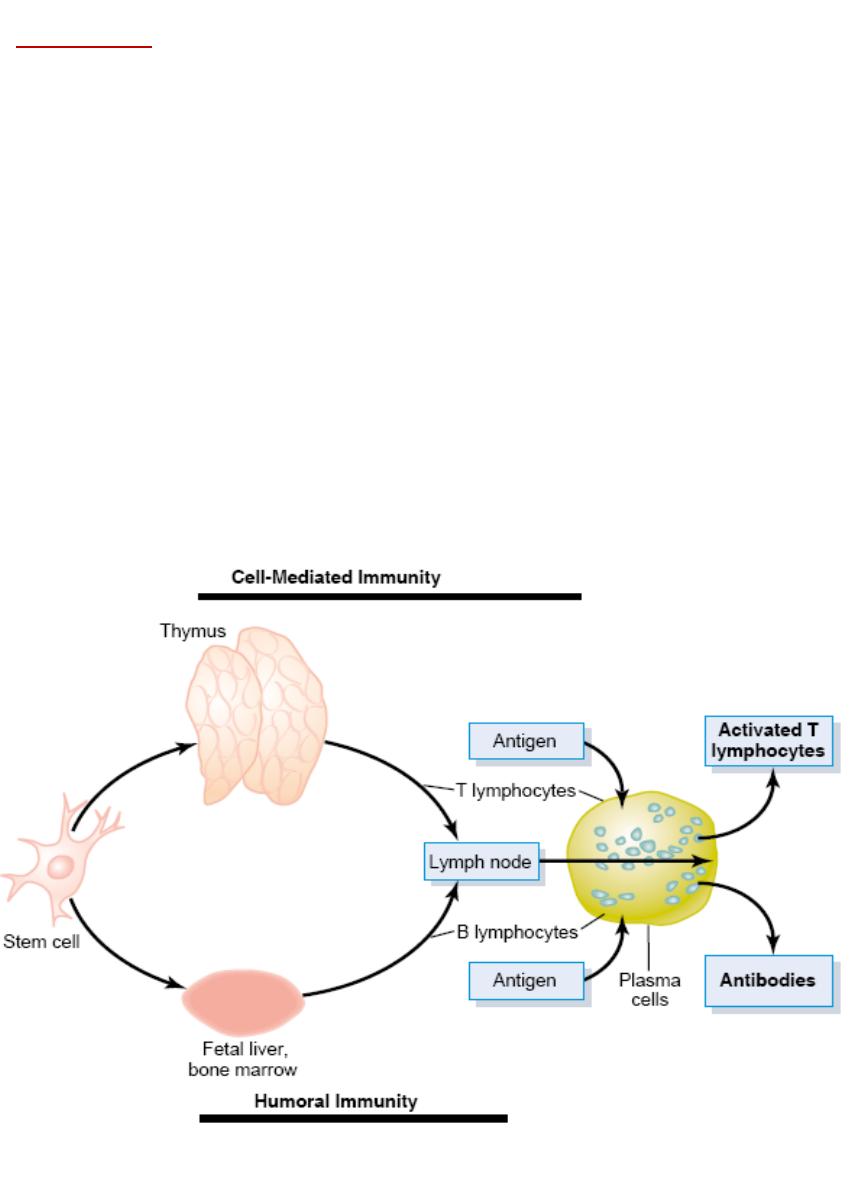
Lymphocytes
•
Are Responsible for Acquired Immunity
•
The lymphocytes are
located
most extensively in the
lymph
nodes
, but they are also found in
special
lymphoid
tissues such as the
spleen
,
submucosal areas of the gastrointestinal tract
,
thymus
, and
bone marrow
.
•
The invading agent
first
enters
the tissue fluids and then is
carried
by way of lymph vessels
to the lymph node or other lymphoid tissue.
•
Two Types of Lymphocytes Promote “Cell-Mediated” Immunity or “Humoral”
Immunity—the T and the B Lymphocytes.
•
Both types of lymphocytes are
derived
originally in the embryo from
pluripotent
hematopoietic
stem cells
that form lymphocytes
•
The lymphocytes that are
destined
to
eventually
form
activated T lymphocytes first migrate
to and are preprocessed in the
thymus
gland, and thus they are called
“T” lymphocytes
to
designate the role of the thymus.
•
B
lymphocytes that are destined to form antibodies—are preprocessed in the
liver
during
midfetal
life and in the bone
marrow
in late fetal life and after birth , they are responsible for
humoral immunity
.

Thymus Gland Preprocesses the T Lymphocytes.
•
1. The T lymphocytes, after origination in the bone marrow, first
migrate
to the thymus gland.
Here they
divide
rapidly and at the same time develop extreme
diversity
for reacting against
different
specific antigens. That is,
one
thymic lymphocyte develops specific reactivity
against one antigen. Then the
next
lymphocyte develops specificity against another antigen.
•
These different types of preprocessed T lymphocytes now
leave
the thymus and spread by
way of the blood throughout the body to
lodge
in lymphoid tissue everywhere.
•
2. The thymus also makes certain that any T lymphocytes leaving the thymus will
not
react
against proteins or other antigens that are present in the body’s own tissues; otherwise, the T
lymphocytes would be
lethal
to the person’s own body in only a few days.
•
The thymus selects
which
T lymphocytes will be
released
by first
mixing
them with virtually
all the specific “
self-antigens
” from the body’s own tissues. If a
T
lymphocyte
reacts
, it is
destroyed
and phagocytized instead of being released.
•
Liver and Bone Marrow
Preprocess
the B Lymphocytes during midfetal life and late
fetal life and after birth respectively.
•
B lymphocytes are
different
from T lymphocytes in two ways:
•
First
, instead of the
whole
cell
developing reactivity against the antigen, as occurs for the T
lymphocytes, the B lymphocytes actively
secrete
antibodies
that are the reactive agents.
These agents are
large
protein molecules that are capable of
combining
with and
destroying
the antigenic substance,
•
Second
, the B lymphocytes have even
greater
diversity
than the T lymphocytes, thus
forming
many
millions
of types of B-lymphocyte antibodies with different specific reactivities.
Role of Macrophages in the Activation Process.
•
millions of macrophages are also
present
in the lymphoid tissue. These
line
the sinusoids of
the lymph nodes, spleen, and other lymphoid tissue, and they lie in apposition to many of the
lymph node lymphocytes.
•
1. Most
invading
organisms are
first
phagocytized
and partially digested by the macrophages,
and the antigenic products are
liberated
into the macrophage cytosol. The macrophages then
pass
these
antigens
by cell-to-cell contact directly to the lymphocytes, thus leading to
activation
of the specified lymphocytic clones.
•
2. The macrophages, in addition,
secrete
a special activating substance that
promotes
still
further growth and reproduction of the specific lymphocytes. This substance is called
interleukin-1
.
The
plasma
cell which develops after B cell activation
produces
gamma globulin antibodies
Antibodies
•
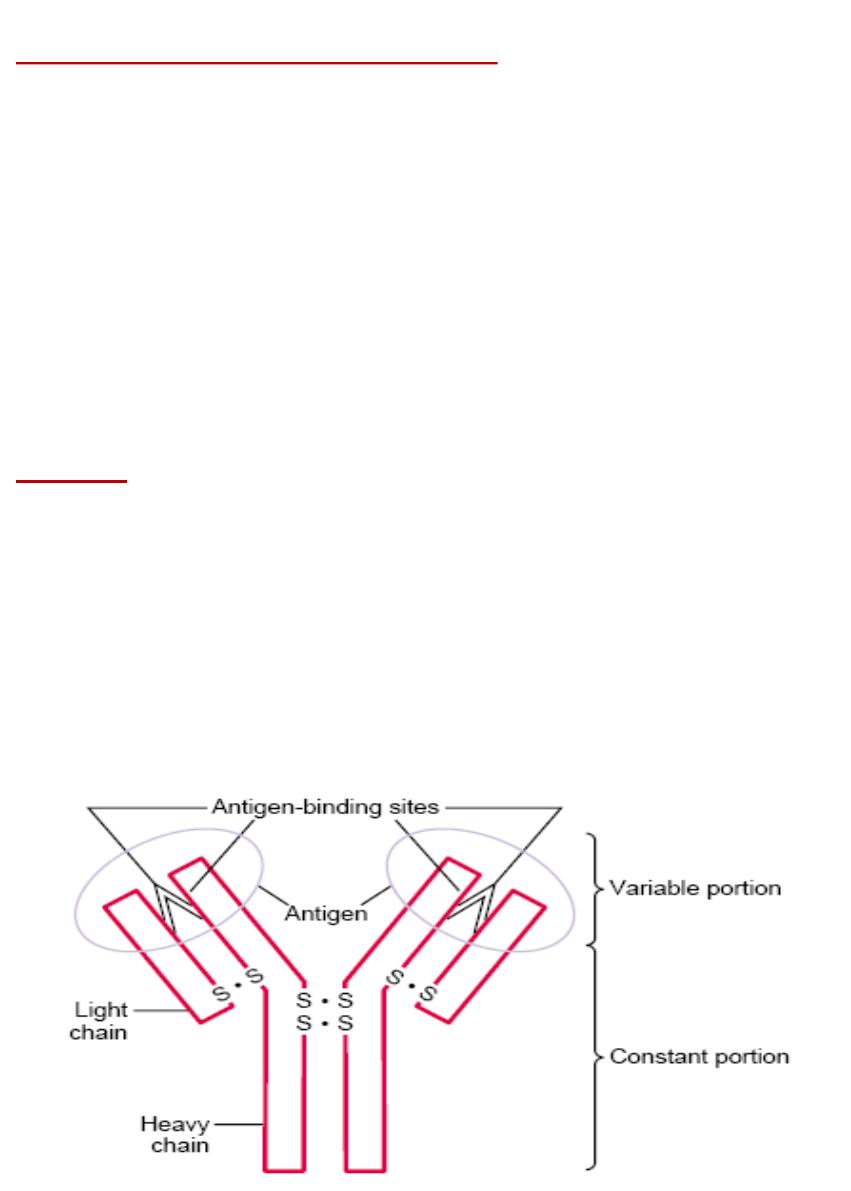
Most are a combination of two light and two heavy chains,
•
The end of each light and heavy chain, called the
variable
portion
; the remainder of each
chain is called the
constant
portion
. The variable portion is different for each
specificity
of
antibody, and it is this portion that attaches specifically to a particular type of antigen. The
constant
portion
of the antibody determines other properties of the antibody, establishing
such factors as
diffusivity
of the antibody in the tissues,
adherence
of the antibody to specific
structures within the tissues, attachment to the
complement
complex
.
Specificity of Antibodies.
Each antibody is specific for a particular antigen; this is caused by its unique structural
organization of amino acids in
the variable portions of both the light and heavy chains.
Classes of Antibodies.
There are five general classes of antibodies, respectively named
IgM, IgG, IgA, IgD,
and
IgE
.
Mechanisms of Action of Antibodies
a. Direct Action of Antibodies on Invading Agents.
1.
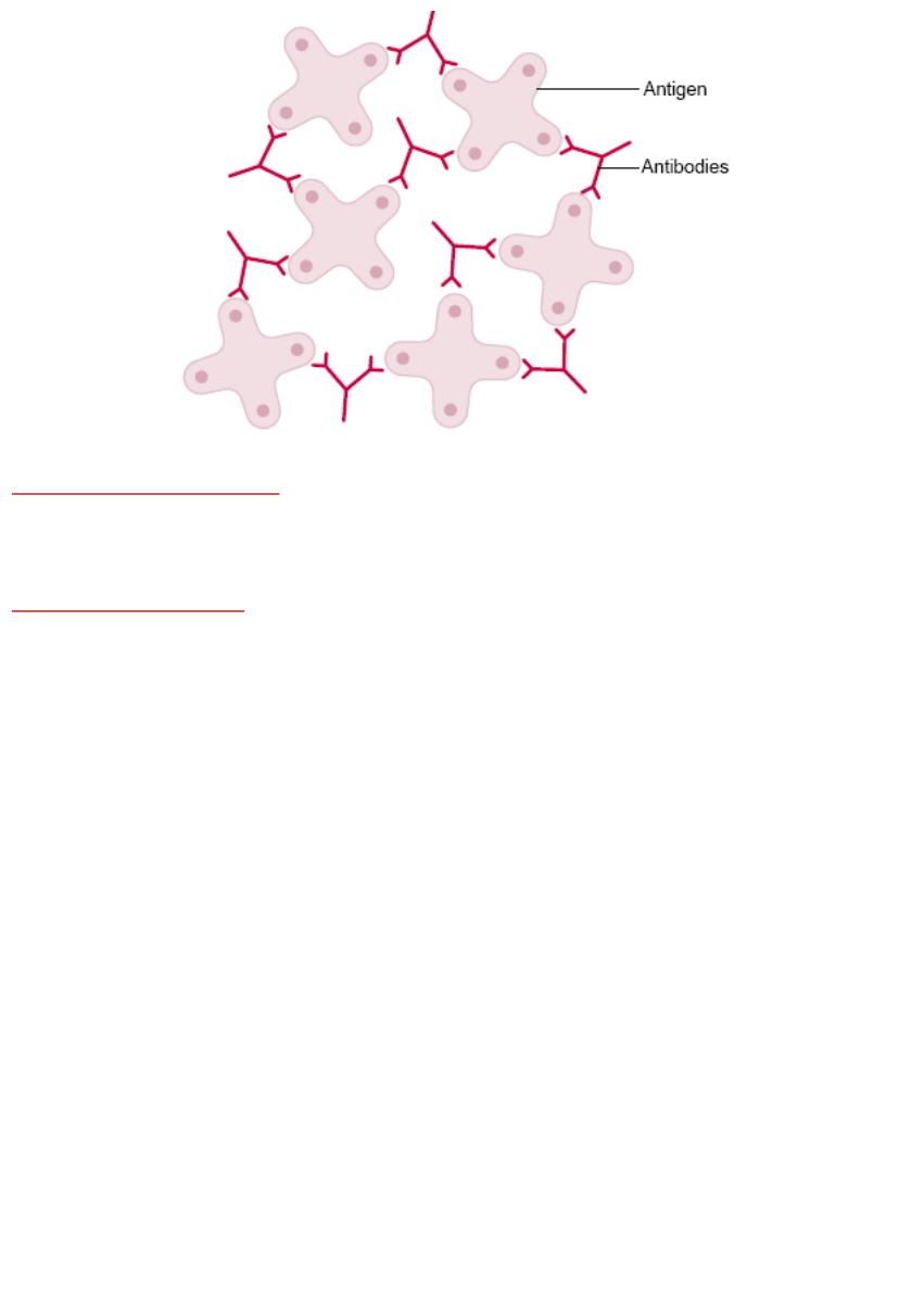
Agglutination
, in which multiple large particles with
antigens
on
their
surfaces
, such as bacteria
or red cells, are bound together into a clump
2.
Precipitation
, in which the molecular complex of soluble antigen (such as
tetanus
toxin) and
antibody becomes so
large
that it is rendered
insoluble
and precipitates
3.
Neutralization
, in which the antibodies cover the
toxic
sites
of the antigenic agent
4.
Lysis
, in which some potent antibodies are occasionally capable of directly
attacking
membranes
of cellular agents and thereby cause rupture of the agent.
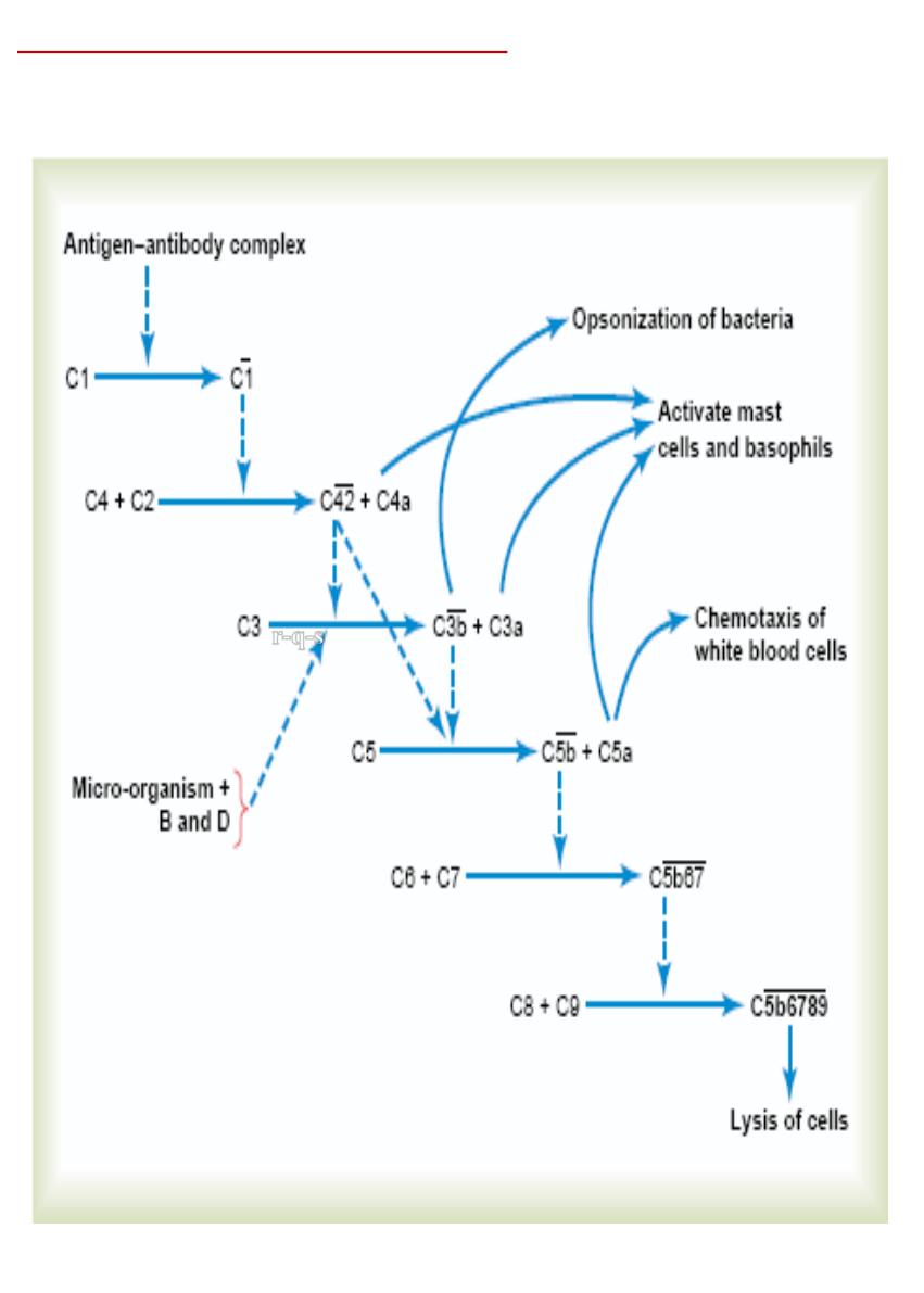
B- Complement System for Antibody Action
•
The principal actors in this system are
11
proteins
designated C1 through C9, B, and D. All
these are present normally among the
plasma
proteins
in
the blood .
r-q-s

•
Classic
Pathway
. The classic pathway is initiated by an
antigen-antibody
reaction
with
activation
of the proenzyme C1, then
activate successively increasing quantities of enzymes
in the later stages of the system, Among the more important effects
of enzymes
are the
following:
•
1.
Opsonization
and
phagocytosis
.
One of the products of the complement cascade,
C3b
,
strongly activates phagocytosis by both neutrophils and macrophages, causing these cells to
engulf the bacteria to which the antigen antibody complexes are attached. This process is
called
opsonization
.
•
2.
Lysis
.
One of the most important of all the products of the complement cascade is the
lytic
complex
, which is a
combination
of
multiple
complement
factors and designated
C5b6789
.
This has a direct effect of
rupturing
the cell membranes of bacteria or other invading
organisms.
•
3.
Agglutination
.
The complement products also
change
the
surfaces
of the invading
organisms, causing them to
adhere
to one another, thus promoting agglutination.
•
4.
Neutralization
of
viruses
.
The complement enzymes and other complement products can
attack the
structures
of some viruses and thereby render them
nonvirulent
.
•
5.
Chemotaxis
.
Fragment
C5a
initiates chemotaxis of neutrophils and macrophages,
•
6.
Activation
of
mast
cells
and
basophils
.
Fragments
C3a
,
C4a
,
and C5a
activate mast cells
and basophils, causing them to release
histamine
,
heparin
, and several other substances into
the local fluids. These substances in turn cause
increased local blood flow
, increased leakage
of fluid and plasma protein into the tissue, and other local tissue reactions that help inactivate
or immobilize the antigenic agent.
•
7.
Inflammatory
effects
.
In addition to inflammatory effects caused by activation of the
mast
cells and
basophils
, several other complement products contribute to local inflammation.
These products cause
•
(1) the already
increased
blood flow to increase still further,
•
(2) the capillary
leakage
of proteins to be increased, and
•
(3) the
interstitial
fluid
proteins
to
coagulate
in the tissue spaces, thus preventing movement
of the invading organism through the tissues.

Several Types of T Cells and Their Different Functions
They are classified into three major groups:
•
(1)
helper
T cells, (2)
cytotoxic
T cells, and (3)
suppressor
T cells. The functions of each
of these are distinct.
Helper T Cells
•
they serve as the major regulator of virtually all immune functions, They do this by
forming
a series of protein mediators, called
lymphokines
, that act on other cells of the
immune system as well as on bone marrow cells. As:
•
Interleukin-2
•
Interleukin-3
•
Interleukin-4
•
Interleukin-5
•
Interleukin-6
•
Granulocyte-monocyte colony-stimulating factor
•
Interferon-ϫ
•
Specific Regulatory Functions of the Lymphokines.
•
In the absence of the lymphokines from the helper T cells, the remainder of the
immune
system is almost
paralyzed
. AIDS virus acts on these cell.
•
1. Stimulation of
Growth
and
Proliferation
of
Cytotoxic
T Cells and
Suppressor
T Cells.
•
2. Stimulation of
B-Cell
Growth and Differentiation to Form Plasma Cells and Antibodies.
•
3. Activation of the
Macrophage
System.
•
4.
Feedback
Stimulatory Effect on the
Helper
Cells Themselves.

Cytotoxic T Cells
•
The
receptor
proteins on the surfaces of the cytotoxic cells cause them to
bind
tightly
to those
organisms or cells that contain the appropriate binding-specific antigen.
•
After binding, the cytotoxic T cell secretes
holeforming
proteins
,
called
perforins
, that
literally punch round holes in the membrane of the attacked cell. Then fluid
flows
rapidly
into the cell from the interstitial space.
•
In addition, the cytotoxic T cell
releases
cytotoxic
substances directly into the attacked cell.
•
Almost immediately, the attacked cell becomes
greatly
swollen
, and it usually dissolves
shortly thereafter.
Suppressor T Cells
•
capable of suppressing the functions of both
cytotoxic
and
helper
T cells.
Immunization by Injection of Antigens (vaccinization)
•
Immunization
has been used for many years to
produce
acquired
immunity against specific
diseases. A person can be immunized by injecting
dead
organisms that are no longer capable
of causing disease but that still have some of their chemical antigens.
Passive Immunity
•
A
temporary
immunity can be achieved by
infusing antibodies
,
activated
T
cells
,
or both
obtained from the blood of someone else or from some other animal that has been actively
immunized against the antigen.
•
Antibodies
last
in the body of the recipient for
2
to
3
weeks
•
Activated T cells last for a few weeks
ALLERGY AND HYPERSENSITIVITY
•
An important undesirable
side
effect
of immunity.
1. Allergy Caused by Activated T Cells:
•
Delayed-reaction allergy
is caused by activated
T cells
and
not
by
antibodies
. toxins may in
itself does not cause much
harm
to the tissues. However, on
repeated
exposure, it does cause
the
formation
of
activated
helper and cytotoxic T cells. And these T
cells
elicit
a
cell-
mediated
type of immune reaction causing release of
many
toxic
substances
from the
activated T cells as well as
extensive invasion
of the tissues by macrophages with
serious
tissue
damage
. such as in the skin or in the lungs to cause lung edema or asthmatic attacks in
the case of some airborne antigens.

2. Allergies in the “Allergic” Person,
•
Who Has Excess IgE Antibodies
•
Some people have an “allergic” tendency. Their allergies are called
atopic
allergies
because
they are caused by a
nonordinary response of the immune system. The
allergic tendency is
genetically
passed from parent
to child and is characterized by the presence of
large
quantities of IgE antibodies
in the blood. These antibodies are called
reagins
or
sensitizing
antibodies
to distinguish them from the more common IgG antibodies.
•
When an
allergen
(defined as an antigen that reacts specifically with a specific type of IgE
reagin antibody) enters the body, an allergen-reagin reaction lakes place, and a subsequent
allergic reaction occurs.
•
A special characteristic of the IgE antibodies (the reagins) is a strong propensity to
attach
to
mast
cells
and
basophils
. this causes immediate
change
in the membrane of the mast cell or
basophil,. At any rate, many of the mast cells and
basophils
rupture
; others release special
agents immediately or shortly thereafter, including
histamine
,
protease
,
slow-reacting
substance of anaphylaxis,
eosinophil
chemotactic substance
,
neutrophil
chemotactic
substance
,
heparin
, and
platelet activating factors
.
•
These substances cause such effects as
dilation
of the local blood vessels;
attraction
of
eosinophils and neutrophils to the reactive site; increased
permeability
of the capillaries with
loss of fluid into the tissues; and
contraction
of local smooth muscle cells.
•
Anaphylaxis
. When a specific allergen is
injected
directly into the circulation, the allergen
can react with basophils of the blood and mast cells in the tissues located immediately
outside the small blood vessels if the basophils and mast cells have been sensitized by
attachment of IgE reagins. Therefore, a widespread
allergic reaction occurs throughout the
vascular system and closely associated tissues.
