HISTOLOGY
BY DR. ZANA
LECTURE -3-
MUSCULAR SYSTEM

INTRODUCTION
•
Although all cells are capable of some sort of movement, the dominant
function of several cell types is to generate motile forces through
contraction.
•
In these specialized contractile cells, motile forces are generated by
the interaction of the proteins actin and myosin (contractile proteins).
3
PROPERTIES OF MUSCLE
•
Contractility
•
Ability of a muscle to shorten with force
•
Excitability
•
Capacity of muscle to respond to a stimulus
•
Extensibility
•
Muscle can be stretched to its normal resting length and beyond to a
limited degree
•
Elasticity
•
Ability of muscle to recoil to original resting length after stretched

•
Certain forms of contractile cell function as single-cell
contractile units.
•
Myoepithelial cells are an important component of certain secretory
glands where they function to expel secretions from glandular acini.
•
Pericytes are smooth muscle-like cells that surround blood vessels
•
Myofibroblasts are cells that have a contractile role in addition to
being able to secrete collagen. This type of cell is generally
inconspicuous in normal tissues but comes to be a dominant cell type
when tissues undergo repair after damage in the formation of a scar
9-5
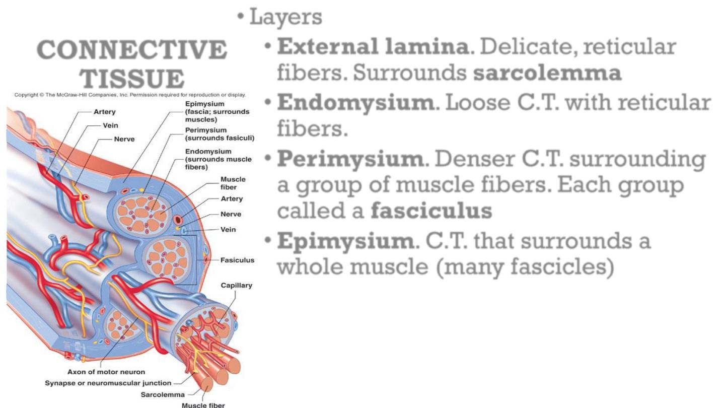
CONNECTIVE
TISSUE
•
Layers
•
External lamina. Delicate, reticular
fibers. Surrounds sarcolemma
•
Endomysium. Loose C.T. with reticular
fibers.
•
Perimysium. Denser C.T. surrounding
a group of muscle fibers. Each group
called a fasciculus
•
Epimysium. C.T. that surrounds a
whole muscle (many fascicles)
•
Fascia: connective tissue sheet
•
Forms layer under the skin
•
Holds muscles together and separates them into functional
groups.
•
Allows free movements of muscles.
•
Carries nerves (motor neurons, sensory neurons), blood
vessels, and lymphatics.
•
Continuous with connective tissue of tendons and
periosteum.
•
Other forms of contractile cell function by forming
multicellular contractile units termed muscles. Such muscle
cells can be divided into three types:
1. Skeletal muscle.
2. Smooth muscle.
3. Cardiac muscle.

SKELETAL MUSCLE
•
responsible for the movement of the skeleton and organs such
as the globe of the eye and the tongue. Skeletal muscle is
often referred to as voluntary muscle since it is capable of
voluntary (conscious) control.
•
The arrangement of the contractile proteins gives rise to the
appearance of prominent cross-striations in some histological
preparations and so the name striated muscle is often
applied to skeletal muscle.

•
The highly developed functions of the cytoplasmic
organelles of muscle cells has led to the use of a
special
terminology
for
some
muscle
cell
components: plasma membrane or plasmalemma =
sarcolemma;
cytoplasm
=
sarcoplasm;
endoplasmic reticulum = sarcoplasmic reticulum.

•
Skeletal muscles have a wide variety of morphological forms
and modes of action; nevertheless all have the same basic
structure.
•
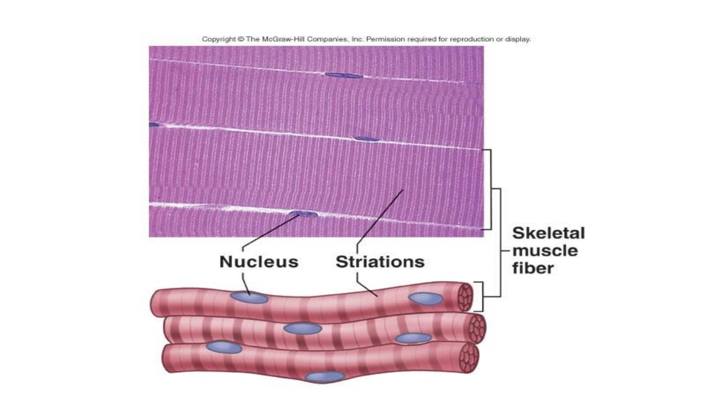
Skeletal muscle is composed of extremely elongated,
multinucleate contractile cells, often described as muscle
fibers, bound together by collagenous supporting tissue.
•
Individual muscle fibers vary considerably in diameter from
10 to 100 μm and may extend throughout the whole length of
a muscle reaching up to 35 cm in length.
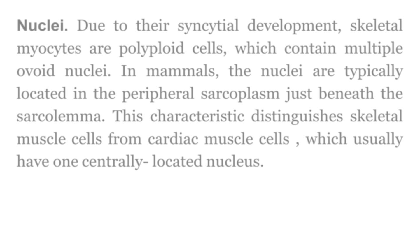
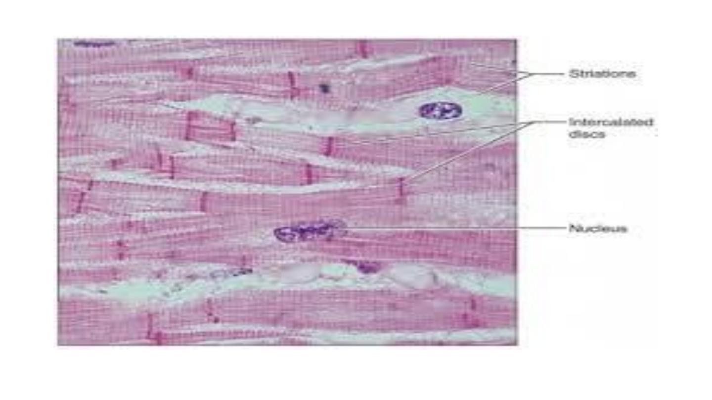
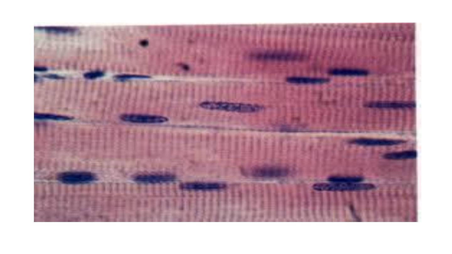
Nuclei.
Due to their syncytial development, skeletal
myocytes are polyploid cells, which contain multiple
ovoid nuclei. In mammals, the nuclei are typically
located in the peripheral sarcoplasm just beneath the
sarcolemma. This characteristic distinguishes skeletal
muscle cells from cardiac muscle cells , which usually
have one centrally- located nucleus.
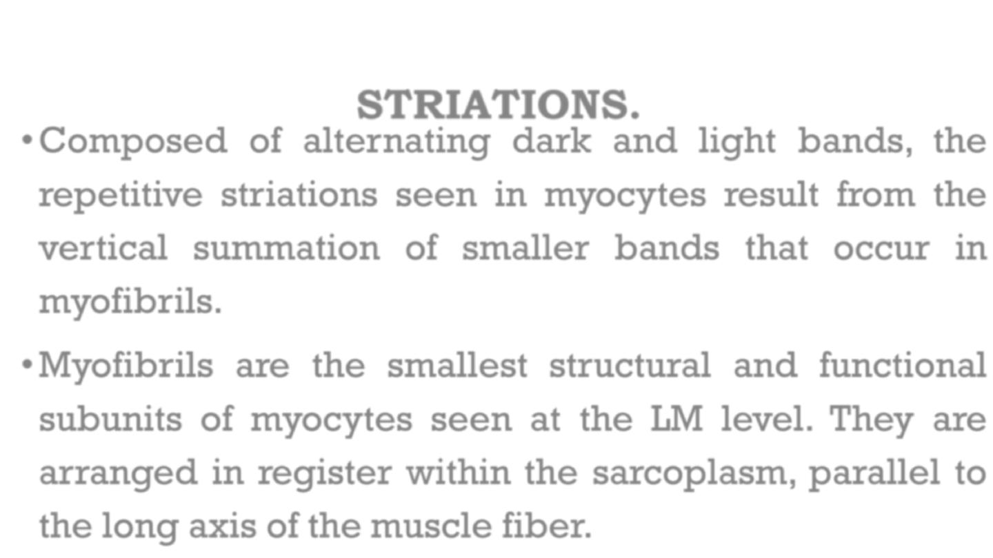
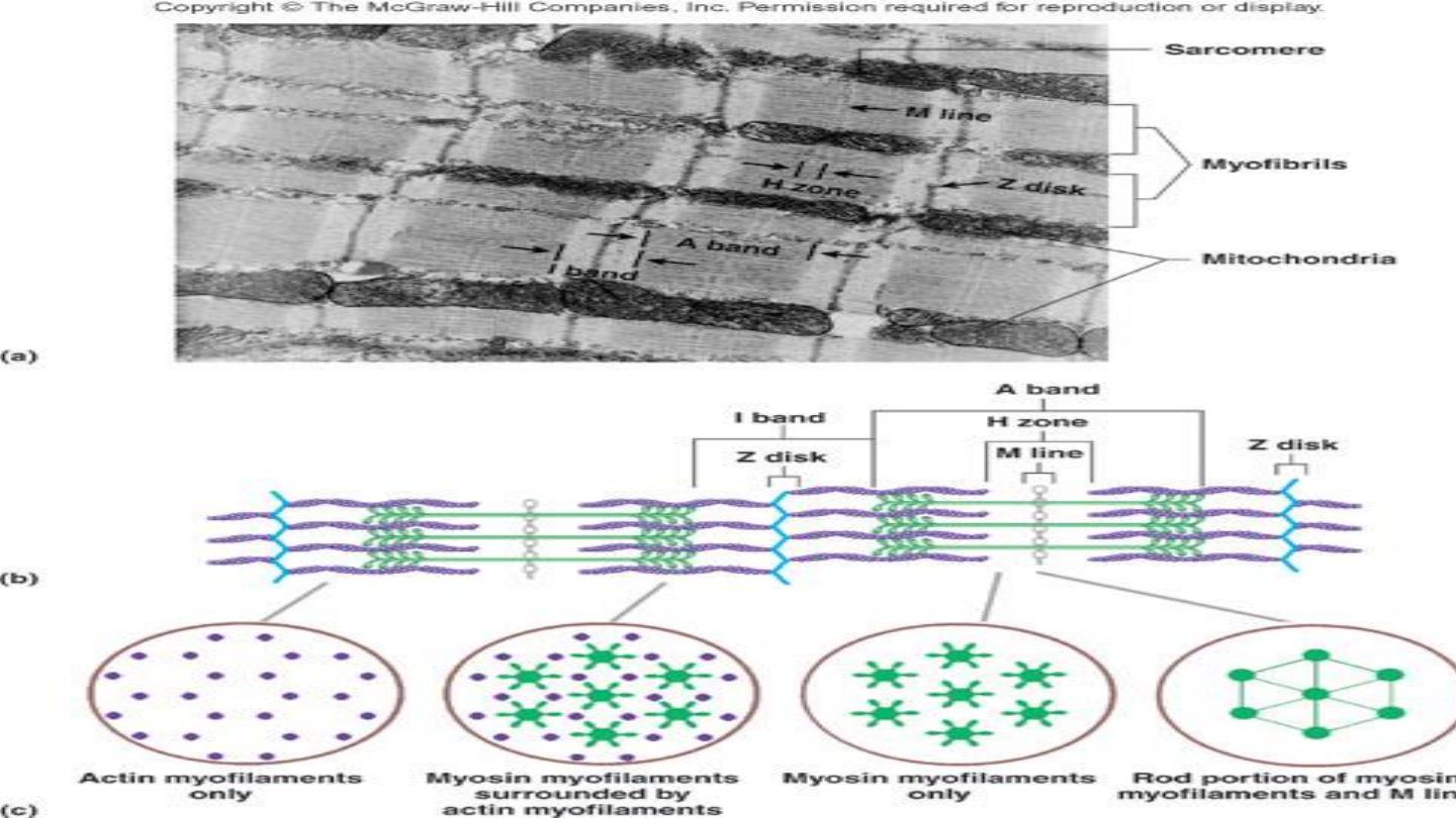
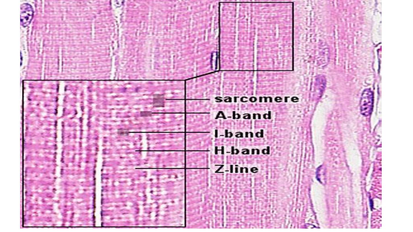
STRIATIONS.
•
Composed of alternating dark and light bands, the
repetitive striations seen in myocytes result from the
vertical summation of smaller bands that occur in
myofibrils.
•
Myofibrils are the smallest structural and functional
subunits of myocytes seen at the LM level. They are
arranged in register within the sarcoplasm, parallel to
the long axis of the muscle fiber.
Dark bands are called A bands because they are anisotropic
in polarized light (i.e., they rotate plane polarized light). The
parallel array of thick filaments are mainly responsible for the
LM appearance of the A band. In the center of this band a
paler region, called the H band [formerly Hensen disk], can be
seen in relaxed muscle.
It represents an area of thick filaments that are not overlapped
by thin filaments. At the middle of the A band (and the H
band) is a middle line called the M line.
Light bands are called I bands because they are isotropic
in polarized light (i.e., they do not rotate plane polarized
light). A dark transverse line, called the Z line (Z disk)
bisects each I band.
•
Thin filaments are
composed of several proteins, but
primarily of actin and two important regulatory
proteins called tropomyosin and troponin.
•
Thick filaments are composed primarily of
composites of the protein myosin II.
Sarcomeres are defined as the basic units of contraction in
striated muscles. A single sarcomere extends from one Z line to
the next and contains one A band separating two semi-I bands.
Thus, myofibrils are composed of a series of tandomly-arranged
sarcomeres consisting of interdigitating polarized thin filaments
(plus end toward Z line; minus end toward A band) and bipolar
thick filaments (myosin heads toward each semi-I band).
Arrangement of filaments in sarcomeres.
Thick filaments occupy central portions of the sarcomere; thin
filaments attach at each end to the Z lines and run parallel to,
and between, the thick filaments.
i. I bands are composed of thin filaments. Each sarcomere has ½
of an I band at its ends. Thus, a whole I band is shared between
adjacent sarcomeres.
ii. A bands are composed mostly of thick filaments and the thin
filaments between them.
iii. H bands are composed of only thick filaments, and in
relaxed muscle represent the area between the ends of
thin filaments that are attached to each Z line at the
other ends. In contracted muscle, when the thin
filaments merge or overlap, the H band disappears.
iv. Actin and myosin together represent approximately
55% of the total proteins in striated muscle.
Electron microscopic appearance. In electron micrographs, the
repeating pattern of bands and sarcomeres is due to the
arrangement of the myofilaments. Myofilaments represent the
thick myosin filaments and the thin actin filaments. These
structures are not resolved at the LM level.

•
Skeletal muscle contraction is controlled by large motor
nerves, individual nerve fibers branching within the muscle to
supply a group of muscle fibers, collectively described as a
motor unit.
•
Excitation of any one motor nerve results in simultaneous
contraction of all the muscle fibers of the corresponding motor
unit.

•
The
structure
of
neuromuscular
junctions
is
described in The vitality of skeletal muscle fibers is
dependent on the maintenance of their nerve supply
which, if damaged, results in atrophy of the fibers.
•
Skeletal muscle contains highly specialized stretch
receptors known as neuromuscular spindles

•
The individual muscle cells (muscle fibers) are grouped
together into elongated bundles called fasciculi with delicate
supporting tissue called endomysium occupying the spaces
between individual muscle fibers.
•
Each fascicle is surrounded by loose collagenous tissue called
perimysium. Most muscles are made up of many fasciculi and
the whole muscle mass is invested in a dense collagenous
sheath called the epimysium. Large blood vessels and nerves
enter the epimysium and divide to ramify throughout the
muscle in the perimysium and endomysium.

•
The size of the fasciculi reflects the function of the
particular muscle concerned. Muscles responsible
for fine, highly controlled movements, e.g. the
external muscles of the eye, have small fasciculi and a
relatively
greater
proportion
of
perimysial
supporting tissue. In contrast, muscles responsible for
gross movements only, e.g. the muscle of the
buttocks, have large fasciculi and relatively little
perimysial tissue.

•
Muscle fibers are anchored to the support tissue so
that contractile forces can be transmitted. The
connective tissue framework contains both collagen
and elastic fibers. This connective tissue becomes
continuous with that of the tendons and muscle
attachments which distribute and direct the motive
forces of the muscle to bone, skin etc.
SKELETAL MUSCLE EMBRYOGENESIS
•
During
embryological
development,
mesenchymal cells in each myotome differentiate
into long, mononuclear skeletal muscle precursors
called myoblasts which then proliferate by mitosis.
Subsequently, the myoblasts fuse end to end
forming elongated multinucleate cells called
myotubes
.

•
Mature muscle cells can regenerate if damaged, by
proliferation of stem cells which remain in adult muscles. These
muscle stem cells resemble myoblasts and are called satellite
cells. They enter mitosis after muscle damage and several fuse
to form differentiated muscle fibers. Muscle fibers which are
the result of regeneration after damage have nuclei in the
center of the fiber rather than at the periphery.
SMOOTH MUSCLE
•
Smooth muscle is so named because, unlike other forms of
muscle, the arrangement of contractile proteins does not give
the histological appearance of cross-striations. This type of
muscle forms the muscular component of visceral structures
such as blood vessels, the gastrointestinal tract, the uterus and
the urinary bladder, giving rise to the alternative name of
visceral muscle. Since smooth muscle is under inherent
autonomic and hormonal control, it is also described as
involuntary muscle.

•
In contrast to skeletal muscle, which is
specialized for relatively forceful contractions of
short duration and under fine voluntary control,
smooth muscle is specialized for continuous
contractions of relatively low force, producing
diffuse movements resulting in contraction of the
whole muscle mass rather than contraction of
individual motor units.

•
Contractility is an inherent property of smooth muscle,
occurring independently of neurological innervation
often in a rhythmic or wave-like fashion. Superimposed
on this inherent contractility are the influences of the
autonomic nervous system, hormones and local
metabolites
which
modulate
contractility
to
accommodate changing functional demands.

•
For example, the smooth muscle of the intestinal wall
undergoes continuous rhythmic contractions which result in
waves of constriction passing along the bowel, propelling the
luminal contents distally.
•
This activity is enhanced by parasympathetic stimulation and
influenced by a variety of hormones released in response to
changes in the nature and volume of the gut contents.

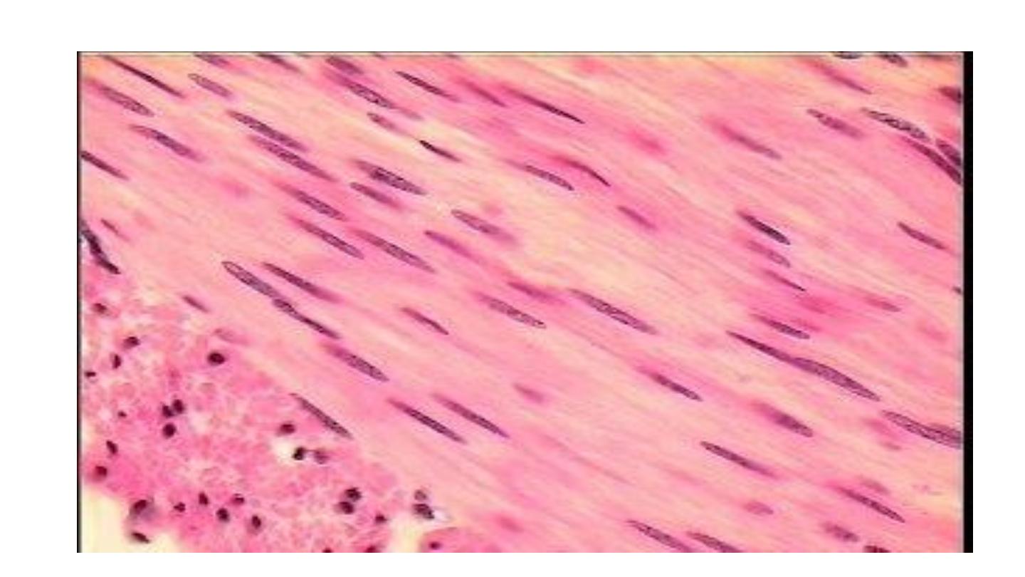
•
The cells of smooth muscle are relatively small with
only a single nucleus. The fibers are bound together
in irregular branching fasciculi, the arrangement
varying considerably from one organ to another
according to functional requirements.

•
smooth muscle fibers are elongated, spindle-shaped cells with
tapered ends which may occasionally be bifurcated. Smooth
muscle fibers are generally much shorter than skeletal muscle
fibers and contain only one nucleus which is elongated and
centrally located in the cytoplasm at the widest part of the cell;
however, depending on the contractile state of the fibers at
fixation, the nuclei may sometimes appear to be spiral-shaped.

•
Smooth muscle fibers are bound together in irregular,
branching fasciculi and these fasciculi, rather than individual
fibers, are the functional contractile units.
•
Within the fasciculi, individual muscle fibers are arranged
roughly parallel to one another with the thickest part of one
cell lying against the thin parts of adjacent cells.
•
The contractile proteins of smooth muscle are not arranged
in myofibrils as in skeletal and cardiac muscle, and thus
visceral muscle cells are not striated.
CARDIAC MUSCLE
•
Cardiac muscle has many structural and functional
characteristics intermediate between those of skeletal and
smooth muscle and provides for the continuous, rhythmic
contractility of the heart. Although striated in appearance,
cardiac muscle is readily distinguishable from skeletal
muscle and should not be referred to by the term 'striated
muscle'

•
Cardiac muscle or myocardium exhibits many
structural and functional characteristics intermediate
between those of skeletal and visceral muscle.
•
Like the former, its contractions are strong and utilize a
great deal of energy, and like the latter the contractions
are continuous and initiated by inherent mechanisms,
although they are modulated by external autonomic and
hormonal stimuli.

•
Cardiac muscle fibers are essentially long cylindrical cells
with one or at most two nuclei, centrally located within the cell.
The ends of the fibers are split longitudinally into a small
number of branches, the ends of which abut onto similar
branches of adjacent cells giving the impression of a
continuous three-dimensional cytoplasmic network; this was
formerly described as a syncytium before the discrete
intercellular boundaries were recognized

•
Between the muscle fibers, delicate collagenous tissue
analogous to the endomysium of skeletal muscle
supports
the
extremely
rich
capillary
network
necessary to meet the high metabolic demands of
strong continuous activity.
•
Cardiac muscle fibers have an arrangement of
contractile proteins similar to that of skeletal muscle
and are consequently striated in a similar manner.
However, this is often difficult to see with light
microscopy due to the irregular branching shape of the
cells and their myofibrils

•
Cardiac muscle fibers also have a system of T tubules
and sarcoplasmic reticulum analogous to that of
skeletal muscles. In the case of cardiac muscle,
however, there is a slow leak of calcium ions into the
cytoplasm from the sarcoplasmic reticulum after
recovery from the preceding contraction; this causes a
succession of automatic contractions independent of
external stimuli. The rate of this inherent rhythm is
then modulated by external autonomic and hormonal
stimuli.

•
Between the ends of adjacent cardiac muscle cells are
specialized intercellular junctions, called intercalated
discs, which not only provide points of anchorage for
the myofibrils but also permit extremely rapid spread of
contractile stimuli from one cell to another.

•
Thus, adjacent fibers are triggered to contract almost
simultaneously, thereby acting as a functional syncytium. In
addition, a system of highly modified cardiac muscle cells
constitutes the pacemaker regions of the heart and ramifies
throughout the organ as the Purkinje system, thus
coordinating contraction of the myocardium as a whole in
each cardiac cycle.
•
Cardiac muscle cells in certain locations in the heart are
responsible for secreting hormones into the bloodstream.