Anthrax
Anthrax is caused by Bacillus anthracis.The spores of these Gram-positive bacilli are extremely hardy and withstand extremes of temperature and humidity.
The organism is capable of toxin production and this property correlates most closely with its virulence.
The disease occurs world-wide.
Epidemics have been reported in The Gambia, in both North and South America and in southern Europe.
Transmission is through direct contact with an infected animal;
infection is most frequently seen in farmers, butchers, and dealers in wool and animal hides.
Spores can also be ingested or inhaled. There have been recent cases in the USA due to the deliberate release of anthrax spores as a bioterrorist weapon
Clinical features
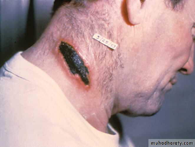
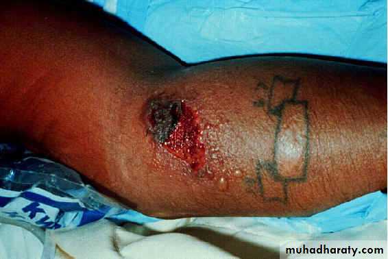
The incubation period is 1-10 days.Cutaneous anthrax is the most common. The small, erythematous, maculopapular lesion is initially painless. It may subsequently vesiculate and ulcerate, with formation of a central black eschar.
The illness is self-limiting in the majority of patients, but occasionally perivesicular oedema and regional lymphadenopathy may be marked, and toxaemia can occur
Inhalational anthrax (woolsorter's disease) follows inhalation of spores, and bioterrorism should be suspected.
A febrile illness is accompanied by non-productive cough and retrosternal discomfort; pleural effusions are common.
Gastrointestinal anthrax is due to consumption of undercooked, contaminated meat.
It presents as severe gastroenteritis; haematemesis and bloody diarrhoea can occur. Toxaemia, shock and death may follow.
Diagnosis
The diagnosis is established by:demonstrating the organism in smears from cutaneous lesions or
by culture of blood and other body fluids.
Serological confirmation can be made using ELISAs detecting antibodies to both the organism and a toxin.
Management
Ciprofloxacin is considered the best treatment.In mild cutaneous infections, oral therapy for 2 weeks is adequate but therapy for 60 days was used in the outbreaks.
In more severe infections high doses of intravenous antibiotics are needed, along with appropriate supportive care.
Control
Any infected animal that dies should be burned and the area in which it was housed disinfected.mass vaccination of animals may prevent widespread contamination, but needs to be repeated annually.
A human vaccine is available for those at high risk,
and prophylactic antibiotics may be indicated following exposure.
Some countries are establishing public health policies to deal with the deliberate release of anthrax spores
Q fever
Q (query) fever is a zoonosis caused by the rickettsia-like organism Coxiella burnetii.
Infection is widespread in domestic, farm and other animals, birds and arthropods: spread is mainly by ticks.
Modes of transmission to humans are by dust, aerosol, and unpasteurized milk from infected cows.
The formation of spores means that C. burnetii can survive in extreme environmental conditions for long periods. The infective dose is very small, so that minimal animal contact is required.
Clinical features
Symptoms begin insidiously 2-4 weeks after infection. Fever is accompanied by flu-like symptoms with myalgia and headache.The acute illness usually resolves spontaneously but pneumonia or hepatitis may develop. Occasionally infection can become chronic, with endocarditis, myocarditis, uveitis, osteomyelitis or other focal infections.
C. burnetii is an obligate intracellular organism, and does not grow on standard culture media.
Diagnosis
is made serologically using an immunofluorescent assay. Antibody tests for two different bacterial antigens allow distinction between acute and chronic infection; a nested PCR assay is now available.
Management
doxycycline 200 mg daily (or a quinolone as an alternative) reduces the duration of the acute illness, but it is not known whether this correlates with eradication of the organism.For chronic Q fever, including endocarditis, doxycycline is often combined with rifampicin or clindamycin. Even prolonged courses of treatment may not clear the infection.