
CARDIOVASCULAR SYSTEM
2
By
Dr. Suhair Majeed

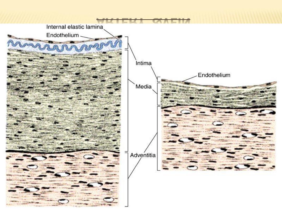
2- BLOOD VESSELS :
blood vessels differ in size, distribution,and
function, structurally they share many common
features. As in the heart, the walls of blood
vessels consist of three major coats or tunics.
From the lumen outward, the wall of a blood
vessel consists of :
1-tunica intima,
2- tunica media, and
3- tunica adventitia

1- TUNICA INTIMA :
The tunica intima corresponds to and is
continuous with the endocardium of the heart.
It consists of an endothelium of flattened
squamous cells resting on a basal lamina and
is supported by a subendothelial connective
tissue.

2- TUNICA MEDIA :
The tunica media is the equivalent of the
myocardium of the heart and is the layer most
variable both in size and structure. Depending on
the function of the vessel, this layer contains
variable amounts of smooth muscle and elastic
tissue.

3- TUNICA ADVENTITIA :
The tunica adventitia also varies in thickness in
different parts of the vascular circuit. It consists
mainly of collagenous connective tissue and
corresponds to the epicardium of the heart, but
it lacks mesothelial cells.
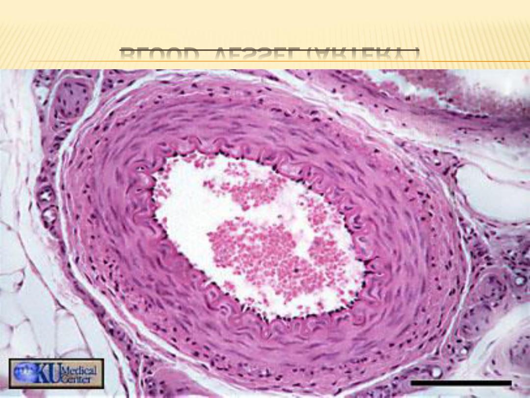
BLOOD VESSEL (ARTERY )

TYPES OF ARTERIES :
three classes of arteries can be distinguished:
large elastic or conducting arteries,
medium-sized muscular or distributing arteries,
and small arteries and arterioles.
A characteristic feature of the entire arterial
side of the blood vasculature system is the
prominence of smooth muscle in the tunica
media.

CONT.
Arteries that leave the heart to distribute
the oxygenated blood exhibit progressive
branching.
With each branching, the luminal diameters
of the arteries gradually decrease, until the
smallest vessel, the capillary, is formed.
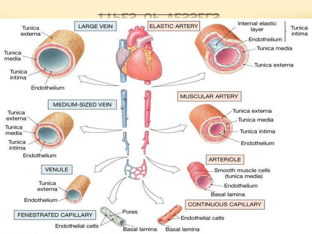
TYPES OF VESSELS
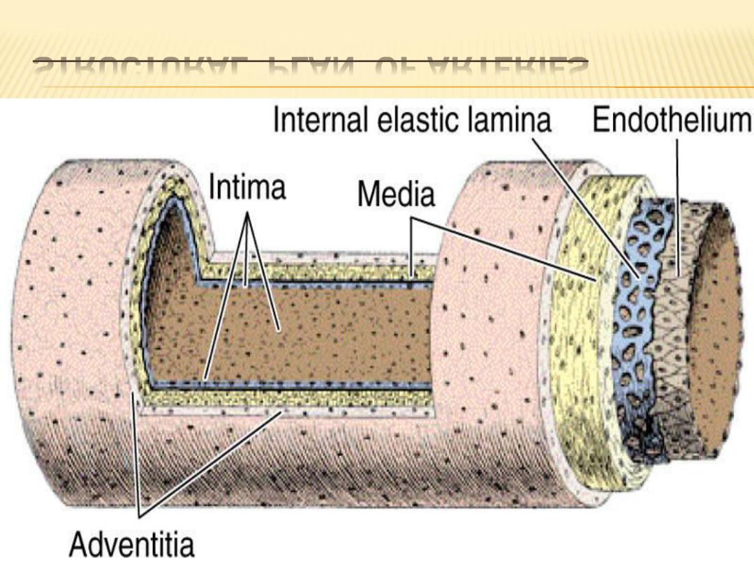
STRUCTURAL PLAN OF ARTERIES
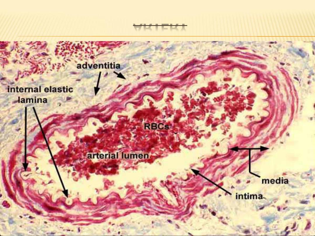
ARTERY

1- ELASTIC ARTERIES :
Elastic arteries are the largest blood vessels in
the body and include the pulmonary trunk and
aorta with their major branches, the
brachiocephalic, common carotid, subclavian,
vertebral, pulmonary, and common iliac arteries.
The walls of these vessels are primarily
composed of elastic connective tissue fibers.
These fibers provide great resilience and flexibility
during blood flow.

CONT.
The tunica intima is relatively thick and is
lined by a single layer of flattened, polygonal
endothelial cells that rest on a complete basal
lamina, about one-fourth of the total thickness of
the intima is formed by the subendothelial layer,
a layer of loose connective tissue that contains
elastic fibers and a few smooth muscle cells

CONT.
The tunica media is the thickest layer and
consists largely of elastic tissue. Smooth muscle
cells are the only cells present in the media of
elastic arteries and synthesize and maintain the
elastic fibers and collagen.
The tunica adventitia is relatively thin and
contains bundles of collagen fibers (type 1) and a
few elastic fibers,
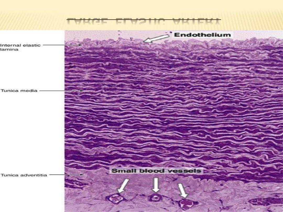
LARGE ELASTIC ARTERY
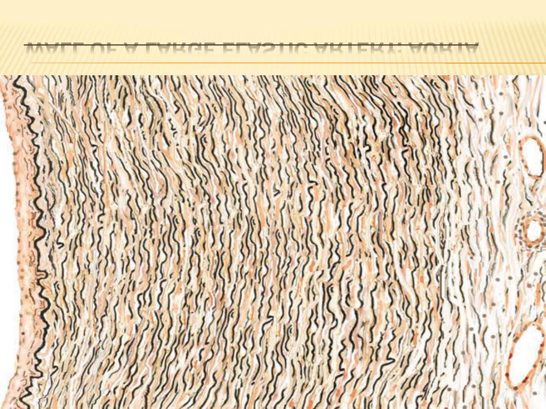
WALL OF A LARGE ELASTIC ARTERY: AORTA

2- MUSCULAR ARTERIES:
The large elastic arteries branch and become
medium-sized muscular arteries, the most numerous
vessels in the body. In contrast to the walls of elastic
arteries, those of muscular arteries contain greater
amounts of smooth muscle fibers. The tunica intima
consists of an endothelium, a subendothelial layer,
and an internal elastic lamina.
The endothelium and subendothelial layers are
similar to those of elastic arteries, but as the size of
the vessel decreases, the subendothelial layer
becomes thinner.
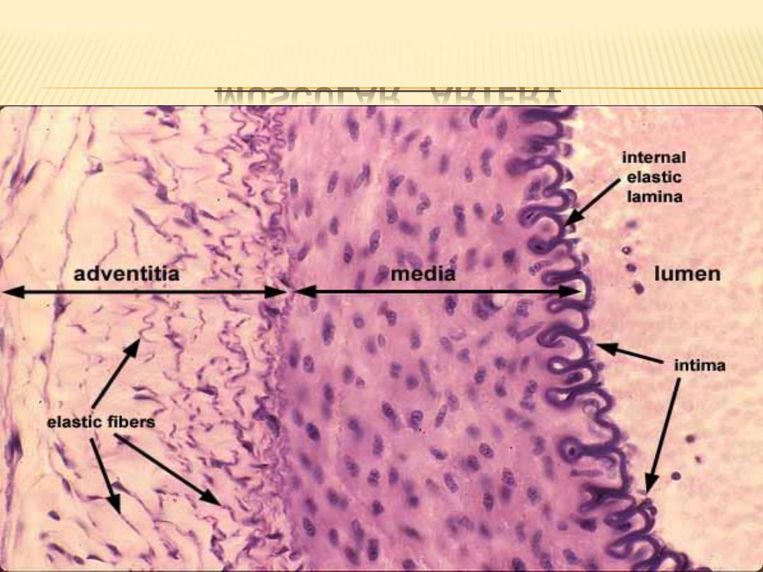
MUSCULAR ARTERY

CONT.
The tunica media is the thickest coat and
consists mainly of smooth muscle cells arranged
in concentric, helical layers. The number of
layers varies from 3 to 4 in smaller arteries to 10
to 40 in the large muscular arteries.
The tunica adventitia is prominent in
muscular arteries and in some vessels may be as
thick as the media. It consists of collagen and
elastic fibers that are longitudinal in orientation

CONT.
The walls of some muscular arteries also
exhibit two thin, wavy bands of elastic fibers.
The internal elastic lamina is located
between the tunica intima and the tunica
media; this layer is not seen in smaller arteries.
The external elastic lamina is located on
the periphery of the muscular tunica media
and is primarily seen in large muscular arteries.
ELASTIC & MUSCULAR ARTERY

FUNCTIONAL CORRELATIONS :
The elastic arteries transport blood from the
heart and move it along the systemic vascular
path. The presence of an increased number of
elastic fibers in their walls allows the elastic
arteries to greatly expand in diameter during
systole (heart contraction), when a large volume
of blood is forcefully ejected from the ventricles
into their lumina

CONT.
In contrast, the muscular arteries control blood
flow and blood pressure through vasoconstriction
or vasodilation of their lumina. Vasoconstriction
and vasodilation, owing to a high proportion of
smooth muscle fibers in the artery walls, are
controlled by unmyelinated axons of the
sympathetic division of the autonomic nervous
system. Similarly, by autonomic constriction or
dilation of their lumina, the smooth muscle fibers
in smaller muscular arteries or arterioles regulate
blood flow into the capillary beds .

CONT.
. During diastole (heart relaxation), the
expanded elastic walls recoil upon the volume
of blood in their lumina and force the blood to
move forward through the vascular channels.
As a result, a less variable systemic
blood pressure is maintained, and blood flows
more evenly through the body during heart
beats.

3- ARTERIOLES :
Arterioles are the smallest branches of the
arterial system. Their walls consist of one to five
layers of smooth muscle fibers. tunica intima
consists only of endothelium and a fenestrated
internal elastic lamina. Tunica adventitia also
decreases in thickness, becoming extremely
thin in the smallest arterioles.
Arterioles deliver blood to the smallest blood
vessels, the capillaries. Capillaries connect
arterioles with the smallest veins or venules.
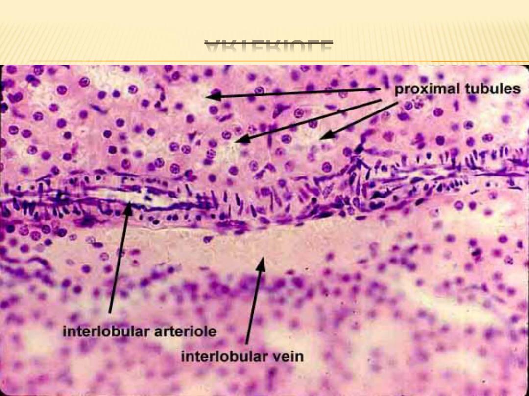
ARTERIOLE
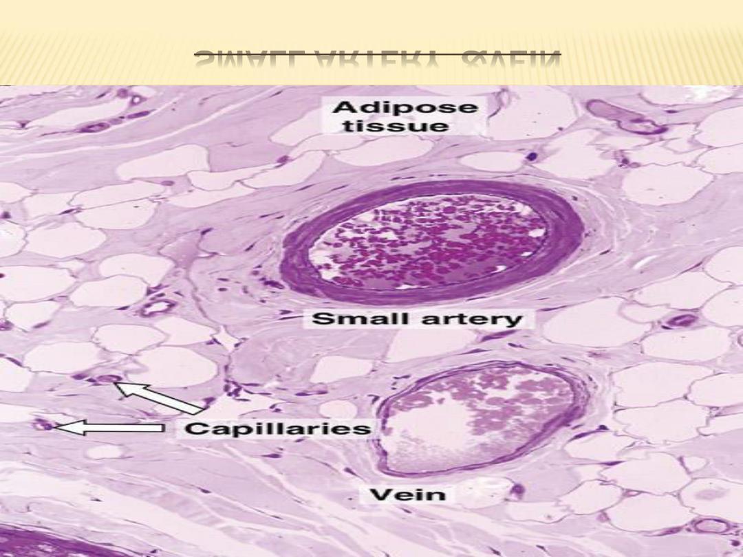
SMALL ARTERY &VEIN

METARTERIOLES
Metarterioles are intermediate between
capillaries and arterioles and regulate the flow of
blood through capillary beds. They also are called
capillary sphincter areas or precapillary arterioles
,their lumina generally are wider than those of the
capillaries .

STRUCTURAL PLAN OF VEINS :
Capillaries unite to form larger blood vessels
called venules; venules usually accompany
arterioles.
The venous blood initially flows into smaller
postcapillary venules and then into veins of
increasing size. The veins are classified as small,
medium, and large.
Compared with arteries, veins typically are
more numerous and have thinner walls, larger
diameters, and greater structural variation.
Small-sized and medium-sized veins,
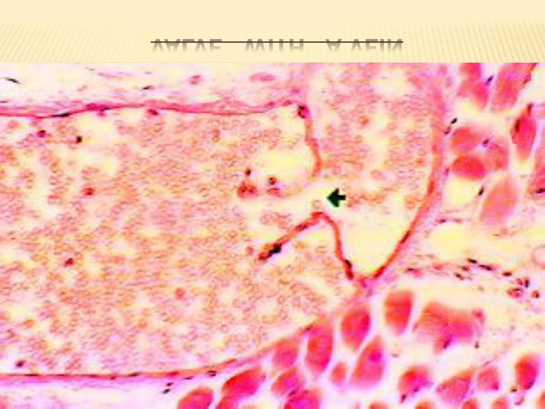
particularly in the extremities, have valves.

CONT.
The presence of valves in veins assists
venous blood flow by preventing back flow.
When blood flows toward the heart, pressure
in the veins forces the valves to open. As the
blood begins to flow backward, the valve flaps
close the lumen and prevent back flow of blood.
Valves are absent in veins of the central
nervous system, the inferior and superior venae
cavae, and viscera.

CONT.
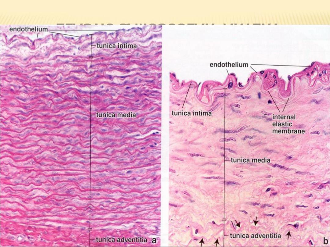
The walls of the veins, like the arteries, also
exhibit three layers or tunics. However, the
muscular layer is much less prominent. The tunica
intima in large veins exhibits a prominent
endothelium and subendothelial connective
tissue.
In large veins, the muscular tunica media is
thin, and the smooth muscles intermix with
connective tissue fibers .
WALL OF A LARGE VEIN: PORTAL VEIN
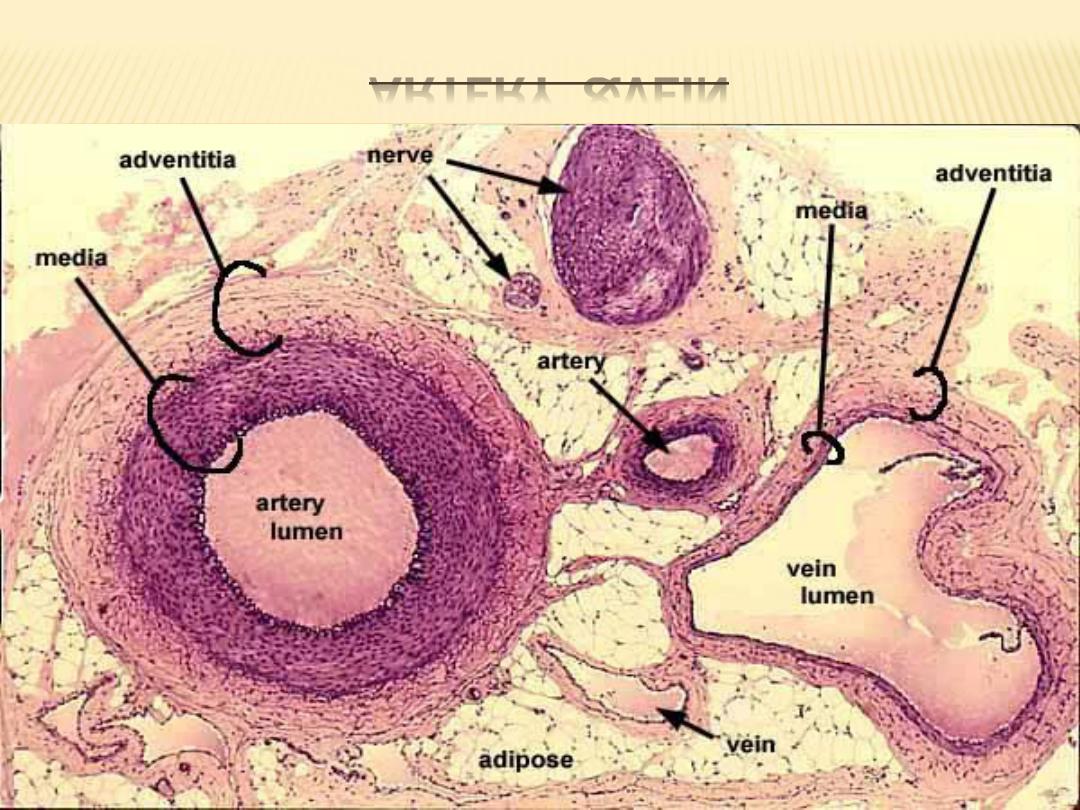
ARTERY &VEIN

CONT.
In large veins, the tunica adventitia is the
thickest and best-developed layer . bundles of
smooth muscle fibers are common in the
connective tissue of this layer. Vasa vasorum
are present and may extend into the Media.
ARTERY &VEIN
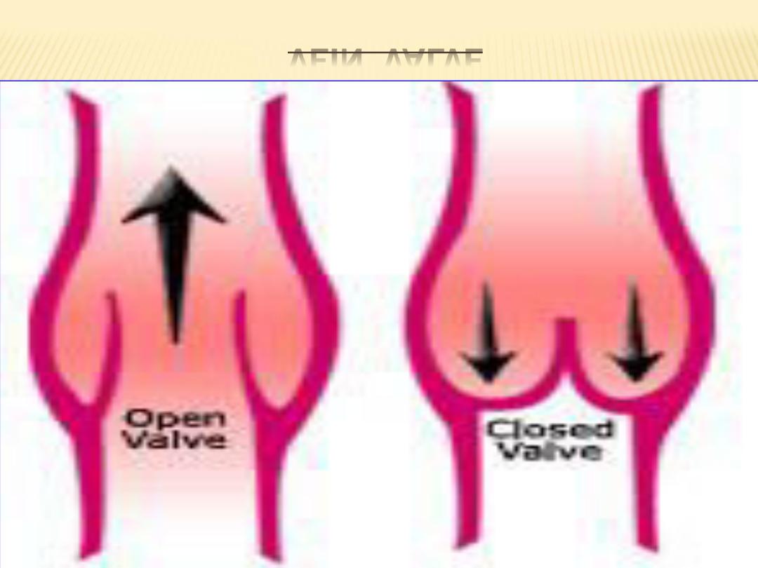
VEIN VALVE
VALVE WITH A VEIN

FUNCTIONAL CORRELATIONS :
In veins, blood pressure is lower than in the
arteries.As a result, venous blood flow is passive.
Venous blood flow in the head and trunk is
primarily owing to negative pressures in the
thorax and abdominal cavities resulting from
respiratory movements. Venous blood return
from the extremities is aided by surrounding
muscle contractions and prevented from flowing
back by numerous valves in the large veins of the
extremities.

MEDIUM VEINS:
The medium size veins, includes most of the
named veins of gross anatomy except for major
trunks. The thin tunica intima consists of
endothelial cells resting on a basal lamina, but a
narrow subendothelial layer may be present. but a
poorly defined internal elastic lamina is formed
only in the larger vessels. In most medium veins,
the tunica media, is thinner than in
corresponding arteries.

CONT.
The thick tunica adventitia forms the bulk of the
wall and is larger than the tunica media. It
consists of collagen and elastic fibers and
contains smooth muscle cells. Vasa vasorum are
present in the larger vessels of this class . Valves
are present in most of the medium size veins .

VENULES :
Venules arise from the union of several
capillaries to form vessels . The junctions
between venules and capillaries are important
sites of fluid exchange between tissues and blood.
The tunica intima consists of a thin,continuous
endothelium. The tunica media is missing in the
smallest venules, and the relatively thin adventitia
contains a few collagen fibers,

VASA VASORUM :
The walls of larger arteries and veins are too
thick to receive nourishment by direct diffusion
from their lumina. As a result, these walls are
supplied by their own small blood vessels called
the vasa vasorum (vessels of the vessel).
The vasa vasorum allows for exchange of
nutrients and metabolites with cells in the
tunica adventitia and tunica media.
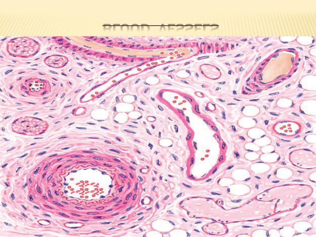
BLOOD VESSELS

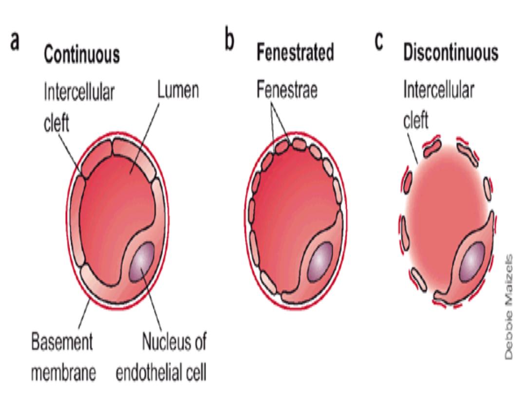
TYPES OF CAPILLARIES :
Capillaries are the smallest blood vessels. Their
size, is about the size of an erythrocyte (red blood
cell). There are three types of capillaries: ---
continuous capillaries,
fenestrated capillaries, and
sinusoids.
These structural variations in capillaries allow for
different types of metabolic exchange between
blood and the surrounding tissues.

CONT.
Regardless of the type, the basic structure
of capillaries is similar and represents an
extreme simplification of the vessel wall. The
tunica intima consists of endothelium and a
basal lamina; the tunica media is absent and
the tunica adventitia is greatly reduced.

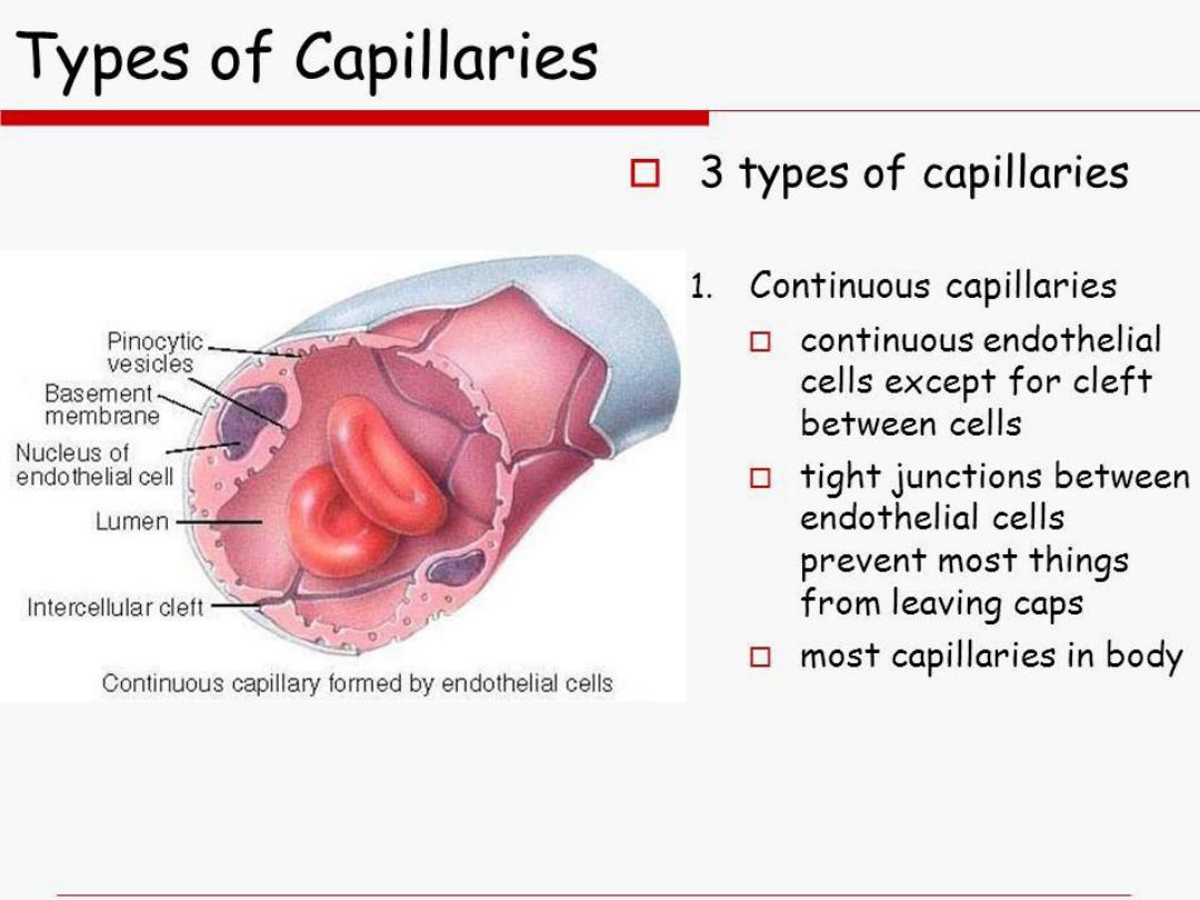
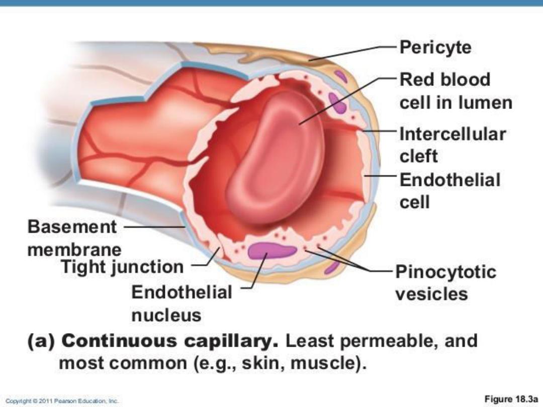
1- CONTINUOUS CAPILLARIES :
Continuous capillaries are the most common.
They are found in muscle, connective tissue,
nervous tissue, skin, respiratory organs, and exocrine
glands. In these capillaries, the endothelial cells are
joined and form an uninterrupted, solid endothelial
lining.
Pericytes are irregular, branched, isolated cells
that occur at intervals along capillaries, enclosed by
the basal lamina of the endothelium. The cells
resemble fibroblasts

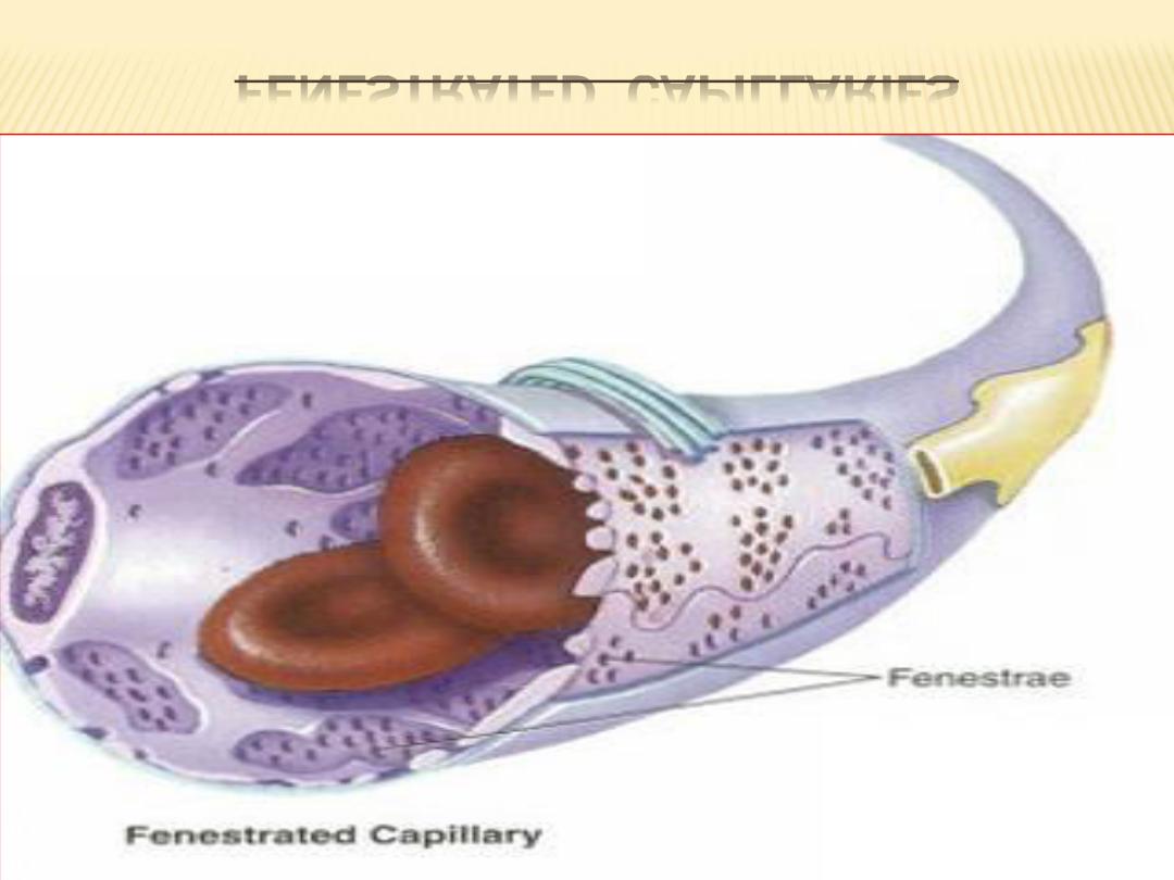
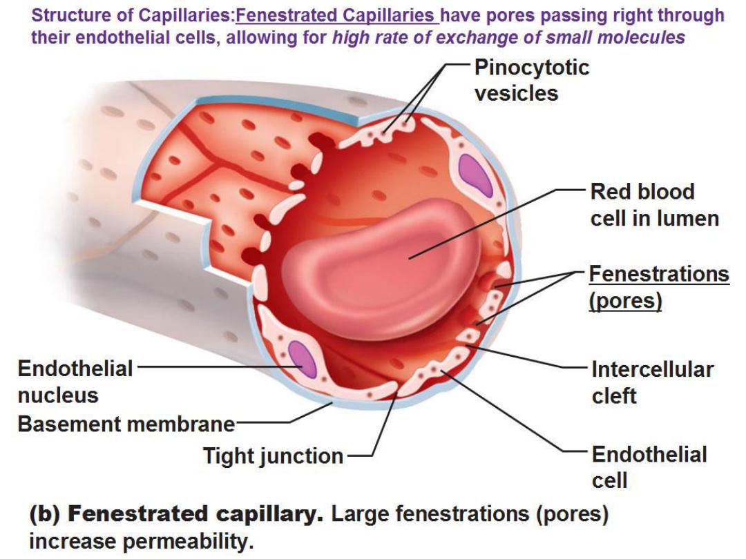
2- FENESTRATED CAPILLARIES :
Fenestrated capillaries are characterized by
fenestrations (pores) in the cytoplasm of
endothelial cells designed for a rapid exchange of
molecules between blood and tissues.
Fenestrated capillaries are found in endocrine
tissues and glands, small intestine, and kidney
glomeruli.
FENESTRATED CAPILLARIES

3- SINUSOIDAL CAPILLARIES :
are blood vessels that exhibit irregular,tortuous
paths . Their much wider diameters slow down the
flow of blood. Also, the cells may be separated by
gaps and rest on a discontinuous basal lamina.
A direct exchange of molecules occurs between
blood contents and cells. Sinusoidal capillaries
are found in the liver, spleen, and bone marrow
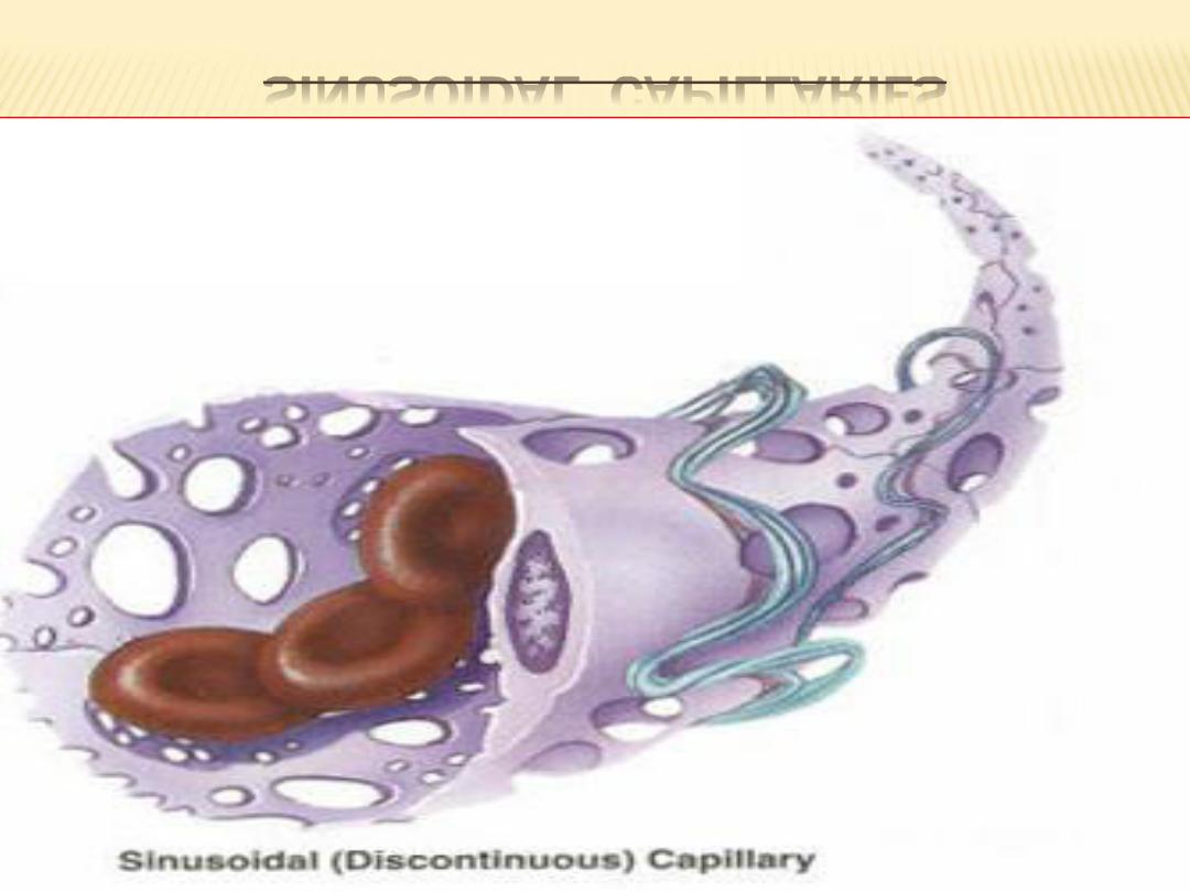
SINUSOIDAL CAPILLARIES


ARTERIOVENOUS ANASTOMOSES :
In addition to their capillary connections,
arteries and veins may unite by shunts
called arteriovenous anastomoses.
Generally these arise from side branches of
arterioles that pass directly to venules. They
are thick-walled, muscular vessels of small
caliber that usually are coiled and
surrounded by a connective tissue sheath.

CONT.
They are plentiful in the plantar and palmar
surfaces, fingertips, toes, lips, and nose and
also occur in the thyroid. When open, the
anastomoses shunt blood around the capillary
bed and thus regulate the blood supply to many
tissues. In the skin they function primarily in the
regulation of body temperature.
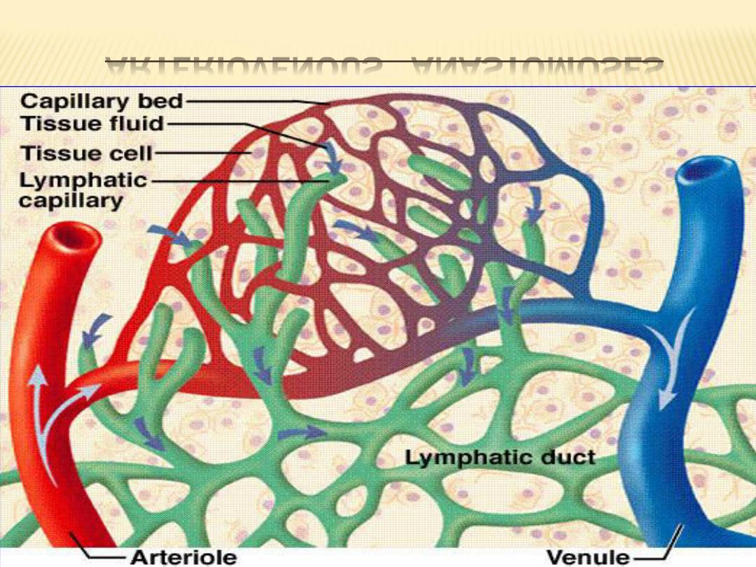
ARTERIOVENOUS ANASTOMOSES

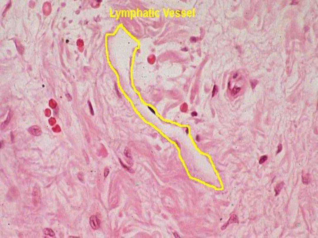
THE LYMPH VASCULAR SYSTEM :
The lymphatic system consists of lymph
capillaries and lymph vessels. This system starts
as blind-ending tubules or lymphatic capillaries in
the connective tissue of different organs.
These vessels collect the excess interstitial
fluid (lymph) from the tissues and return it to the
venous blood via the large lymph vessels, the
thoracic duct and right lymphatic duct.

CONT.
Also, to allow greater permeability, the
endothelium in lymph capillaries and vessels is
extremely thin.
The structure of larger lymph vessels is similar
to that of veins except that their walls are much
thinner. Lymph movement in the lymphatic
vessels is similar to that of blood movement; that
is, the contractions of surrounding skeletal
muscles forces the lymph to move forward.

CONT.
Also, the lymph vessels contain more valves to
prevent a backflow of collected lymph. Lymph
vessels are found in all tissues except the central
nervous system, cartilage, bone and bone
marrow, thymus, placenta,and teeth.
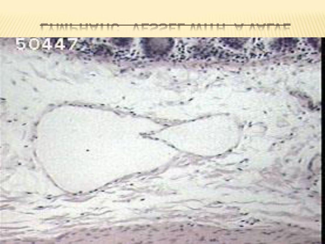
LYMPHATIC VESSEL WITH A VALVE

1- LYMPH CAPILLARIES :
Lymph capillaries are thin-walled, blind tubes
that branch to form a rich network in organs and
tissues. They are wider and more irregular than
blood capillaries.
The wall of a lymph capillary consists only of a
thin continuous endothelium and a discontinuous
basal lamina that is present only in patches or
may even be absent. Externally, the endothelium
is surrounded by a small amount of collagenous
connective tissue.

2- COLLECTING LYMPH VESSELS :
Collecting lymph vessels differ from lymph
capillaries in size and the thickness of their walls.
Although three coats - intima, media, and
adventitia- are described as in blood vessels, they
are not clearly delineated. The tunica intima
consists of an endothelium supported by a thin
network of elastic fibers. tunica media is
composed of smooth muscle cells ,with a few fine
elastic fibers.

CONT.
The tunica adventitia is the thickest coat
and consists of bundles of collagen fibers,
elastic fibers, and some smooth muscle cells.

THANK YOU