Urogenital system
Special embryology

Functionally the urogenital system can be divided into
two entirely different components: the
urinary system
and the
genital system
.
Embryologically and anatomically, however, they are
intimately interwoven. Both develop from a common
mesodermal ridge (
intermediate mesoderm
) along the
posterior wall of the abdominal cavity, and initially the
excretory ducts of both systems enter a common cavity,
the
cloaca
.

Urinary system
Kidney systems
;
Three slightly overlapping kidney systems are
formed in a cranial to caudal sequence during
intrauterine life in humans: the
pronephros
,
mesonephros
, and
metanephros
.
The
first of these systems is rudimentary and
nonfunctional; the second may function for a
short time during the early fetal period; the
third forms the permanent kidney.

1-pronephros
O
At the beginning of the fourth week, the
pronephros is represented by 7 to 10 solid
cell groups in the cervical region. These
groups form vestigial excretory units,
nephrotomes, that regress before more
caudal ones are formed. By the end of the
fourth week all indications of the pronephric
system have disappeared.

2-mesonephros
O
Mesonephros and mesonephric ducts are derived from
intermediate mesoderm from upper thoracic to upper
lumbar (L3) segments.
O
Early in the fourth week of development, during
regression of the pronephric system
, the first excretory
tubules of the mesonephros appear. They lengthen
rapidly, form an S-shaped loop, and acquire a tuft of
capillaries that will form a
glomerulus
at their medial
extremity
O
Around the glomerulus the tubules form
Bowman’s
capsule
,
O
and together these structures constitute a
renal
corpuscle
.
O
Laterally the tubule enters the longitudinal collecting
duct known as the mesonephric or
wolffian duct

O
In the middle of the second month the
mesonephros forms a large ovoid organ on each
side of the midline. Since the developing gonad
is on its medial side, the ridge formed by both
organs is known as the
urogenital ridge
O
While caudal tubules are still differentiating,
cranial tubules and glomeruli show degenerative
changes, and by the end of the second month
the majority have disappeared. In the male a few
of the caudal tubules and the mesonephric duct
persist and participate in formation of the
genital system, but they disappear in the female.
O

Metanephros:
the defenitive kidney
O
It appears in
the fifth week. Its excretory units develop
from metanephric mesoderm in the same manner as in
the mesonephric system. The development of the duct
system differs from that of the other kidney systems.
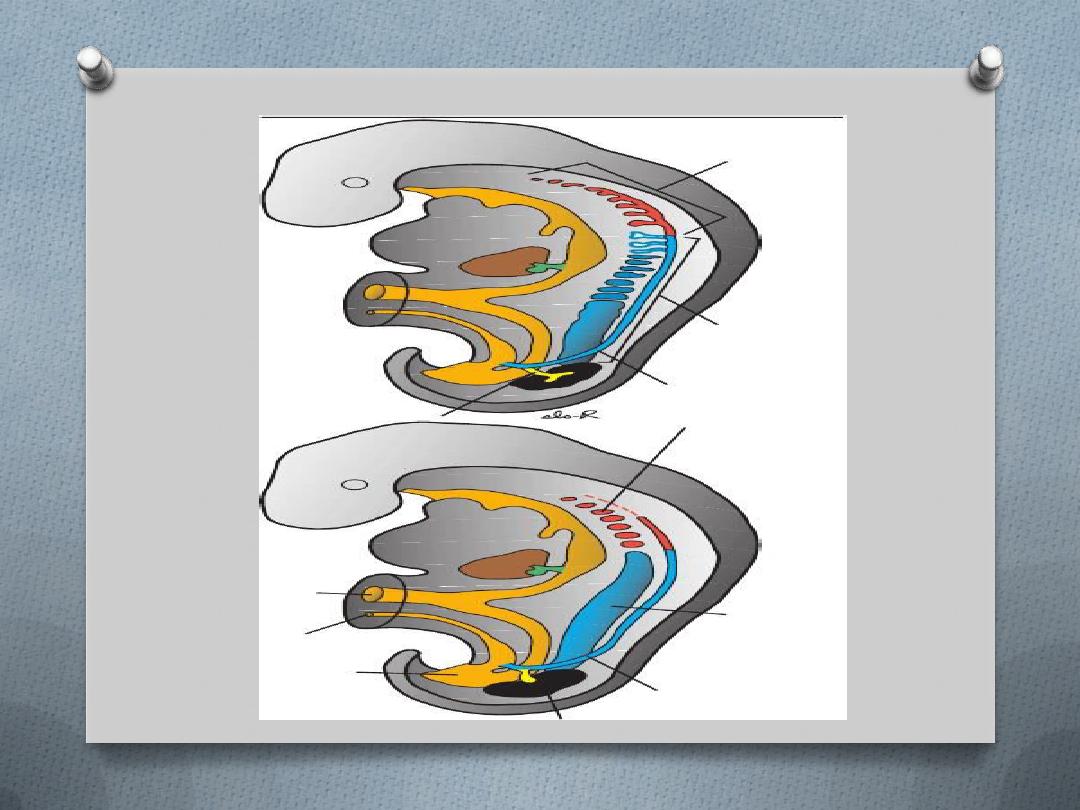
Collecting System
.
O
Collecting ducts of the permanent kidney develop from
the
ureteric bud
, an outgrowth of the mesonephric duct
close to its entrance to the cloaca. The bud penetrates the
metanephric tissue, which is molded over its distal end as
a cap.
O
Subsequently the bud dilates, forming
the primitive renal
pelvis
, and splits into cranial and caudal portions, the
future
major calyces

O
Each calyx forms two new buds while penetrating
the metanephric tissue.These buds continue to
subdivide until 12 or more generations of tubules
have formed.
O
Meanwhile, at the periphery more tubules form
until the end of the 5
th
month, the tubules of the
2
nd
order enlarge and absorb those of the 3
rd
& 4
th
generations, forming the
minor calyces
.
O
During further development, collecting tubules of
5
th
& successive generations elongate & converge
on the minor calyx, forming the
renal pyramid
.
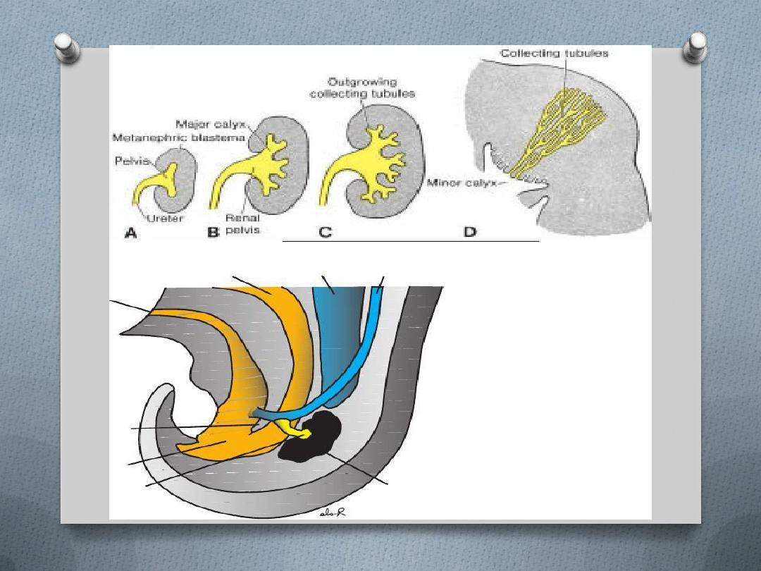

O
The ureteric bud gives rise to the ureter, the
renal pelvis, major & minor calyces, and
approximately 1 to 3 million collecting tubules.
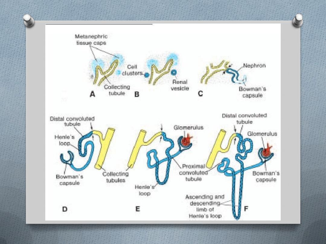
Excretory system:
O
Each newly formed collecting tubule is
covered at its distal end by a
metanephric tissue cap. Under the
inductive influence of the tubule, cells of
the tissue cap form small vesicles, the
renal vesicles
, whichi n turn give rise to
small S-shaped tubules . Capillaries
growinto the pocket at one end of the S
and differentiate into
glomeruli
.

O
These tubules, together with their glomeruli, form
nephrons
, Or
excretory units.
O
The proximal end of each nephron forms
Bowman’s capsule
, form by the which is deeply
indented by a glomerulus
O
The distal end forms an open connection with
one of the collecting tubules, establishing a
passageway
from Bowman’s capsule to the
collecting unit. Continuous lengthening of the
excretory tubule results in formation of the
proximal convoluted tubule, loop of Henle, and
distal convoluted tubule

O
Hence, the kidney develops from two sources:
O
(1) metanephric mesoderm, which provides
excretory units; and
O
(2) the ureteric bud, which gives rise to the
collecting system.
O
Nephrons are formed until birth, at which time
there are approximately1 million in each kidney.
Urine production begins early in gestation, soon
after differentiation of the glomerular capillaries,
which start to form by 10th week.
O
At birth the kidneys have a lobulated
appearance, but the lobulation disappears
during infancy as a result of further growth of
the nephrons, although there is no increase in
their number.