
Part 2--- special embryology
skeletal system
• ^ skeletal system develops from paraxial & lateral plate
(parietal layer) mesoderm,& neural crest.
• ^ paraxial mesoderm forms a segmented series of tissue
blocks on each side of neural tube, known as
somitomeres
in
^ head region, &
somites
from occipital region & caudally.
• Somites differentiate into a ventromedial, ^
sclerotome,
& a
dorsolateral part, ^
dermomyotome.
• At ^ end of 4
th
week, sclerotome cells become polymorphous
& loosely organized tissue , ^ mesenchyme which may be
either: fibroblasts, chondroblasts, or osteoblasts( bone-
forming cells).
1-skull
^ skull is divided into 2 parts:
1- neurocranium, which forms a protective case around ^ brain.
2- viscerocranium which forms ^ skeleton of ^ face.
NEUROCRANIUM
Divided into 2 portions: (1)-^ membranous part, consists of flat
bones, which surround ^ brain as a vault. & (2)- ^ cartilaginous
part or chondrocranium, which forms bones of base of ^ skull.
Membranous neurocranium:
derived from neural crest cells
& paraxial mesoderm. Mesenchyme from these two sources
invests ^ brain & undergoes membranous ossification.

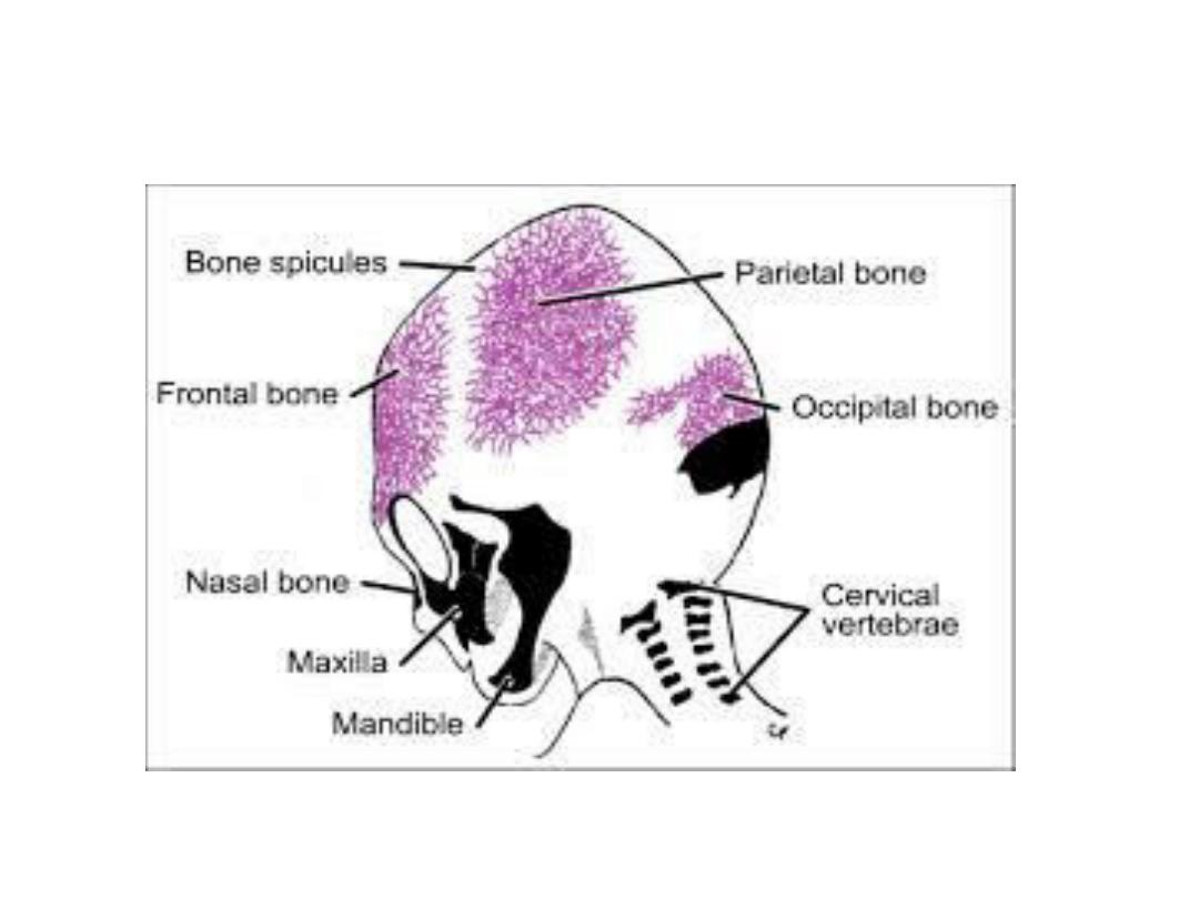
• ^ result is formation of no. of flat bones characterized by ^
presence of needle like bone specules. These specules
progressively radiate from primary ossification centers toward
^ periphery.
• With further growth during fetal & postnatal life,
membranous bones enlarge by apposition of new layers on ^
outer surface & by simultaneous osteoclastic resorption from
^ inside.
• At birth, ^ flat bones of skull are separated from each other by
a narrow seems of connective tissue, ^ sutures.
• Sutures are also derived from 2 sources: neural crest cells
(sagittal sutures) & paraxial mesoderm (coronal sutures).
• At points where more than 2 bones meet, sutures are wide&
are called fontanelles.
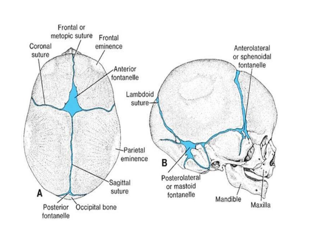

• ^ most prominent of these is ^ anterior fontanelle, which is
found where ^ 2 parietal & 2 frontal bones meet.
• Fontanelles allow ^ bones of ^ skull to overlap(molding)
during birth.
• Several sutures & fontanelles remain membranous for a
considerable time after birth. The bones of ^ vault continue to
grow after birth.
• ^ bones of ^ vault continue to grow after birth, mainly
because ^ brain grows. Although a 5- to 7- year-old child has
nearly all of his or her cranial capacity, some sutures remain
open until adulthood.
• In most cases, ^ ant. fontanelle closes by 18 months of age, &
^ posterior fontanelle closes by 1-2 months of age.

Cartilaginous Neurocranium
• It consists of a no. of separate cartilages. Those that lie in
front of ^ rostral limit of ^ notochord, which ends at ^ level of
^ pituitary gland in ^ center of ^ sella turcica, are derived from
neural crest cells. They form ^ prechordal chondrocranium.
• Those that lie posterior to this limit arise from occipital
sclerotomes formed by paraxial mesoderm & form ^ chordal
chondrocranium.
• The base of skull is formed when these cartilages fuse & ossify
by endochondral ossification.
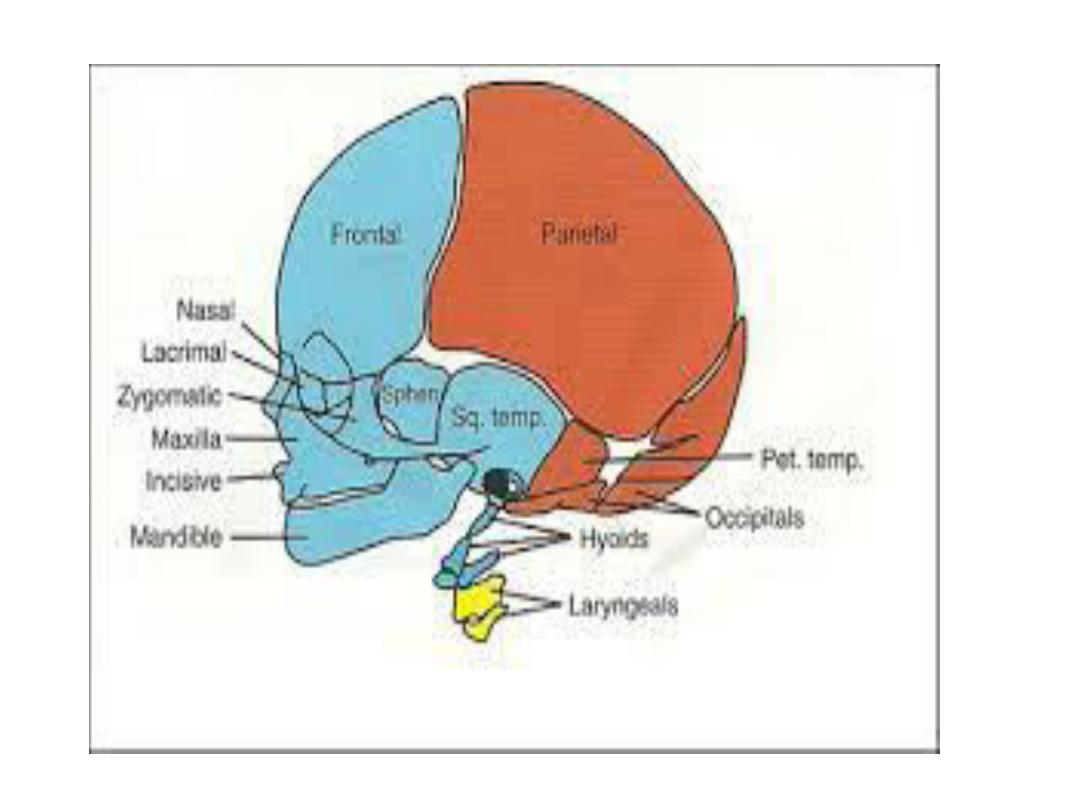
viscerocranium
• It consists of ^ bones of ^ face, is formed mainly from ^ 1
st
2
pharyngeal arches.
• ^ 1
st
arch gives rise to dorsal portion, ^ maxillary process,
which extends forward beneath ^ region of eye & gives rise to
^ maxilla, ^ zygomatic bone, & part of ^ temporal bone.
• ^ ventral portion, ^ mandibular process, contains ^ meckel
cartilage.
• Mesenchyme around ^ meckel cartilage condenses & ossifies
by membranous ossification to give rise to ^ mandible.
• ^ meckel cartilage disappears except in ^ sphenomandibular
ligament.

• ^ dorsal tip of ^ mandibular process, along with that of ^ 2
nd
pharyngeal arch, later gives rise to ^ incus, malleus & stapes.
ossification of these 3 ossicles begins in ^ 4
th
month making
these ^ 1
st
bones to become fully ossified.
• At first, ^ face is small in comparison to neurocranium. This
appearance is caused by:
• 1-virtual absence of ^ paranasal sinuses.
• 2-^ small size of bones, particularly ^ jaws.
• With ^ development of air sinuses, & appearance of teeth, ^
face loses its babyish characteristics.

LIMBS
• AT ^ end of 4
th
week of development, limb buds become
visible as outpocketings from ^ ventrolateral body wall.
• ^ forelimb appears 1
st
followed by hindlimb 1-2 days later.
• Initially, ^ limb buds consists of a mesenchymal core derived
from ^ parietal layer of lateral mesoderm that will form ^
bones & connective tissues of ^ limb, covered by a layer of
cuboidal ectoderm.
• Ectoderm at ^ distal part of ^ limb thickens & forms ^ apical
ectodermal ridge (AER).
• ^ Ridge exerts an inductive influence on adjacent
mesenchyme causing it to remain as a population of
undifferentiated, rapidly proliferating cells,^ progress zone.

• As ^ limb grows, cells farther from ^ influence of ^ AER begin
to differentiate into cartilage & muscle. Thus ^ development
of ^ limb proceeds proximodistally.
• In 6-week-old embryos, ^ terminal portion of ^ limb buds
become flattened to form ^ hand-& footplates & is separated
from ^ proximal segment by a circular constriction.
• Later, a 2
nd
constriction divides ^ proximal portion into 2
segments, & ^ main parts of ^ extremities can be recognized.
• Fingers & toes are formed when cell death in ^ AER separates
this ridge into 5 parts.
• Further formation of ^ digits depends on their continued
outgrowth under ^ influence of ^ 5 segments of ridge
ectoderm, condensation of ^ mesenchyme to form
cartilaginous digital rays, & ^ death of intervening tissue
between rays.

• Development of ^ upper & lower limbs is similar except that
morphogenesis of ^ lower limb is approximately 1-2 days
behind that of ^ upper limb.
• Also during ^ 7
th
week of gestation, ^ limbs rotate in apposite
directions.
• ^ upper limb rotates 90 degree laterally, so that ^ extensor
muscles lie on ^ lat. & post. surface & thumbs lie laterally,
whereas ^ lower limb rotates approximately 90 d medially,
placing ^ extensor muscles on ^ ant. surface & ^ big toe
medially.
• While ^ external shape is established, mesenchyme in ^ buds
begins to condense, & these cells differentiate into
chondrocytes.

• By ^ 6
th
week of development, ^ 1
st
hyaline cartilage models,
foreshadowing ^bones of ^ extremities, are formed by these
chondrocytes.
• Ossification of ^ bones of ^ extremities, endochondral
ossification, begins by ^ end of embryonic period.
• Primary ossification centers, are present in all long bones of
^limbs by ^ 12
th
week of development.
• From ^ primary center in ^ shaft or diaphysis of ^ bone,
endochondral ossification gradually progresses toward ^ ends
of ^ cartilaginous model.
• At birth, ^ diaphysis of ^ bone is usually completely ossified,
but ^ 2 ends, ^ epiphyses, are still cartilaginous, shortly
thereafter, ossification centers arise in ^ epiphyses.

Vertebrate & ^ Vertebral Column
• Vertebrate form from ^ sclerotome portion of somites.
• A typical vertebra consists of a vertebral arch & foramen
(through which ^ spinal cord passes), a body, transverse
processes, & spinous process.
• During ^ 4
th
week , sclerotome cells migrate around ^ spinal
cord & notochord to merge with cells from ^ opposite somite
on ^ other side of ^ neural tube.
• As development continues, ^ sclerotome portion of each
somite undergoes a process called resegmentation.
• Resegmentation occurs when ^ caudal half of each scletome
grows into & fuses with ^ cephalic subjacent sclerotome.

• Thus, each vertebra is formed from ^ combination of ^ caudal
half of one somite cranial half of its neighbor.
• Mesenchymal cells between cephalic & caudal parts of ^
original sclerotome segment, do not proliferate but fill ^ space
between 2 precartilaginous vertebral bodies, lead to
formation of intervertebral disc.
• Although ^ notochord regress in ^ region of vertebral bodies,
it persists & enlarges in ^ region of intervertebral disc. Here it
contributes to ^ nucleus pulposus, which is later surrounded
by circular fibers of ^ annulus fibrosus.
• Combined, these 2 structures form ^ intervertebral disc.

• Resegmentation of sclerotomes into definitive vertebrae
causes ^ myotomes to bridge ^ intervertebral discs, & this
alteration gives them ^ capacity to move ^ spine.
• For the same reason, intersegmental arteries, at first lying
between ^ sclerotomes, now pass midway over ^ vertebral
bodies.
• Spinal nerves, however, come to lie near ^ disc & leave ^
vertebral column through ^ intervertebral foramina.
• As ^ vertebrae form, 2 primary curves established: thoracic &
sacral curvatures.
• later, 2 secondary curves established: cervical & lumbar
curvatures.

Ribs & Sternum
• ^ Bony portion of each rib is derived from sclerotome cells
that remain in paraxial mesoderm.
• Costal cartilages are formed by sclerotome cells that migrate
across ^ lateral somitic frontier into ^ adjacent lateral plate
mesoderm.
• Sternum is formed independently in parietal layer of lateral
plate mesoderm in ventral body wall as 2 sternal bands , &
these later fuse to form cartilaginous models of ^ manubrium,
sternebrae, & xiphoid process.