Cardiovascular system

Establishment of cardiogenic field
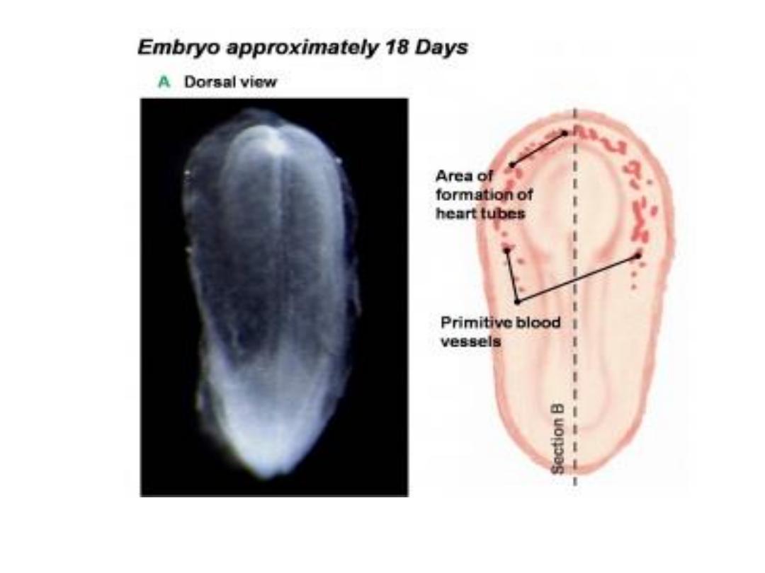
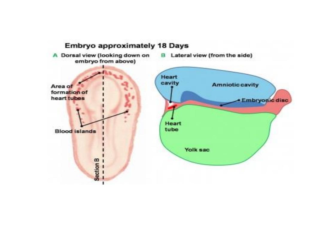
• ^ vascular system appears in ^ middle of ^ 3
rd
week,
when ^ embryo is no longer able to satisfy its
nutritional requirements by diffusion alone.
• Cardiac progenitor cells lie in ^ epiblast immediately
lateral to ^ primitive streak, from there they migrate
through ^ streak .
• Cells destined to form cranial segments of ^ heart,
^
outflow tract,
migrate first, & cells forming ^ caudal
portions,
Rt. Ventricle, Lt. ventricle, & sinus venosus,
respectively, migrate in sequential order.

• This region is known as
^ cardiogenic field;
^
intraembryonic cavity over it later develops
into
pericardial cavity.
• In addition to ^ cardiogenic region, other
blood islands appear bilaterally, parallel &
close to ^ midline of ^ embryonic shield.
These islands form a pair of longitudinal
vessels,
^ dorsal aorta.

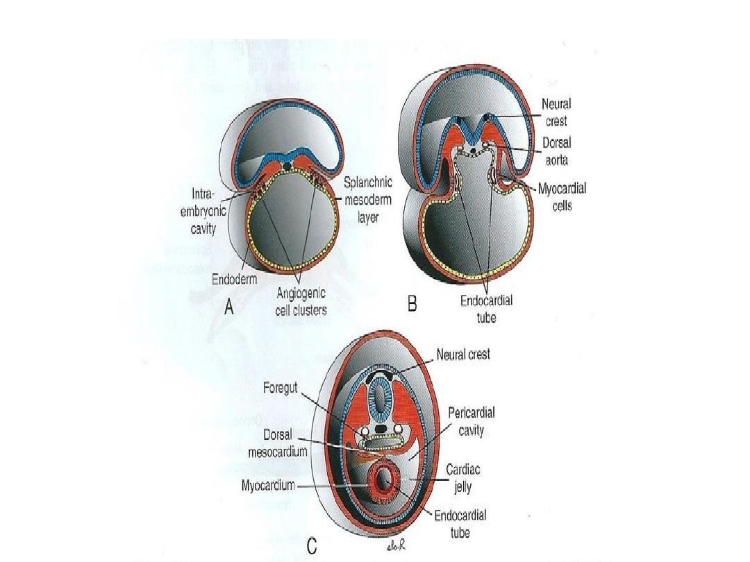
• ^ cells proceed toward ^ cranium & position
themselves rostral to ^ oropharyngeal membrane &
neural folds.
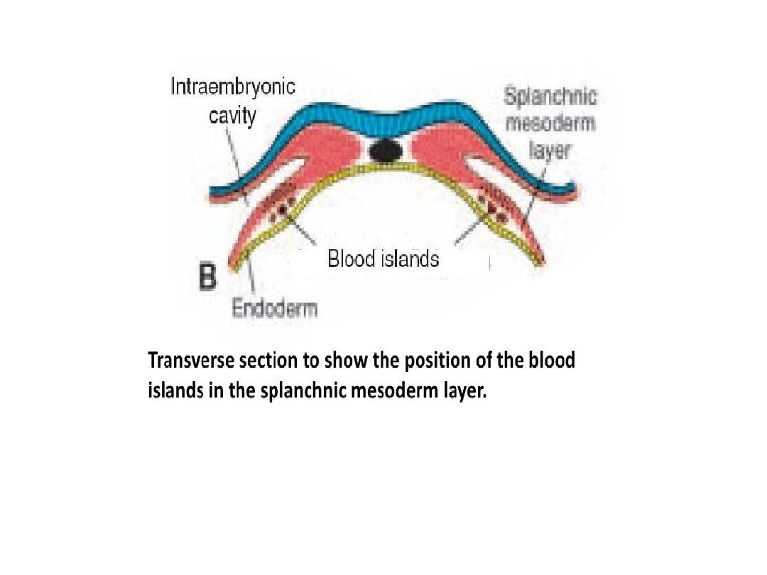
• Here they reside in ^ splanchnic layer of ^ lateral
plate mesoderm .
• At this time (pre-somite stage), they are induced by ^
underlying pharyngeal endoderm to form cardiac
myoblasts.
• Blood islands also appear in this mesoderm, where
they will form blood cells & vessels by process of
vasculogenesis.
• With time, ^islands unite & form a
horseshoe-shaped
endothelial lined tube surrounded by myoblasts.

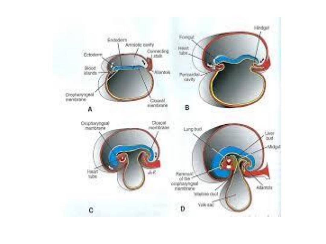
Formation & position of heart tube
• Initially, central portion of ^ cardiogenic area is
anterior to ^ oropharyngeal membrane & ^
neural plate. With closure of ^ neural tube &
formation of ^ brain vesicles, however, ^ CNS
grows cephalad so rapidly that it extends over ^
central cardiogenic area & ^ future pericardial
cavity.
• As a result of growth of ^ brain & cephalic folding
of ^ embryo,
^ oropharyngeal membrane
is
pulled forward, while ^ heart & pericardial cavity
move first to ^ cervical region & finally to ^ thorax

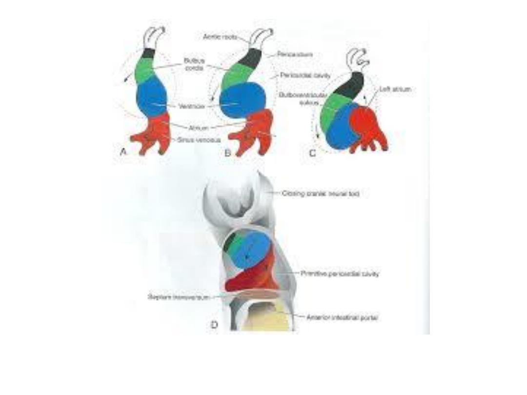
• As ^ embryo folds cephalocaudally, it also
folds laterally. As a result. ^ caudal regions of ^
paired cardiac primordia merge except at their
caudalmost ends.
• Simultaneously, ^crescent part of ^ horseshoe
-shaped area expands to form future outflow
tract & ventricular regions.
• Thus ^ heart becomes a continuous expanded
tube consisting of an inner endothelial lining
& an outer myocardial layer.

• It receives venous drainage at its caudal pole&
begins to pump blood out of ^ 1
st
aortic arch into
^ dorsal aorta & its cranial pole.
• ^ developing heart tube bulges more & more into
pericardial cavity.
• Initially, ^ tube remains attached to ^ dorsal side
of ^ pericardial cavity by a fold of mesodermal
tissue, ^
dorsal mesocardium
. No ventral
mesocardium is ever formed.
• With further development, ^ dorsal mesocardium
disappears, creating ^
transverse pericardial
sinus,
which connects both sides of ^ pericardial
cavity.

• ^ heart is now suspended in ^ cavity by blood
vessels at its cranial & caudal poles.
• During these events, ^ myocardium thickens &
secrets a thick layer of extracellular matrix,
rich in hyaluronic acid that separates it from ^
endothelium.
• In addition, mesothelial cells on ^ surface of ^
septum transversum form ^
proepicardium
near ^ sinus venosus & migrates over ^ heart
to form most of ^
epicardium.
The reminder of
^epicardium is derived from mesothelial cells
originating in ^ outflow tract region.

• Thus ^ heart tube consists of three layers:
(a)^
endocardium,
forming ^internal endothelial
lining of ^ heart.
(b)^ myocardium,
forming ^ muscular wall
(c) ^epicardium
covering ^ outside of ^ tube.
This outer layer is responsible for formation of
^^ coronary arteries, including their endothelial
lining & smooth m.