
1st week of development
Ovulation to implantation

Ovarian cycle
At puberty, the female begins to undergo regular monthly cycle
GnRH (HYPOTHALAMUS) __ Gn (pituitary) FSH & LH __ Estrogen &
progesterone (ovary)
At the beginning 15-20 primary follicles r stimulated to grow under the
influence of FSH, & only one reaches full maturity , during this period
follicular cells when grow they produce estrogen which leads to :
1-^ uterine endometrium enters ^ proliferative phase.
2- thinning of cervical mucus to allow passage of sperm.3- stimulate pit.
Gland to secret LH.
At midcycle, there is an LH surge that:
*stimulates oocytes to complete meiosis I& initiates meiosis II
*Stimulates production of progesterone by follicular cells
*Causes follicular rupture & ovulation.
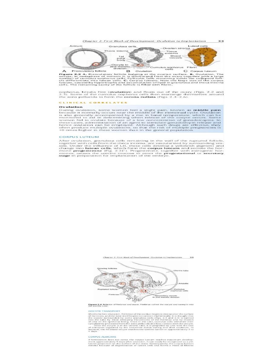
OVULATION
During midcycle, an LH surge leads to digestion of collagen fibers
surrounding ^ follicle, increase in PG level causing muscular contraction in
^ ovarian wall, leading to expulsion of the oocyte with surrounding follicular
cells (cumulus oophorus), then rearrange themselves around zona
pellucida forming (coronaradiata).
Corpus luteum (CL)
After ovulation, the remaining granulosa cells from ^ wall of ^ ruptured
follicle together with cells from theca interna, under influence of LH , these
cells develop a yellowish pigment & change to
lutean cells
, which forms
CL
& secrets estrogens &
progesterone.
In turn these hormons causes
uterine mucosa to enter
secretory stage
in preparation for implantation of ^
embryo.
If fertilization does not occur, CL reaches maximum dev. 9 days after
ovulation, then subsequently, degeneration of lutean cells occur
(luteolysis)& forms a mass of fibrotic scar tissue called
corpus albicans,
progesterne decreases & leads to menstruation.
If fertilization occur, a hormone secreted by ^ syncytiotrophoblast of the
developing embryo called human chorionic gonadotropin ( HCG) which
prevent degeneration of CL & continue to grow & forms CL of pregnancy
(CL graviditatis)& continue to produce progesterne till ^ end of ^ 4th month.
Oocyte transport
Before ovulation, fimbria of ^ uterine tube sweep over ^ surface of ^ ovary,
& ^ tube itself begins to contract rhythmically. The oocyte with ^
surrounding granulosa cells carried into ^ tube by these sweeping
movements of ^ fibriae & ^ motion of ^ cilia on ^ epithelial lining. Once in ^
tube, ^ cumulus cells lose their contact with ^ oocyte.
Fertilization
^ process by which male & female gametes fuse, occurs in ^ ampullary
region of ^ uterine tube.
Only 1% of ^ sperm deposited in ^ vagina enter ^ cervix, movement of
sperm from ^ cervix to ^ uterine tube occurs by muscular contraction of ^
uterus & uterine tube & by their own propulsion ( 2-7hrs), when reaching ^
isthmus, ^ sperm become less motile& cease their migration.
At ovulation
, sperm again become motile, perhaps because of
chemoattractants produced by cumulus cells, & swim to ^ ampula, where
fertilization occur (widest part of ^ tube & close to ^ ovary.
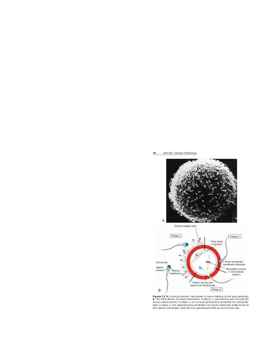
Spermatozoa are not able to fertilize oocyte unless undergo:
1-
capacitation
2- acrosome reaction

capacitation:
a period of conditioning in ^ female genital tract (7hrs) by
which a glycoprotein coat & seminal plasma proteins removed from ^
acrosomal region ( due to interaction bet. ^ sperm & ^mucosa of the uterine
tube).
Acrosomal reaction:
which occur after binding to zona pellucida, induced by
zona proteins, leads to release enzymes (acrosin & trypsin like sub.) which
r needed to penetrate zona pell.
(only capacitated sperms can pass through ^ corona cells & undergo
acrosome reaction).
Phases of fertrilization
Phase1 :
penetration of ^ CR:
200-300 million sperm deposited in female
genital tract, only 300-500 reach ^ site of fertilization. Only one of these
fetilizes ^ egg, only cpacitated sperm pass through CR, ^ other sperms aid
the fertilizing sperm.
Phase2 :
penetration of ^ zona pellucida:
Zona pell. Facilitates &
maintains sperm binding & acrosome reaction by a zona protein . An
enzyme (acrosin) produced by ^ sperb facilitate its penetration ^ zona, &
coming in contact with ^ plasma membrane of ^ oocyte . This contact leads
to release a lysosomal enzymes from the cortical granules lining ^ PM of ^
oocyte.
In turn, these enzymes alter properties of ^ zona pell. ( zona reaction) to
prevent other sperm penetration .
Phase 3
(fusion of oocyte & sperm cell membrane):
Actual fusion is
accomplished between ^ oocyte membrane & ^ membrane covers ^
posterior part of ^ sperm head because the PM covering ^ acrosomal head
cap disappears during ^ acrosome reaction. Both ^ head & ^ tail of ^ sperm
enter ^ cytoplasm of ^ oocyte , but ^ plasma membrane is left behind on ^
oocyte surface.
When ^ spermatozoa enters ^ oocyte responds in 3 ways:
1- cortical & zona reactions.
2-resumption of ^ 2nd meiotic division, leads to a mature ovum ,in which its
chromosomes arrange themselves in a vesicular nucleus called
female
pronucleus
.
3-metabolic activation of ^ egg
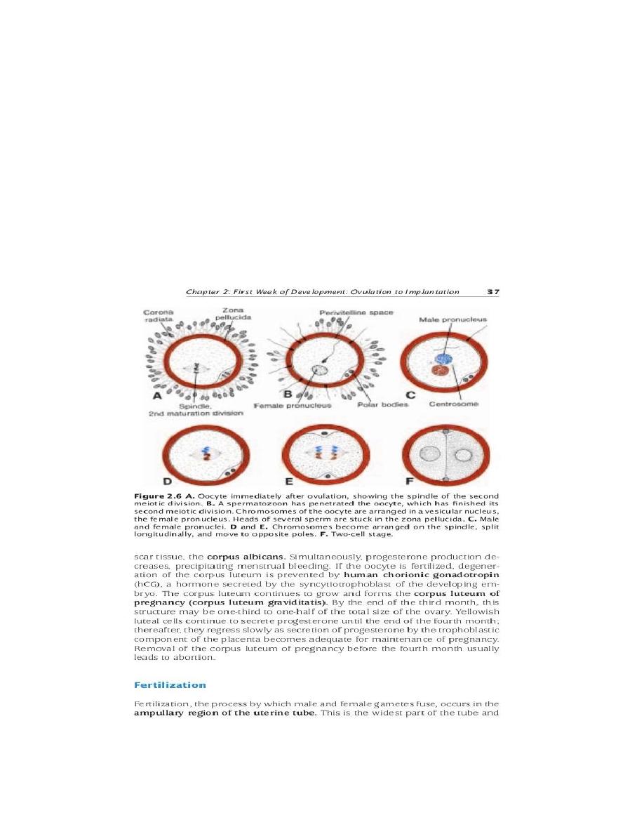
The spermatozoon, meanwhile moves forward until it lies close to ^
female pronucleus, its nucleus become swollen & forms ^
male pronucleus.
The male & female pronucli lose their nuclear envelopes & fuse forming a
zygote.
The main results of fertilization are:
1- restoration of ^ diploid no. of chromosomes.
2-determination of ^ sex of ^ new individual (XX, or XY).
3-Initiation of cleavage.
Without fertilization, ^ oocyte usually degenerates 24 hrs after ovulation.
CLEAVAGE
once ^ zygote has reached ^ 2 cell stage, it undergoes a series of mitotic
divisions, increasing in ^ no. of cells (blastomeres) which become smaller
with each division, until 8 cells stage, they r loosely arranged, after 3rd
cleavage, they arrange together by tight junctions, (
compaction
) ,
segregates inner cells from outer cells.
Approximately 3 days after fertilization, cells of ^ compacted embryo
divide again to form (
morula
) 16 cells, composed of Inner cell mass (
embryo proper), & outer cell mass (trophoblast___ placenta).
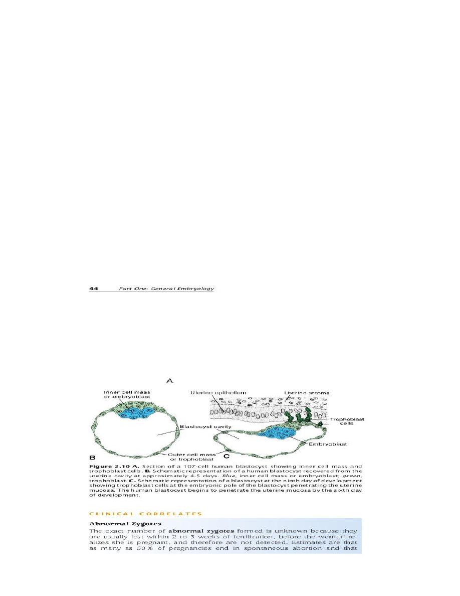
BLASTOCYST FORMATION
About ^ time of ^ morula enters ^ uterine cavity, fluid begins to penetrate
through ^ zona pell. Into ^ intercellular spaces of ^ inner cell mass, which
gradually become confluent &forms a single cavity (blastocele), at this time
^ embryo is a
blastocyst.
The inner cell mass now called embryoblast, & ^ outer cell mass called
trophoblast.
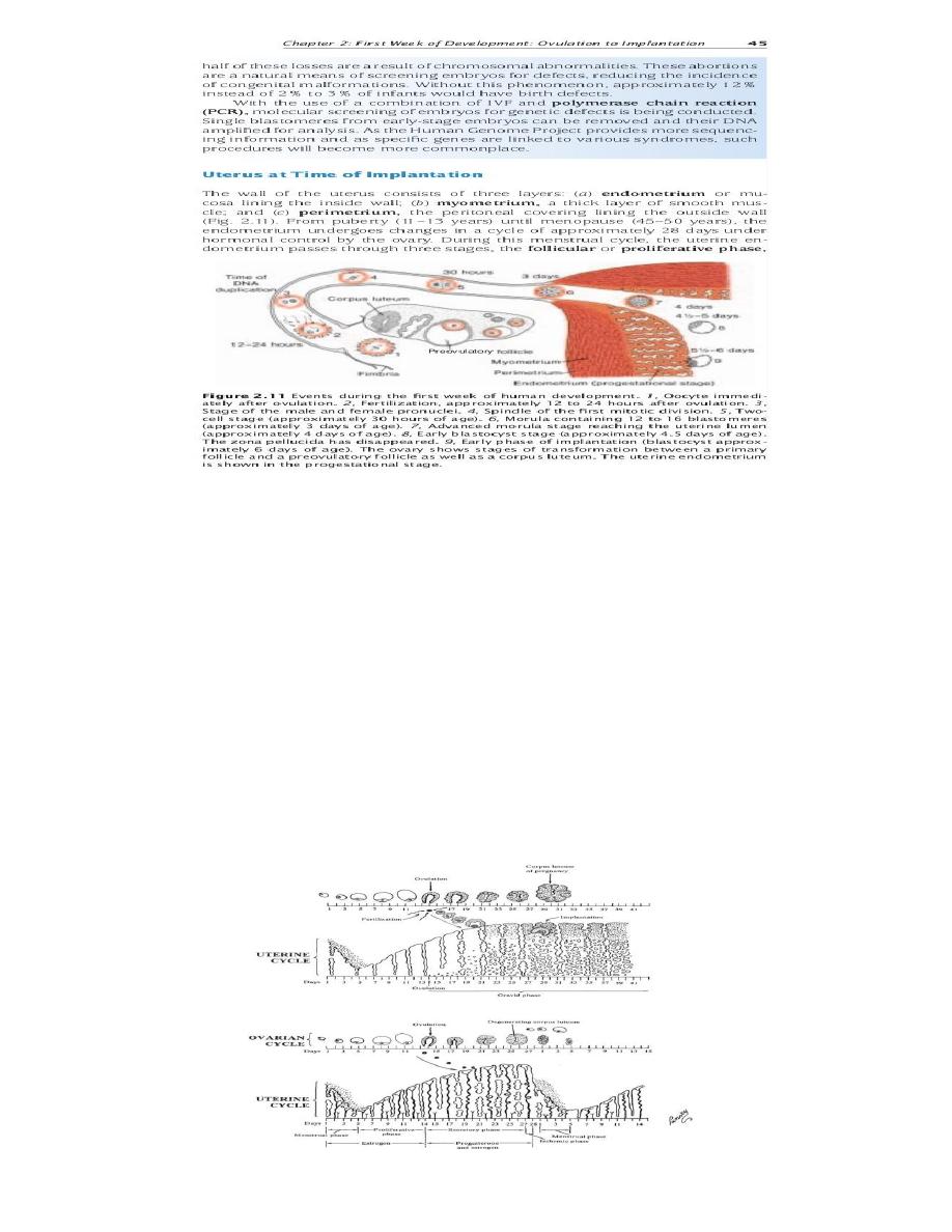
UTERUS AT TIME OF IMPLANTATION
^ wall of ^ uterus consist of :1- endometrium 2- myometrium 3-
perimetrium.
The endometrium undergoes cyclic changes under hormonal control.
1- menstrual phase
when fertilization not occur leads shedding of
endometrium
2-proliferative (follicular) phase
begins after menstruation (estrogen)
parallel to ^ growth of ovarian follicles.
3- secretory (progestational) phase
begins 2-3 days after ovulation
(progesterone) become spongy , compact & basal layers, ready for
implantation of embryo ,if fertilization does not occur, shedding of spongy &
compact layers occur.