
Rheumatology
Final Med revision programme 2007
Rheumatology
Final Med revision programme 2007
Dr. David Kane
Consultant Rheumatologist
AMNCH & TCD

Regional examination of the
Lower Limb
Regional examination of the
Lower Limb
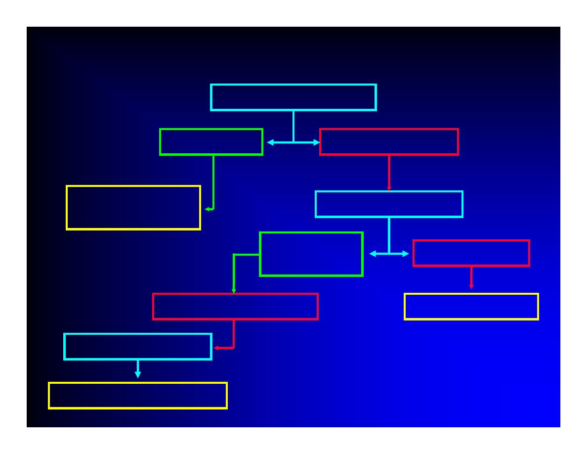
Theory
Theory
Painful / restricted
Active joint movement
Painless / full
Joint / muscle/
tendon all normal
Passive movement
Painless and
full
Painful
Joint problem
muscle/ tendon problem
Resisted movements
Test individual muscles

General Principles of Joint
Examination
General Principles of Joint
Examination
z
Inspection……………..
Look
z
Palpation………………
Feel
z
Active motion…………
Move
patient puts joint through full range of movement
z
Passive motion
when active range of motion is reduced
z
Resisted movements (provocative tests)
z
Special tests……………
Special Tests

The Lower Limb
The Lower Limb
z
Foot
z
Ankle
z
Knee
z
Hip
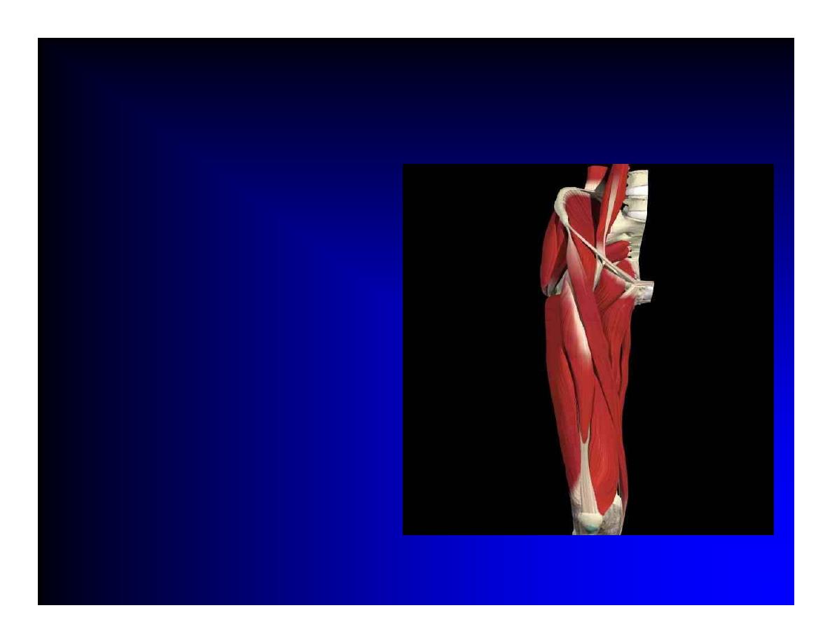
The Hip
The Hip
z
Inspection
z
Palpation
z
Active motion
z
Passive motion
z
Resisted movements
z
Special tests

Examination of the Hip
Examination of the Hip
z
LOOK
z
Deeply buried joint
z
Inspection usually unhelpful (but do it anyway!)
Muscle wasting
}
Pelvic tilt
}
Patient standing
Iliopsoas bursa
}
z
Inspection
Scars
}
Patient
Lower limb length discrepancy }
lying
Flexion deformity of hip
}
supine

Examination of the Hip
Examination of the Hip
z
FEEL
z
Palpation may elicit tenderness
Anterior groin – true hip joint
Lateral aspect hip – greater trochanteric bursitis

Examination of the Hip
Examination of the Hip
z
MOVE
z
Flexion
135º with knee flexed; 90º with knee extended
NB FFD contralateral hip, Thomas’s Test
z
Internal and External Rotation
With hip and knee flexed 90º
With knee extended (roll leg)
z
Abduction and Adduction
Trendelenberg test

z
FUNCTION
z
Antalgic gait
z
Trendelenburg test
Pelvic brim drops when patient stands on one leg
Indicates weakness of gluteus medius
Normal action is to tilt pelvis whilst walking to enable leg
to swing freely
Trendelenburg gait
Bilateral positive Trendelenburg sign
Trunk swings as patient walks to compensate for
dysfunctional pelvic tilt
Examination of the Hip
Examination of the Hip
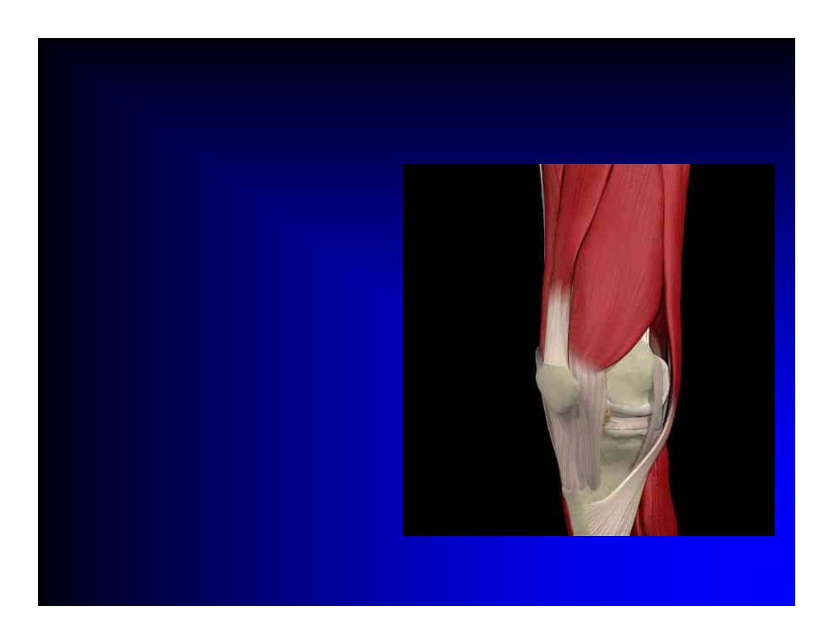
The Knee
The Knee
z
Inspection
z
Palpation
z
Active motion
z
Passive motion
z
Special tests

Examination of the Knee
Examination of the Knee
z
LOOK
z
Symmetry and Alignment
z
Deformity
Fixed flexion
Varus
Valgus
z
Operation scars
z
Quadriceps muscle bulk
z
Swelling/ heat / colour change
z
Psoriasis

Examination of the Knee
Examination of the Knee
z
FEEL
z
Temperature: Back of hand
z
Tenderness
Joint margins
Tibial tuberosity
z
Effusion
Patellar tap
Cross-fluctuation
Baker’s cyst
z
Patellofemoral crepitus / grind

Examination of the Knee
Examination of the Knee
z
MOVE
z
Flexion
z
Extension
z
Compare both sides
z
Active and Passive movements
z
Feel for crepitus on movement

Examination of The Knee
Examination of The Knee
z
Special Tests
z
Anterior / Posterior Cruciate ligament stability
Knee flexed to 90
0
Anterior and Posterior Drawer sign
z
Medial / Lateral Collateral ligament stability
Knee flexed to 15
0
Stress each side of knee joint

Examination of The Knee
Examination of The Knee
z
Special Tests
z
Menisceal tests: only for experienced examiner
McMurray’s test
Flex/ extend the knee while the internally rotating and
then externally rotating the tibia
Pain and an associated click felt over the joint line
diagnostic of a menisceal tear
Apley’s grinding test

Examination of The Knee
Examination of The Knee
z
FUNCTION
z
Walk a few steps
z
Observe gait and deformity
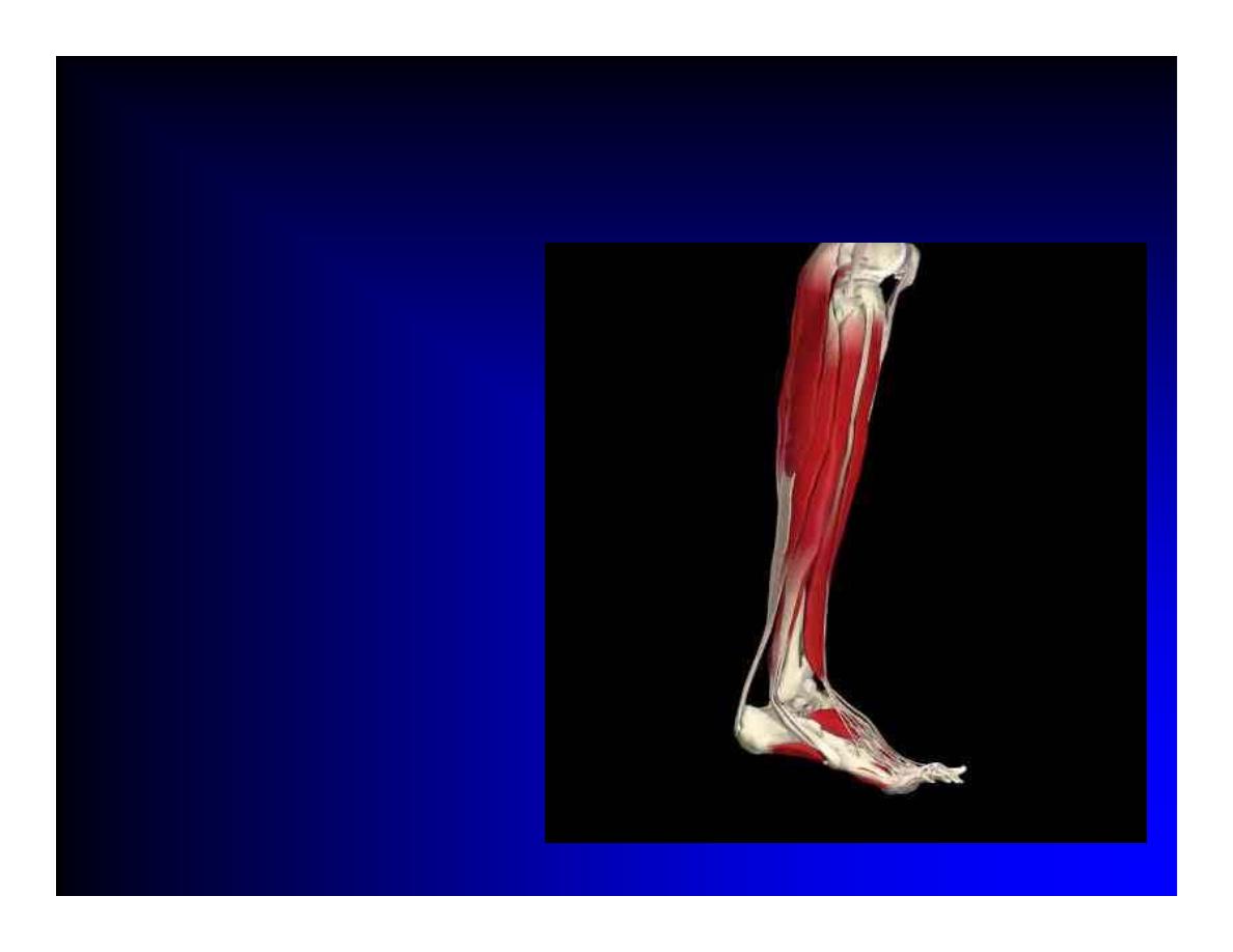
The Foot and Ankle
The Foot and Ankle
z
Inspection
z
Palpation
z
Active motion
z
Passive motion

Examination of the Foot and Ankle
Examination of the Foot and Ankle
z
Patient on couch
z
LOOK
z
Symmetry / Deformity / Swelling / colour change
Toes
Midfoot (Midtarsal joint)
Hindfoot (Subtarsal joint)
Arches (flat foot, pes planus)
z
Nail changes / Psoriasis
z
Examine soles for Calluses / keratoderma
blenorrhagica / pustular psoriasis
z
Inspect Shoes for abnormal wear

Examination of the Foot and Ankle
Examination of the Foot and Ankle
z
FEEL
z
Temperature
z
Metatarsal squeeze
then palpate individual metatarsals and web spaces if positive
z
Palpate ankle, subtalar and midfoot
z
Achilles tenderness
Achilles tendonitis
z
Heel tenderness
Plantar fasciitis

Examination of the Foot and Ankle
Examination of the Foot and Ankle
z
MOVE
z
Active and Passive
z
Ankle Dorsiflexion / Plantarflexion
z
Inversion / Eversion
tibialis posterior
Peroneal
z
MTP 1 Dorsiflexion / Plantarflexion

Completing the examination of
the lower limb
Completing the examination of
the lower limb
z
Leg length discrepancy
Measure anterior superior iliac spine to lateral
malleolus
z
Gait
Antalgic
Trendelenburg