1
Ante partum feral surveillance
Background and objectives
Fetal hypoxia and acidosis can result from any cause of placental insufficiency.
Placental insufficiency is commonly associated with hypertensive disorders during
pregnancy, diabetes mellitus and any cause of placental insufficiency. Ante partum
fetal surveillance tests are group of procedures to identify fetal hypoxia before the
onset of labor.
Kick count
Kick count is simply defined as the number of fetal movements felt by the pregnant
woman during 12 hours. It is still the most reliable ante partum fetal distress. Simply
the pregnant woman is asked to record the number of fetal movements per 12 hours.
In well oxygenated fetus, at least 10 movements is felt. Should the number is less that
10, further evaluation is required.
Non stress test
Definition
Correlation between fetal heart rate changes compared with fetal movement
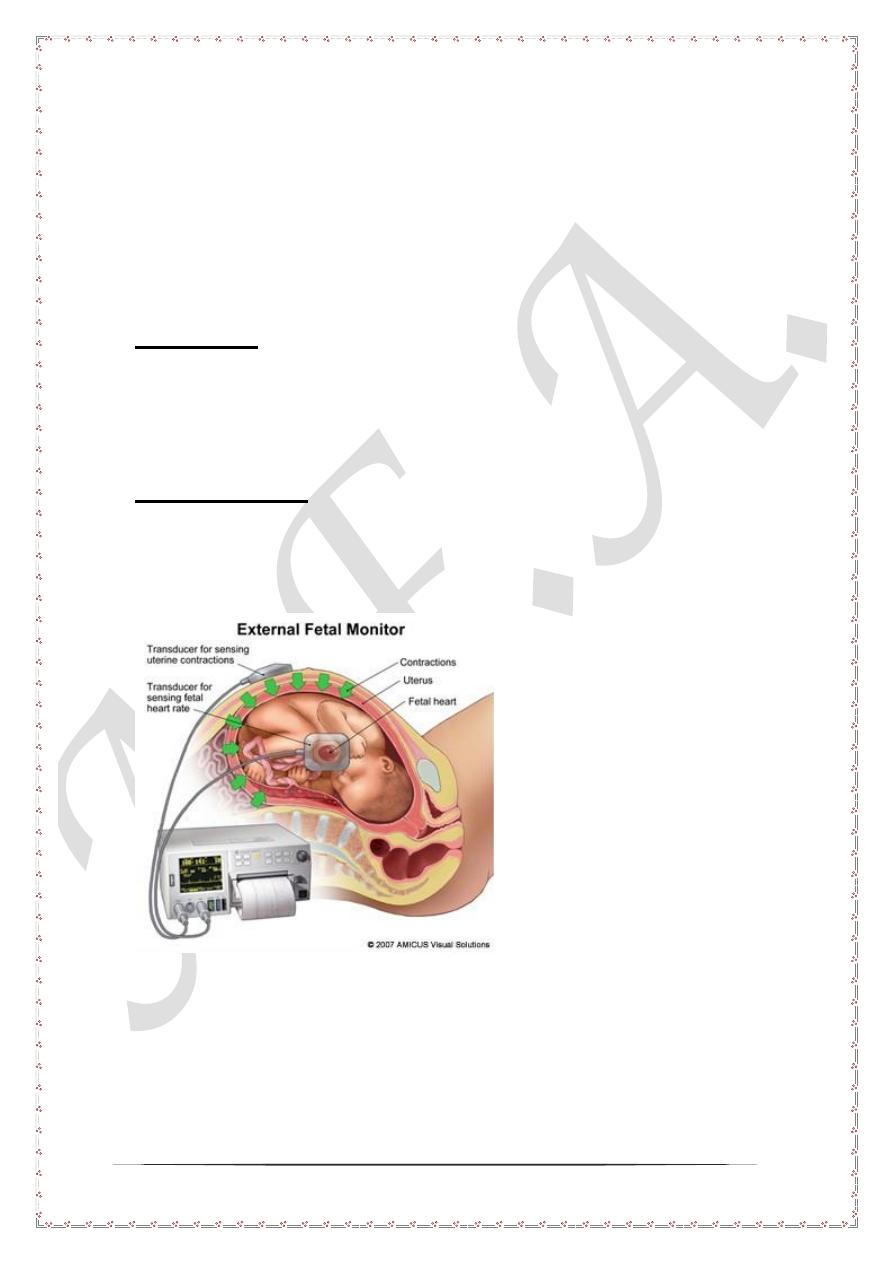
Technique
Ultrasound device is usually used to record fetal heart sound over 30 minutes. The
results are plotted in a strip paper which is called NST strip. During this 30 minutes
the fetal heart rate is usually plotted while fetal movements are plotted whenever they
occur
2
Finding in NST strip
For each NST strip the followings should be evaluated
1- Baseline fetal heart which should be 110 to 150 beats per minutes.
2- Beat to beat variability which shown as zigzag shaped line.
3- Fetal heart rate acceleration associated with fetal movement. With each fetal
movement the fetal heart rate should increase 15 beats above the base line and
endure at least15 seconds.
Interpretation
1- In well oxygenated fetus there should be at least 2 movements during 30
minutes tracing. Each movement is associated with rise in the fetal heart rate
by 15 beat per minutes and last for 15 seconds. In such cases the tracing is
assuring and called reactive non stress test
2- Otherwise in the absence of fetal of fetal movement, beat to beat variability or
fetal heart acceleration the strip is called non reactive NST and the fetus
requires further assessment.
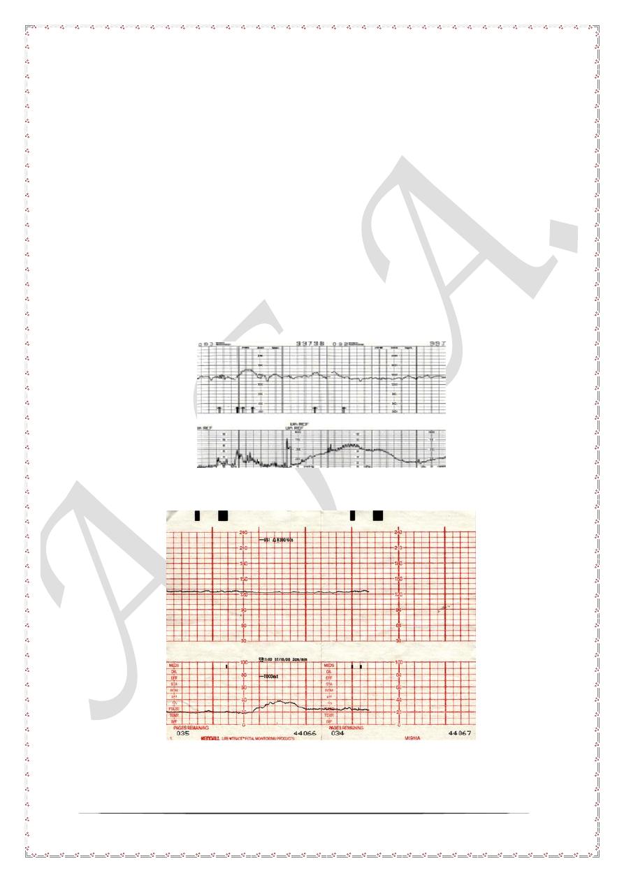
Example of reactive non stress test
Example of non reactive NST strip
3
Biophysical profile of the fetus
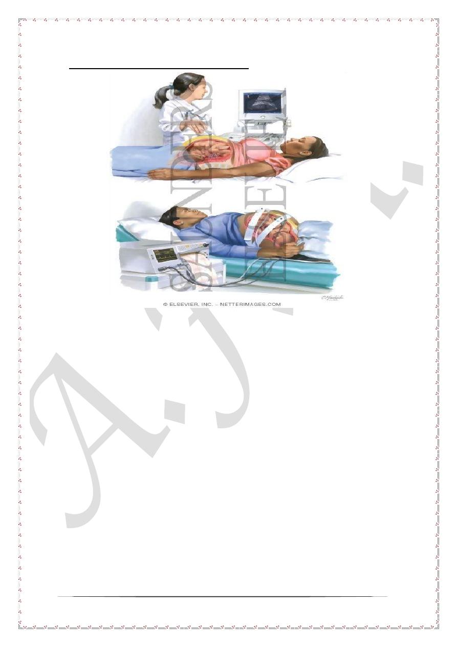
Background and technique
An ultrasound based investigation which last for 30 minutes. During this 30
minutes the followings should be noticed and scored
1- Fetal attitude which should be in flexion position. Extended fetus indicates
fetal hypoxia.
2- Fetal gross body movements. During 30 minutes observation the fetus
should move at least once. The fetal movement should swing from fetal
flexion, extension and back to flexion.
3- Fetal respiratory movements. During 30 minutes observation the fetus
should have one episode of fetal breathing movement which last for 30
seconds.
4- Amount of liquor. The amount of liquor can be easily assessed by
ultrasound scan. Should the amount of liquor is normal this indicates well
oxygenated fetus. Oligohydramnios indicates fetal a risk for hypoxia.
5- Fetal heart rate changes with fetal movement. Should the fetal heart rate
increase with fetal movement this indicates well oxygenated fetus.
Otherwise absence of fetal movement indicates fetal hypoxia

4
Scoring of biophysical profile
parameter
present
absent
Gross body
movement
2
0
Respiratory
movement
2
0
Fetal tone
2
0
oligohydramios
0
2
Fetal heart rate
variability with
movement
2
0
Score may be 0, 2,4,6,8 and 10
Interpretation and clinical management
1- Score 8- 10 repeat after 1 week.
2- Score 6 repeat after 24 hours.
3- Scores 0, 2, 4 are indication for immediate delivery
5
Doppler of the umbilical artery
Background
Doppler of the umbilical artery is defined simply as measuring the blood velocity in
the artery by the use of ultrasound probes. The red blood cells in the blood reflect the
ultrasound wave from the probe. With this reflection there frequency of the ultrasound
wave is shifted or changed. The degree of change is dependent on the velocity of
blood. And from this frequency change the blood velocity can be calculated and
plotted as strip paper.
Technique
The procedure is usually done by special ultrasound probe which is equipped with
Doppler facility. First the umbilical artery is visualized and insinuated with ultrasound
wave at 60 degree. The results are plotted on strip paper as well as the on the screen.
6
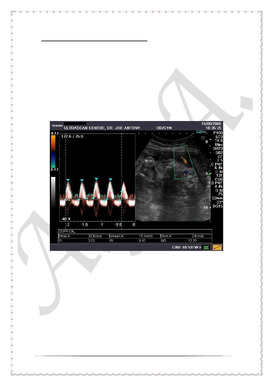
Clinical interpretation of Doppler
Normal Doppler
In normal Doppler strips with assuring adequate blood flow to the fetus, both systolic
and diastolic waves are shown. The peak systolic wave represents the maximum
blood flow velocity during systole.
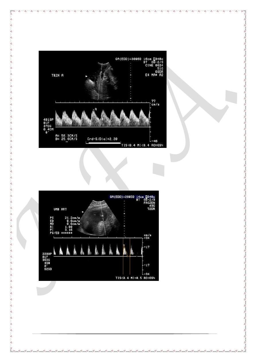
Non assuring Doppler
None assuring Doppler is said to be present when the diastolic wave is absent a
pattern with is called
Absent end diastolic wave
As shown in the picture above. The absence of diastolic wave indicates high
resistance in placental blood flow. A pattern commonly associated with severe feta,
acidosis and hypoxia. In such cases prompt delivery is required.