
Lecture 2
النسائية
د. أحمد جاسم
Benign disorder of cervix
Page 1 of 12
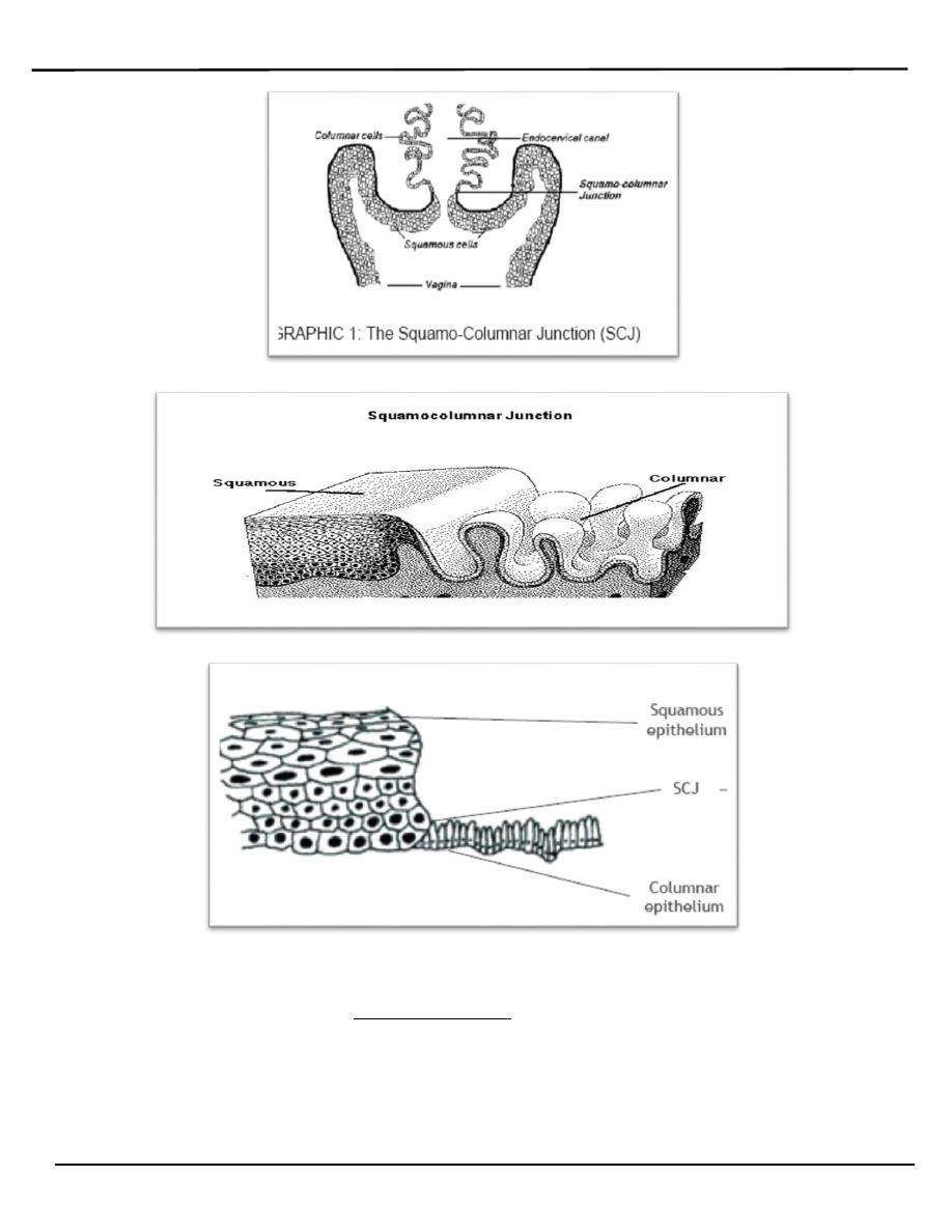
Anatomy
o The cervix is fibromuscular inferior part of the uterus protruding into the
vagina. It measures 2.5-3 cm in diameter and 3-5 cm in length. The
normal anatomic position of the cervix is angulated slightly downward
and backward.
o The external os is usually small and round in nulliparous women but can
be seen as a transverse slit in those who have had cervical dilation during
labour. The anterior and posterior fornices limit the portio (exocervix).
The cervical canal measures approximately 8 mm wide and contains
longitudinal ridges. The opening of the cervical canal into the uterus is
called the internal cervical os. The area between the endocervical and
endometrial cavity is called the isthmus or lower uterine segment.
o Ectocervix is covered by a pink stratified squamous epithelium,
consisting of multiple layers of cells. It is smooth, pearly, opaque
appearance.
o Endocervix is lined by a reddish columnar epithelium consisting of a
single layer of cells. it appears reddish in colour because the thin single
cell layer allows the coloration of the underlying vasculature in the stroma
to be seen more easily. It covers a variable extent of the ectocervix,
depending upon the woman’s age, reproductive, hormonal and
menopausal status.
o The columnar epithelium is normally visible with the speculum during
o Ovulatory phase of the menstrual cycle
o Pregnancy
o Women taking the combined oral contraceptive pill
o Where estrogen levels are elevated.
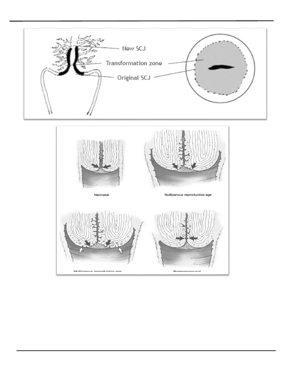
o The point where these two epithelia meet is called the squamo-columnar
junction.
:العدد
6
22/2/2014
Lecture 2
النسائية
د. أحمد جاسم
Benign disorder of cervix
Page 2 of 12
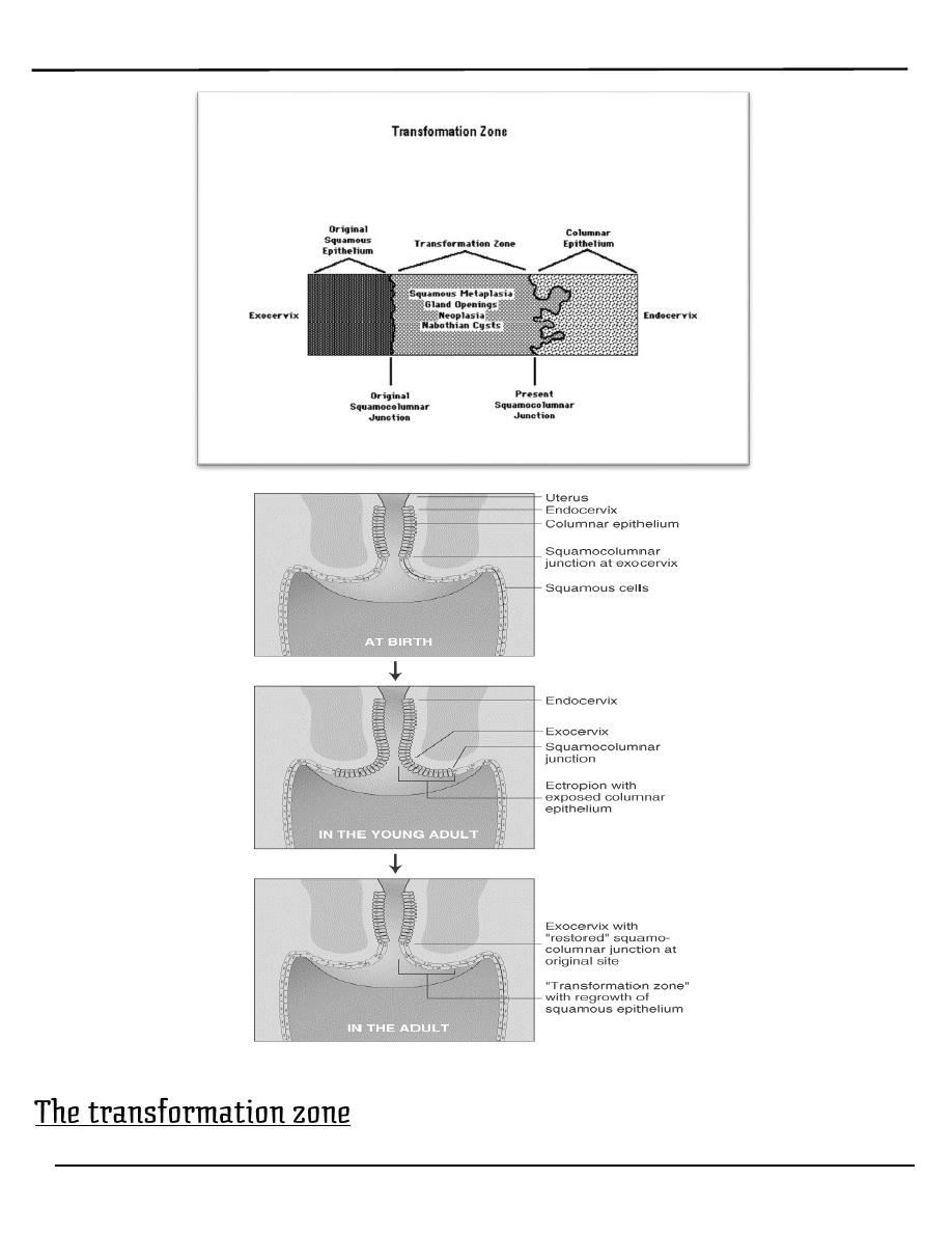
o Squamous metaplasia is physiological replacement of the everted
columnar epithelium by a newly formed squamous epithelium. It is an
irreversible process; the transformed epithelium (now squamous in
character) cannot revert to columnar epithelium.
o The region of the cervix where squamous metaplasia occurs is referred to
Lecture 2
النسائية
د. أحمد جاسم
Benign disorder of cervix
Page 3 of 12
as the transformation zone. Identifying the transformation zone is of great
importance in colposcopy, as almost all manifestations of cervical
carcinogenesis occur in this zone.
o The point where these two epithelia meet is called the squamo-columnar
junction.
o The location of squamocolumnar junction in relation to the external os
varies depending upon age, menstrual status, and other factors such as
pregnancy and oral contraceptive use. The epithelium of the
transformation zone is identified by the variegation of the color between
the two native epithelia.
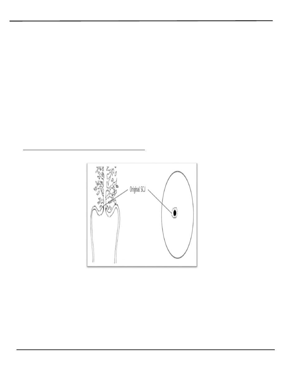
o The anatomical site of the squamo-columnar junction fluctuates under
hormonal influence through out the reproductive life.
o During childhood and perimenarche, the original squamocolumnar
junction is located at, or very close to, the external os
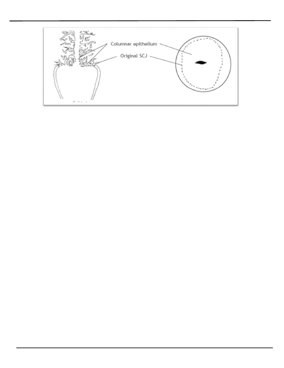
o After puberty and during the reproductive period, the female genital
organs grow under the influence of estrogen. Thus, the cervix swells and
enlarges and the endocervical canal elongates. This leads to the eversion
of the columnar epithelium of the lower part of the endocervical canal on
to the ectocervix (This condition is called ectropion).
Lecture 2
النسائية
د. أحمد جاسم
Benign disorder of cervix
Page 4 of 12
o At menarche (With acidification of a vagina), the ectocervix undergoes an
accelerated rate of squamus metaplasia which produces the
transformation zone.
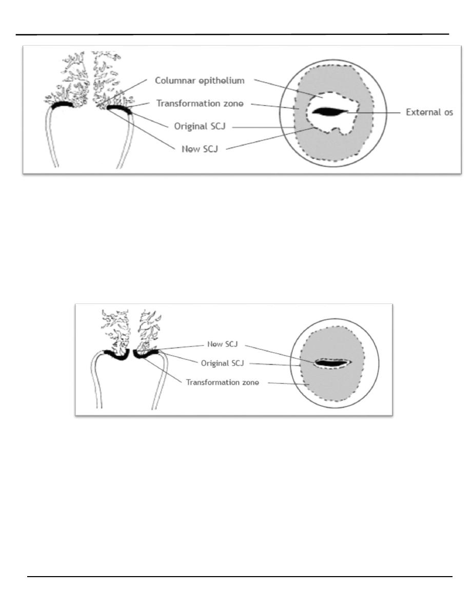
o The metaplastic process mostly starts at the original squamocolumnar
junction and proceeds towards the external os through the reproductive
period to perimenopause. Thus, a new squamocolumnar junction is
formed between the newly formed metaplastic squamous epithelium and
the columnar epithelium remaining everted onto the ectocervix.
o The buffer action of the mucus covering the columnar cells is interfered
with when the everted columnar epithelium in ectropion is exposed to the
acidic vaginal environment. This leads to the destruction and eventual
replacement of the columnar epithelium by the newly formed metaplastic
squamous epithelium. Metaplasia refers to the change or replacement of
one type of epithelium by another. The metaplastic process mostly starts
at the original squamocolumnar junction and proceeds centripetally
towards the external os through the reproductive period to
perimenopause. Thus, a new squamocolumnar junction is formed between
the newly formed metaplastic squamous epithelium and thecolumnar
epithelium remaining everted onto the ectocervix.
Lecture 2
النسائية
د. أحمد جاسم
Benign disorder of cervix
Page 5 of 12
o As the woman passes from the reproductive to the perimenopausal age
group, the location of the new squamocolumnar junction progressively
moves on the ectocervix towards the external os.
o From the perimenopausal period and after the onset of menopause, the
cervix shrinks due the lack of estrogen, and consequently, the movement
of the new squamocolumnar junction towards the external os and into the
endocervical canal is accelerated.
o In postmenopausal women, the new squamocolumnar junction is often
invisible on visual examination.
Lecture 2
النسائية
د. أحمد جاسم
Benign disorder of cervix
Page 6 of 12
Lecture 2
النسائية
د. أحمد جاسم
Benign disorder of cervix
Page 7 of 12
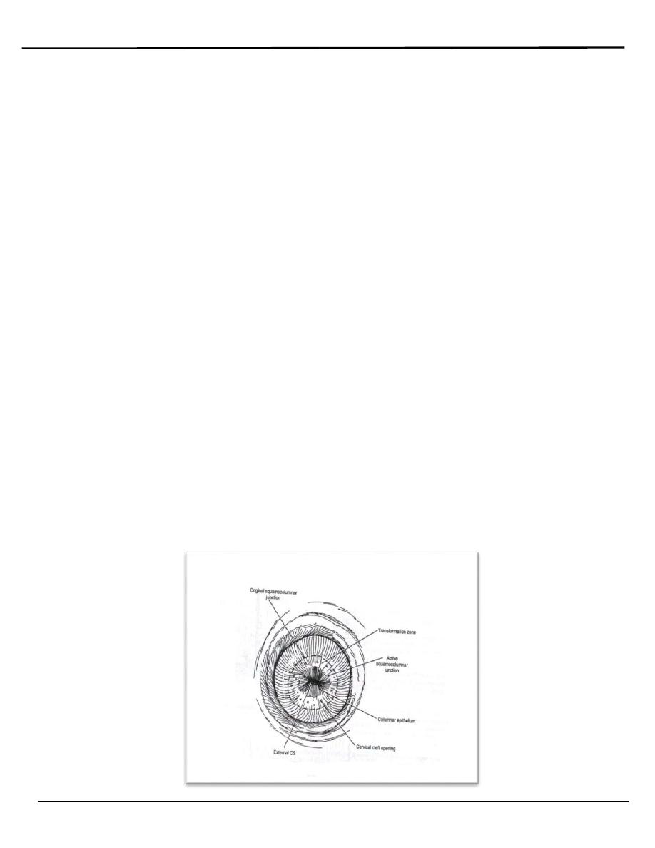
o The transformation zone is a dynamic area, usually located on the
Lecture 2
النسائية
د. أحمد جاسم
Benign disorder of cervix
Page 8 of 12
ectocervix. At times, the distal edge of the transformation zone extends
into the upper vagina. The transformation zone, by definition, is the area
between the original squamocolumnar junction and the current
squamocolumnar junction. The transformation zone is that portion of the
cervix that originally was columnar epithelium and through a process of
squamous metaplasia is now squamous epithelium. Squamous metaplasia
occurs continuously; however, this process is most active during fetal
development, around the time of menarche, and during pregnancy. Local
hormonal changes, as reflected by vaginal pH, influence this process.
o It consists of endocervical stroma and glands covered by squamous
epithelium.
o The position of the transformation zone varies according to age. In
women during the childbearing age, the transformation zone is fully
located on the ectocervix.
o In post-menopausal women the transformation zone is often located
within the endocervical canal as the cervix shrinks with the decreasing
levels of estrogen and Consequently, the transformation zone may move
into the cervical canal.
o Identifying the transformation zone is of great importance in colposcopy,
as almost all manifestations of cervical carcinogenesis occur in this zone
due to the high cell turnover of this tissue which is important in the
pathogenesis of cervical carcinoma
Lecture 2
النسائية
د. أحمد جاسم
Benign disorder of cervix
Page 9 of 12
Cervical ectropion
o Is the presence of everted endocervical columnar epithelium on the
ectocervix.
o It is in reality an area of columnar epithelium that has not yet undergone
squamous metaplasia. it occurs when the cervix grows rapidly and
enlarges under the influence of estrogen, after menarche and during
pregnancy. And also seen in women under the influence of estrogen
(combined oral contraceptive pills COCP, pregnancy).
o It is a normal, physiological occurrence in a woman’s life.
o It is previously called Cervical erosion which is very inappropriate name
and best to be avoid as it conveys quite the wrong impression of what is
really a normal phenomenon.
o Most patients have no complaint. (Seen during speculum examination), or
it can be associated with:
1. Excessive Mucoid vaginal discharge.
2. Brown intermenstrual discharge.
3. Slight postcoital bleeding.(should investigated)
4. During pregnancy slight bleeding (could be a cause of early
pregnancy bleeding or Anti-Partum Haemorrhage APH).
o Pain is never caused by an ectropion nor is it a cause of backache or
dysparunia.
o Speculum examination:
o *Bright red area is seen around the external os continuous with the
endocervix with clearly defined outer edge. The eversion of the columnar
epithelium is more pronounced on the anterior and posterior lips of the
ectocervix and less on the lateral lips.
o *It is not tender.
o *It bleeds from multiple pinpoint areas when touched.

Lecture 2
النسائية
د. أحمد جاسم
Benign disorder of cervix
Page 10 of 12
o Carcinoma.
o Tuberculosis.
o Syphlytic ulcer
o Other ulcer.
Cervical smear must be taken in all cases.
A. No treatment
o Ectropion found on routine examination should not be treated unless they
are causing troublesome discharge.
o Ectropion are not treated during pregnancy, most of them resolve after
delivery.
o Change the oral contraceptive contraception if patient complain of
discharge to other contraceptive method.
B. Treatment needed
o When a patient has a trouble some discharge, the Ectropion is treated by :
o Thermal cauterization.
o Cryosyrgery (freezing)
o Laser.
o The resulting raw area takes 6-8 weeks to become covered with squamous
epithelium
Nabothian cysts (follicles):
o Nabothian follicles (cysts) are retention cysts that develop as a result of
the occlusion of an endocervical crypt opening or outlet by the overlying
metaplastic squamous epithelium and it is located in the transfusion zone.
The underlying (buried) columnar cells continuo to secret mucus, and a
mucous retention cyst is created on the ectocervix.
o It is so common that they are considered a normal variant and it is of no
pathological significance.
Lecture 2
النسائية
د. أحمد جاسم
Benign disorder of cervix
Page 11 of 12
Nabothian cysts
o Nabothian cysts are opaque, ivory-white to yellowish on visual
examination.
o They vary generally in size from 2mm -3 cm.
o It needs no treatment.
Cervical polyp
o Ectocervical and endocervical polyps are the most common benign
neoplastic growths of the cervix.
o It may be isolated or multiple and vary in diameter from a few millimeters
to several centimeters.
A. Asymptomatic
B. symptomatic:
they most commonly cause:
1. Post coital bleeding.
2. Menorrhagia & irregular vaginal Bleeding.
3. Pregnancy slight bleeding (could be a cause of early pregnancy
bleeding or APH (antepartum Haemorrhage).
:
o Removal of polyp.
o All specimens must be sent for pathologic examination, because
squamous cell carcinoma and adenocarcinoma can be present as polyps.
Pyometra and Haematometra
o Haematometra: It is a collection of blood in the uterine cavity, caused by
obstruction in the genital tract at or below the level of cervix.
o Pyometra: It is a collection of pus in the uterine cavity, caused by
obstruction in the genital tract at or below the level of cervix.
Lecture 2
النسائية
د. أحمد جاسم
Benign disorder of cervix
Page 12 of 12
1.Congenital
2.Vaginal atresia or absent causing haematocolpos which rarely associated
with haematometra.
3.Functioning rudimentary horn.
4.One half of double uterus not communicate with vagina.
stenosis of cervix caused by operation :
1.a Amputation of cervix.
2.Cone biopsy.
3.Cervical cauterization
4.Vigorous curettage.
5.Carcinoma
a. of cervix
b. lower part of body of uterus.
o Suggestive feature in the history are amenorrhoea associated with severe
cyclical dysmenorrhoea-like pain, with a previous history of cervical
surgery in reproductive years.
o In post menopausal women it may give rise to pyometra where
accumulated secretions become a focus of infection.
Evacuation of uterus & treatment of the cause.
By: Mu’taz Fathi