Developmental (Congenital) Abnormalities of the Nervous System
Developmental (Congenital) Abnormalities of the Nervous SystemArachnoid Cysts
Encephaloceles
Craniosynostosis
Chiari malformation
Spinal Dysraphism
Arachnoid Cysts:
These are fluid filled intracranial mass lesions formed by splitting of the arachnoid membrane.Arachnoid Cysts:
The sites of Arachnoid Cysts:• Around one-half are located in the Sylvian fissure.
• Cerebellopontine angle.
• Suprasellar region.
• Posterior fossa regions.
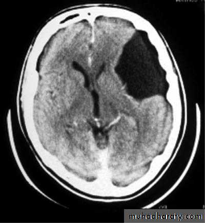
Sylvian Fissure Arachnoid Cyst
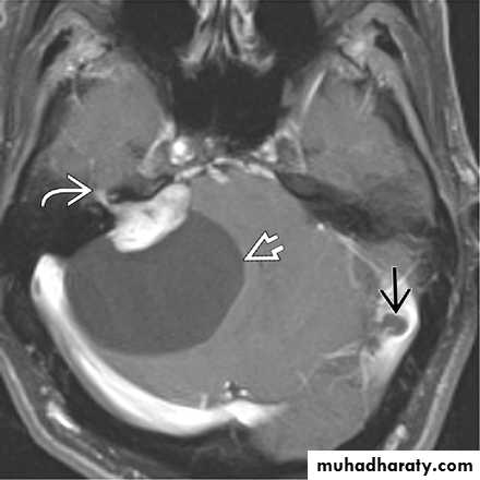
Cerebellopontine angle arachnoid cyst
Suprasellar Arachnoid Cyst
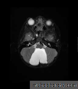
Posterior Fossa Aracnoid cyst
Arachnoid Cysts:
Presentations:• They often present in paediatric age group.
• Some remain asymptomatic.
• Others enlarge in size causing a mass effect.
• Others may present with haemorrhage.
Arachnoid Cysts:
Investigations:
CT Brain
MRI Brain
Arachnoid Cysts:
Treatment:• Conservative: by observation and follow up.
• Surgery: reserved only for symptomatic cases.
• Surgical options include:
• Endoscopy and fenestration into a cistern or ventricle.
• Shunting: e.g. cystoperitoneal shunt.
Encephaloceles:
These are developmental herniation of cerebral tissue through a cranial defect.They occur with a frequency of approximately 1-4 in 1000 births.
Encephaloceles:Sites: they may be:
• Occipital
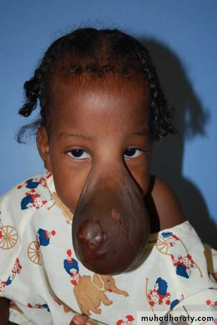
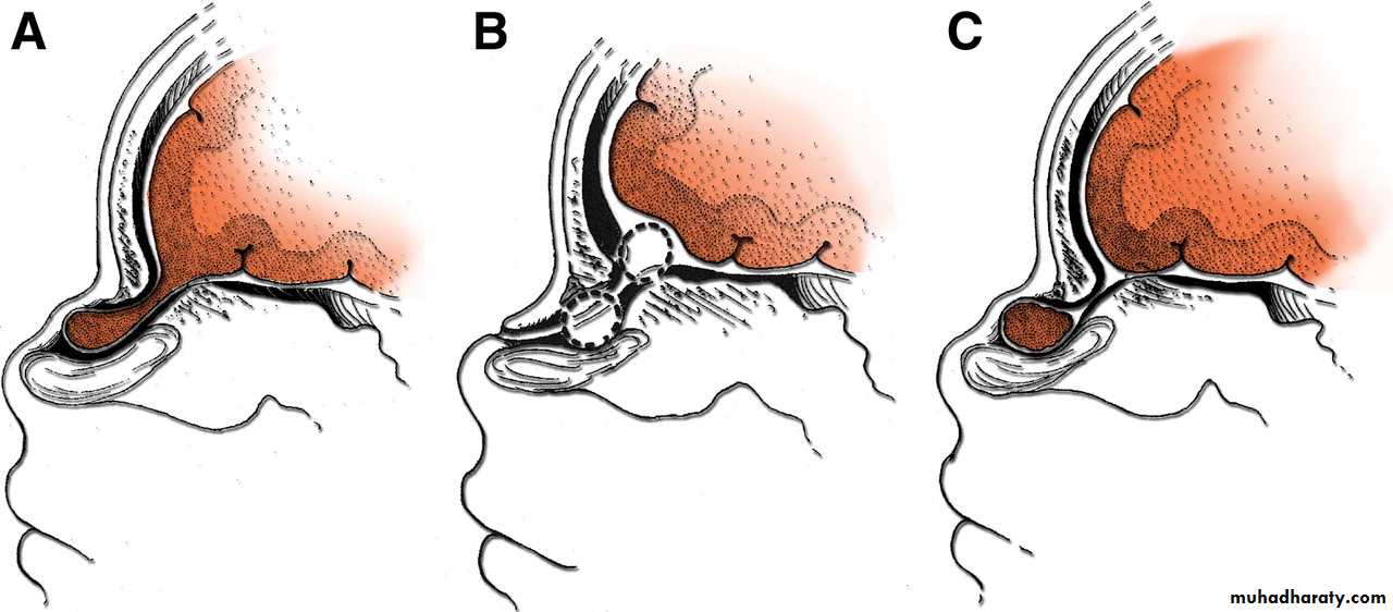
• Nasofrontal
• Frontoethmoidal
• Basal.
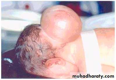
Occipital Encephalocele
Occipital Encephalocele
Nasofrontal Encephalocele
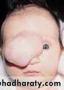
Nasoethmoidal Encephalocele
Basal Encephalocele
Encephaloceles:
Treatment: Surgical repair in order to:• Prevent infection.
• Reconstruct the skull.
Craniosynostosis:
These are premature fusion of the cranial sutures.They may affect single or multiple sutures.
Premature fusion leads to restricted growth of the head resulting in presentation with an abnormal head shape.Craniosynostosis:
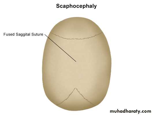
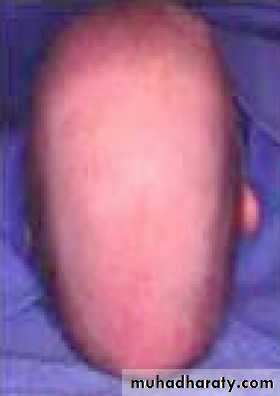
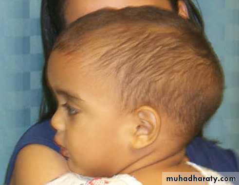
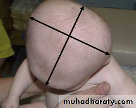
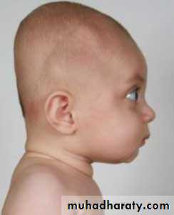
Types of Craniosynostosis:• Sagittal synostosis causes a narrow boat shaped head with frontal and occipital bossing (Scaphocephaly).
• Bicoronal synostosis causes a shortened forehead (Brachycephaly).
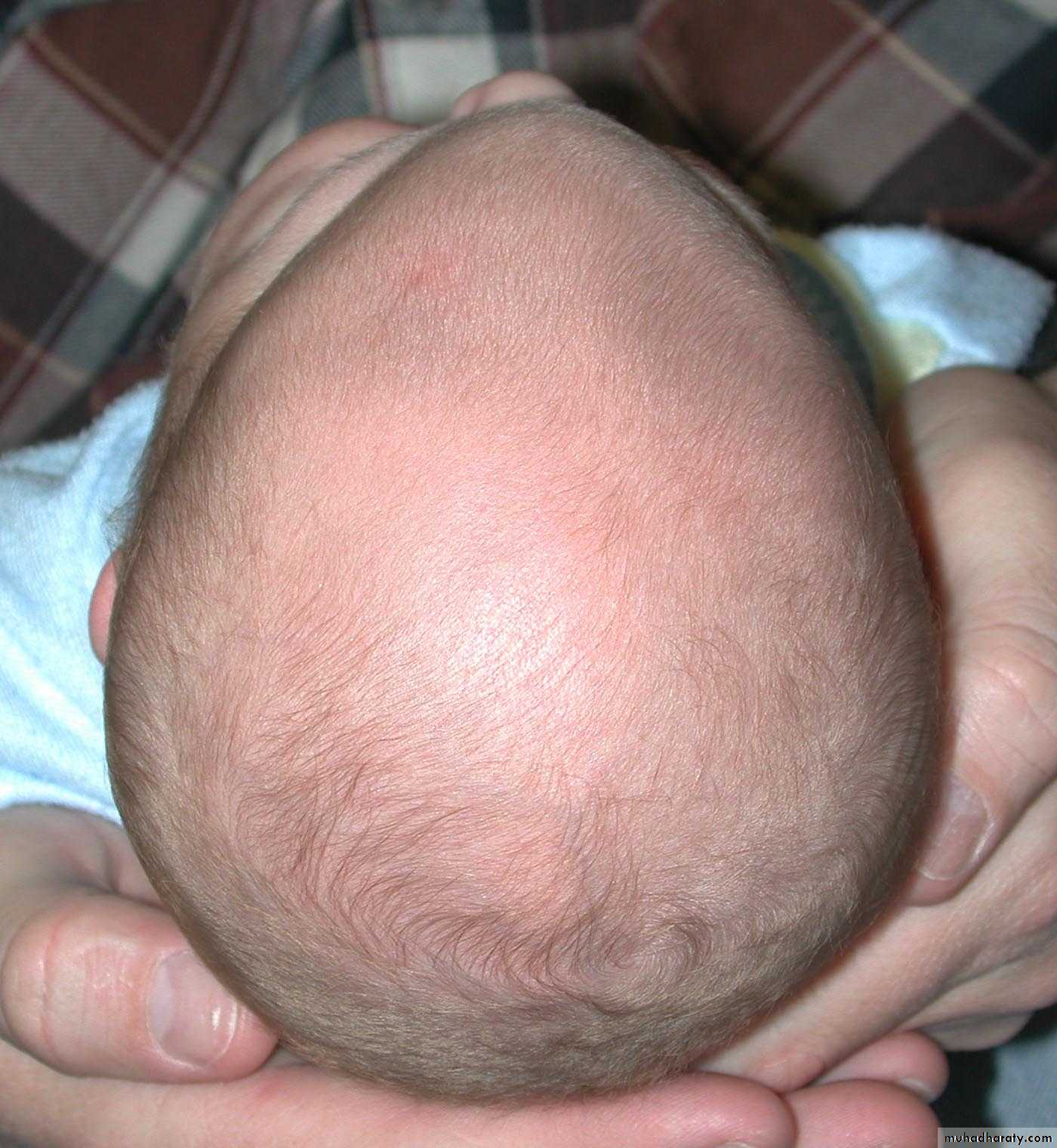
• Metopic synostosis causes Trigonocephaly.
Scaphocephaly
Scaphocephaly
Scaphocephaly
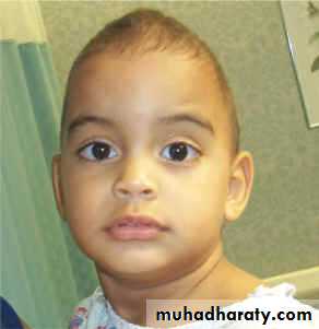
Brachycephaly
Brachycephaly
Trigonocephaly
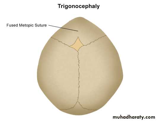
Trigonocephaly
Craniosynostosis:
Diagnosis by CT brain with bone window.Surgical Treatment aimed at:
• Correction of deformity.• Prevent the development of raised intracranial pressure.
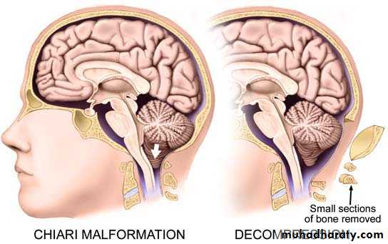
Chiari malformation:
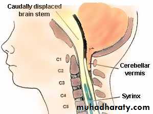
Called also Arnold-Chiari Malformations:Herniation of posterior fossa contents (e.g. cerebellar tonsils) through the foramen magnum.
Normally up to 5mm of tonsillar descent through the foramen magnum.
Chiari malformation:
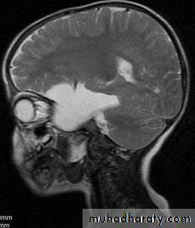
Normally up to 5mm of tonsillar descent through the foramen magnum.Chiari I: >5 mm of tonsillar descent.
Chiari II: descent of the tonsils and cerebellar vermis.
Chiari malformation Type I
Chiari malformation Type II
Chiari malformation:
Presentations:Chiari I Malformation present in young adults with headache, exacerbated by coughing and straining.
Chiari II malformation present in infancy with signs of brainstem compression such as poor feeding, stridor and apnoeic spells.
Chiari malformation:
Treatment of Chiari malformation include:• Treatment of associated Hydrocephalus, and then
• Foramen magnum decompression.DEVELOPMENTAL (CONGENITAL) ANOMALIES OF THE SPINESSPINA BIFIDA (SPINAL DYSRAPHISM)
SPINA BIFIDADefinition:
This is the most common developmental anomaly affecting the spinal column, and it means failure of the neural tube to close fully, so there is split or open spine.
SPINA BIFIDA
SPINA BIFIDA
Types of spina bifida:Spina bifida Occulta
Spina bifida CysticaSpina bifida Aperta
Normal Spine
SPINA BIFIDA
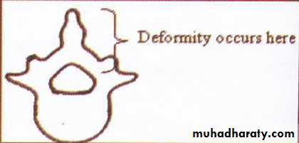
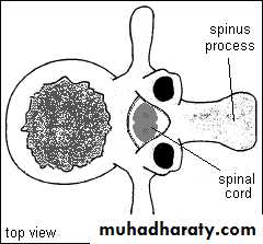
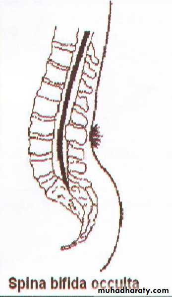
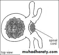
• Spina bifida Occulta:• The posterior vertebral arch has a defect within it, but there is no herniation of the neural tube.
• This defect is found in 10% of the population.
• On the skin over the defect various skin changes may be seen, e.g. hairy patch, an area of pigmentation, a fatty lump or a dermal sinus.
Spina bifida occulta

Spina bifida Occulta
SPINA BIFIDA
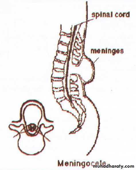
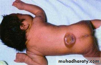
• 2. Spina bifida Cystica:• In this situation there is skin covering the defect making a cyst like.
• If this cyst contains CSF only it is called MENINGOCELE.
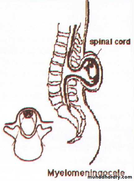
• If there is neural tissue within the sac, it is called MYELOMENINGOCELE.
Meningocele
MENINGOCELE
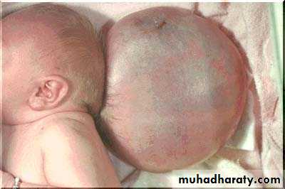
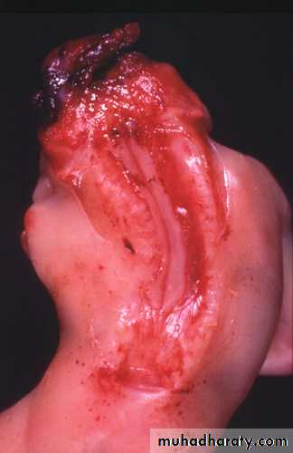
MYELOMENINGOCELE
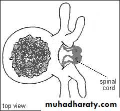
MYELOMENINGOCELE
SPINA BIFIDA
• 3. Spina bifida Aperta:• The neural tube is open with no skin coverage, through a defect in the posterior vertebral arch.
• CSF leakage usually occurs so there is a high risk of meningitis.
Spina bifida Aperta
Spina bifida Aperta
SPINA BIFIDA
Aetiology:90% of cases occur sporadically.
Hereditary factors: children of parents with spina bifida have a 5% risk of having the condition.
Dietary factors: folic acid administration during pregnancy may lower its incidence.
Some Anticonvulsants medications.
SPINA BIFIDA
Clinical Features:Antenatal screening:
Post-delivery: General features
Spina bifida occulta:
Meningocele:
Myelomeningocele:
SPINA BIFIDA
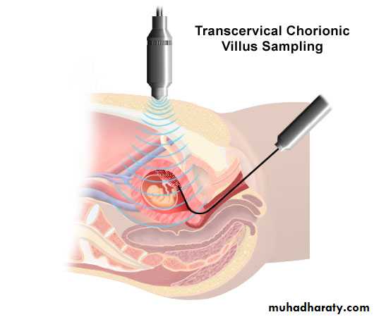
• Antenatal screening: by using:• Detection of alpha-fetoprotein in blood or in amniotic fluid obtained by amniocentesis.
• The use of ultrasound.
• Both tests allow the detection of such defects in over 80% of embryos with open neural tube .
Antenatal screening
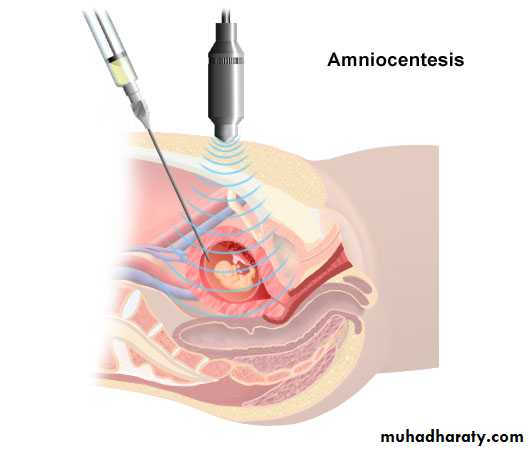
Antenatal screening
SPINA BIFIDA
• b. Post-delivery: General features include:• The incidence is ranging from 1 to 8 per 1000 of the population.
• Most problems tend to occur in the lumbosacral area and are associated with changes in bladder and bowel functions.
• The higher the lesion the more sever the defect.
• If the lesion is open then there is associated risk of meningitis.
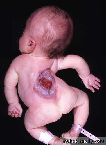
• Other associated disorders include Arnold-Chiari malformations type II, and in up to 80% hydrocephalus.
Myelomeningocele+ Hydrocephalus
SPINA BIFIDA
• c. Spina bifida occulta:• Usually asymptomatic. Accidental finding in X-rays.
• Lipoma, skin dimple or a tuft of hair over the bifid spine.
• Rarely urinary incontinence starting at adolescence.
SPINA BIFIDA
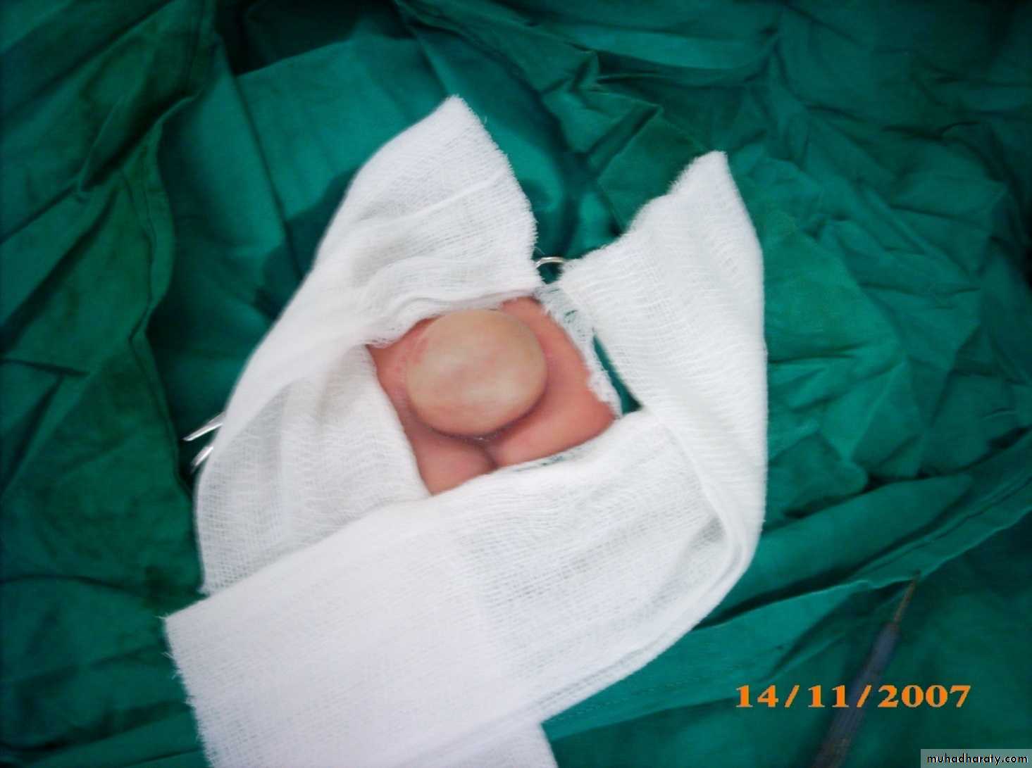
• d. Meningocele:
• No neurological manifestations.
• Cystic translucent swelling with an expansible impulse on coughing.
• The swelling is compressible.
Meningocele
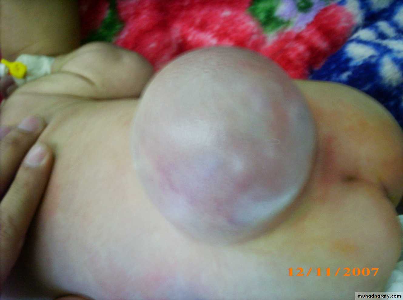
Meningocele
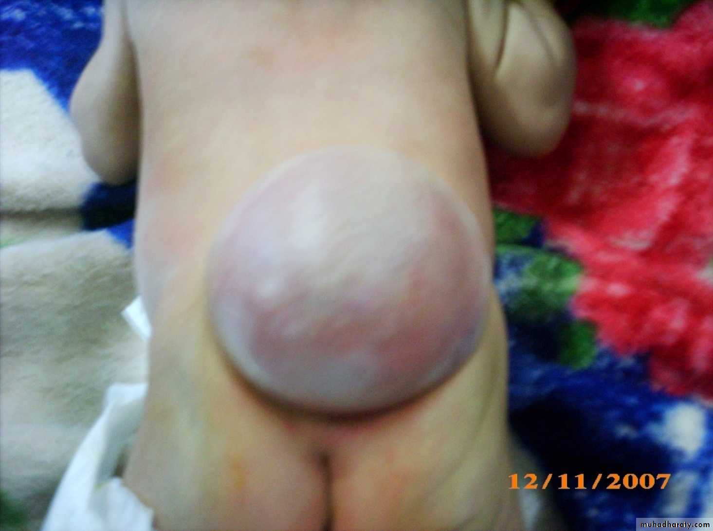
Meningocele

Meningocele
SPINA BIFIDA
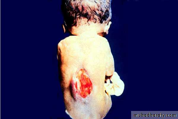
• e. Myelomeningocele:• Paraplegia, wasting, or contractures, with sensory loss.
• Trophic disturbances are marked, especially perforating ulcers of foot.
• Other congenital anomalies associated, mainly hydrocephalus.
Myelomeningocele
Myelomeningocele
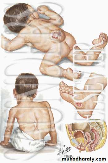
Myelomeningocele
SPINA BIFIDA
Investigations:Head circumference measurement to check for an associated hydrocephalus.
Plain radiograph of the spine.
Ultrasound of the brain to exclude hydrocephalus.
CT and/or MRI brain to rule out hydrocephalus.
SPINA BIFIDA
Treatment:
Spina bifida occulta requires no treatment in the majority of cases.
In the other forms of open spina bifida surgery should be performed as early as possible, and include:
Closure of the defect as early as possible to avoid meningitis.
The hydrocephalus needs immediate shunting even prior to closure of the defect of spina bifida.