• Subarachnoid hemorhhage
• Cerebral vasospasm• Traumatic aneurysm
• Mycotic aneurysm
Vascular neurosurgery
Subarachnoid hemorrhage
Trauma is the most common cause of SAHSpontaneous SAH can be due to
Ruptured aneurysm
Ruptured AVM (arteriovenous malformation)
Vasculitis
Tumor
Coagulopathy
Dural sinus thrombosis
Idiopathic
10 % die before reaching the hospital
10 % die within the first few daysOverall mortality 45 %
Peak age for spontaneous SAH is 55-60 years
Patients more than 70 years having higher incidence and severe neuro deficit
Risk factors
Hypertension
Cigarette and alcohol consumption
Oral contraception , pregnancy and parturition
Advancing age
Clinical feature
SymptomsSudden onset of severe headache (the most common symptom 97% , usually severe described as the worst headache in my life, this type of headache may clear and called sentinel hemorrhage or warning headache
Vomiting, photophobia, diplopia due to 3rd nerve palsy from aneurysmal compression and with more severe cases neurological complication and impaired consciousness
Signs
1- Meningismus: positive meningeal signs that often develop in 6-24 hrs and includeNuchal rigidity (neck stiffness) on flexion
Kernig sign ( flex the thigh and knee joint to 90 degree, then straighten the knee, pain in the hamstring muscles will develop)
Brudzinski ( involuntary hip flexion on flexing the neck )
2- Hypertension, focal neurological deficit and impaired consciousness
3- Ocular hemorrhage in form of
Intraretinal hemorrhage
Preretinal hemorrhage
Vitreal hemaorrhage ( terson syndrome ) occur in 6-26%
diagnosis
Clinical features
Investigations
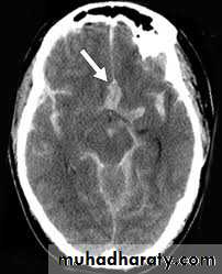
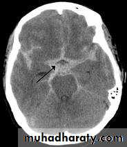
Non contrast high resolution CT scan which can demonstrate the amount of blood in the cisterns and fissure, ventricular size, associated intracranial hemorrhage, infarction and predict the location of aneurysm.
LP (lumbar puncture) if CT scan negative (the most sensitive test for SAH)
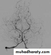
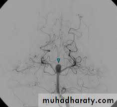
Cerebral angiography (the gold standard for evaluation of cerebral angiography) and can also demonstrate cerebral vasospasm
SAH versus traumatic LP
Opening pressure : elevatedNon-clotting bloody fluid that does not clear with sequential tubes
Xanthochromia : pink or yellow coloration of CSF supernatant due to broken RBC , the most important point needing 2-4 hrs to develop
RBC count more than 100.000 RBC/ml
Protein elevated due to broken RBC
Glucose : normal or reduced
complications
SeizureRebleeding, the major concern initially
Hydrocephalus ( usually obstructive by blood clot or communicating type which develop often late due to toxic effect of blood breakdown)
Cerebral vasospasm
Hunt and Hess grading of SAH
• Grade• Features
• 1
• Asymptomatic, mild headache and slight neck stiffness
• 2
• Moderate to severe headache, neck stiffness
• 3
• Mild focal deficit, lethargy , confusion
• 4
• Stuper, moderate to severe hemiparesis
• 5
• Deep coma, decerebrate, moribund
WFNS grading of SAH
• WFNS grade
• Glascow coma scale
• Major focal deficit
• 1
• 15
• -
• 2
• 13-14
• -
• 3
• 13-14
• +
• 4
• 7-12
• + 0r -
• 5
• 3-6
• + or -
management
ICU with arterial line, endotracheal intubation for comatose patient, ECG monitor ,folley catheter and intraventricular catheterVital signs with neuro check
Head elevation 30 degree
fluid input and output
Pneumatic compression stocking
Prophylactic anticonvulsant, phenytoin 17mg/kg loading then 100mg TID or luminal 10 mg/kg loading then 5 mg/kg/day
Sedation with propofol
Analgesia e.g. phentanyl
Decadron
Stool softener
Antiemetic, avoid phenothiazines that lower seizure threshold, use zofran (ondansetron) 4 mg slow I.V TID
H2-blocker , ranitidine ampule 150mg BID or omeprazole or lansaprazole 30mg PO q day)
Calcium channel blocker with Nimodipine 60 mg 6 times a day PO within 96 hrs
Treatment of underlying aneurysm by
Surgical clipping
Endovascular coils application
Endovascular trapping
Endovascular balloon therapy
Cerebral vasospasm
Angiographic vasospasm: asymptomatic arterial narrowing seen on angiography
Clinical vasospasm : delayed ischemic neurological deficit, manifested clinically as
increasing headache
alteration in the level of consciousness
Disorientation
meningismus
a feature of anterior or middle cerebral artery syndrome
• Most significant cause of morbidity,
• almost never before day 3 post hemorrhage• usually the onset is between day 6-8 and resolve by day 12
Risk factors for vasospasm include
Increasing age
Female with middle cerebral artery aneurysm
Hypertension
Pial enhancement on CT scan
Hypovolemia
Low GCS on admission
Higher amount of blood within subarachnoid space
• diagnosis
Clinical deterioration
Ruling out rebleeding, hydrocephalus, seizure, cerebral oedema, hyponatremia
Transcranial Doppler, high blood velocity with spasmodic artery , more than 120 cm/s
Cerebral angiography
treatment
Triple H therapy, hypertension, hypervolemia and hemodilusion
Calcium channel blocker, nimodipine
Removing blood clot surgically
Intra-arterial Balloon dilatation
Intra-arterial papaverine injection
Traumatic aneurysm
Compromise less than 1 % of intracranial aneurysmMost are false aneurysm (pseudo aneurysm)
Rare in children
Caused by
Penetrating trauma
Closed head injury (more common)
Iatrogenic , following surgery
Can be presented by
Progressive headache
Delayed intracranial hemorrhage
Progressive cranial nerve palsy
Treated by
Balloon trapping or balloon embolization
Mycotic aneurysm (infectious or bacterial )
Compromise 4 % of intracranial aneurysm
Occur in 4-14 % of patient with subacute bacterial endocarditis (SBE)
Usually caused by streptococcus followed by staphylococcus
Treated by antibiotic for 4-6 weeks with angiographic follow up if failure treated surgically