Genetics
objectives1- human genome project
2- DNA structure
3- genetic disorders
4- mendelian inheritance
5- autosomal dominant inheritance
The Human Genome Project, With DNA technology rapidly progressing, a group of visionary scientists in the USA persuaded Congress in 1988 to fund a coordinated international program to sequence the entire human genome. The program would run from 1990 to 2005 and 3 billion US dollars were initially allocated to the project
The draft DNA sequence of 3 billion base pairs was successfully completed in 2000 It is now estimated that the human genome contains 30 000-35 000 genes, although the function of many of these genes remains unknown
Clinical application of these advances is available to families through specialist genetic centres that offer diagnosis, investigation, counselling and antenatal diagnosis for an ever-widening range of disorders. Gene therapy trials are also underway, bringing hope of improved treatment in the future.
DNA
DNA consists of long chains of molecules called nucleotides. Each nucleotide consists of a nitrogenous base(pyrimidine and purine ), deoxyribose sugar and a phosphate group. DNA structure is in the form of two polynucleotide chains arranged in a double helix.
The arrangement of the bases in the double helix is organised such that adenine always pairs with thymine, and cytosine always pairs with guanine. Thus the arrangement of nucleotides in the two chains is complementary. Thymine is not present in RNA but is substituted by uracil.
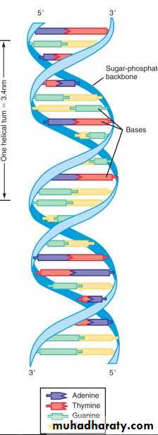
The pyrimidines are cytosine (C) and thymine (T); the purines are guanine (G) and adenin(A
DNA exists not as one long molecule, but as multiple fragments that together with a protein skeleton (chromatin) form chromosomesEvery human cell has 23 pairs of chromosomes. One copy of each chromosome is inherited from each parent. Twenty-two pairs of chromosomes are autosomes; the remaining pair is called the sex chromosomes. Females have two X chromosomes; males have one X and one Y.
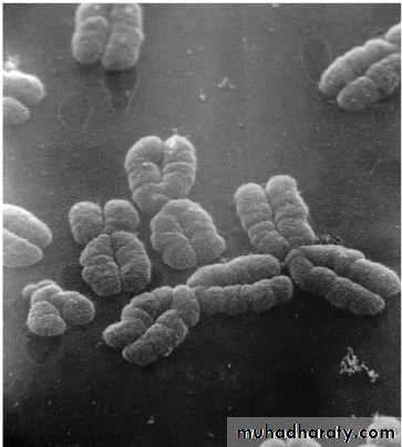
DNA is packaged into chromosomes and at a very simple level these can be considered as being made up of tightly coiled long chains of genes. Unlike DNA, chromosomes can be visualized during cell division using a light microscope, under which they appear as thread-like structures or 'colored bodies'. The word chromosome is derived from the Greek chroma (= color) and soma (= body).
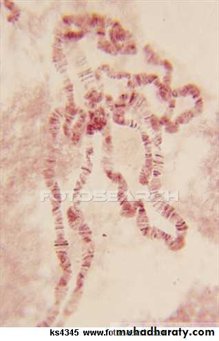
Under the electron microscope chromosomes can be seen to have a rounded and rather irregular morphology
Genetic disorders
common, with 2% of live-born babies having a significant congenital malformation and about 5% a genetic disorderburdensome to the affected individual, family and society, as many are associated with severe and permanent disability
Genetically determined diseases
1. Single gene mutations,((Mendelian disorders)2. Chromosomal disorders
3. Multifactorially inherited conditions
4. Disorders that show an unusual pattern of inheritance
5. Teratogenically caused conditions
Mendelian inheritance
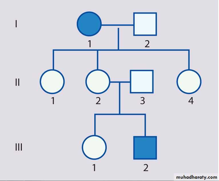
Disorders with these patterns of inheritance, described by Mendel in 1865, are rare individually, but collectively numerous, with over 15 000 single gene traits or disorders describedThere are three classic forms of genetic inheritance: autosomal dominant, autosomal recessive, and X-linked
Autosomal dominant inheritance
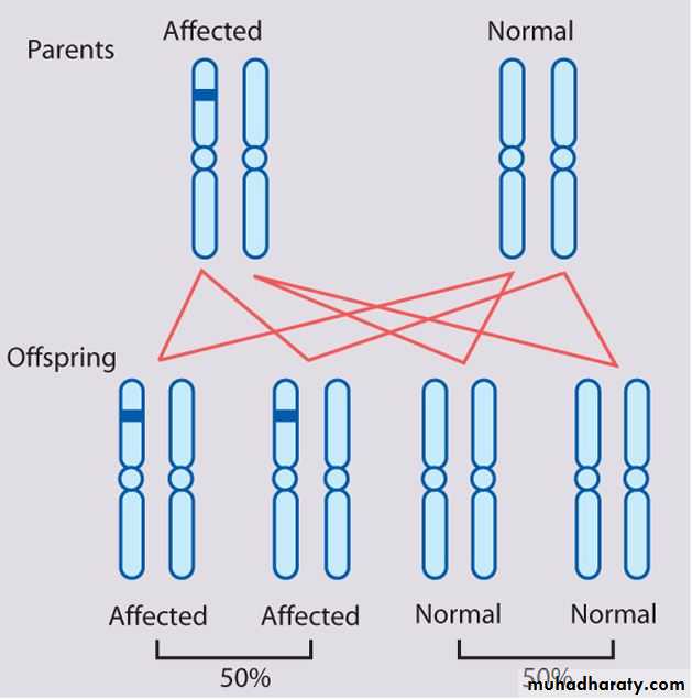
• This is the most common mode of Mendelian inheritance• Autosomal dominant inheritance is determined by the presence of one abnormal gene on one of the autosomes (chromosomes 1–22).
• Male and female offspring each have a 1 in 2 (50%) chance of inheriting the abnormal gene from an affected parent
Tuberous sclerosis
Marfan syndromeNeurofibromatosis
Huntington's disease
Retinoblastoma
Waardenburg syndrome
Myotonic dystrophy
Familial hypercholestrolemia (LDL receptor defect Type IIa)
Adult polycystic kidney disease
von Hippel Lindau
Familial adenomatous polyposis and Peutz Jeghers Syndrome
Hereditory spherocytosis
Achondroplasia
Ehlor's Danlos (vascular type)
Acute intermittent porphyria
Hypertrophic Obstructive Cardiomyopathy (HOCM)
Von Willebrand Disease
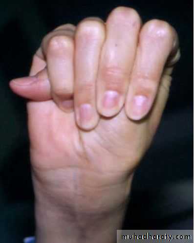
Polydactyly
Osteogenesis Imperfecta (Except Type VII)
Hereditary hemorrhagic telengiactasia (Osler-weber-rendu syndrome)
Osteopetrosis Type II (Adult type)
Hypokalemic Periodic Paralysis
Variation in expression
Within a family, some affected individuals may manifest the disorder mildly and others more severely. For example, a parent with tuberous sclerosis may have mild skin abnormalities only, but his or her affected child may have, in addition, epilepsy and learning difficulties.

Non-penetrance
Refers to the lack of clinical signs and symptoms in an individual who must have inherited the abnormal gene. An example of this is otosclerosis, in which only about 40% of gene carriers develop deafnessExample of non-penetrance
No family history of the disorder
A new mutation in one of the gametes leading to the conception of the affected person. This is the most common reason for absence of a family history in dominant disorders, e.g. >80% of individuals with achondroplasia have normal parents.Rules of Autosomal Dominant Inheritance
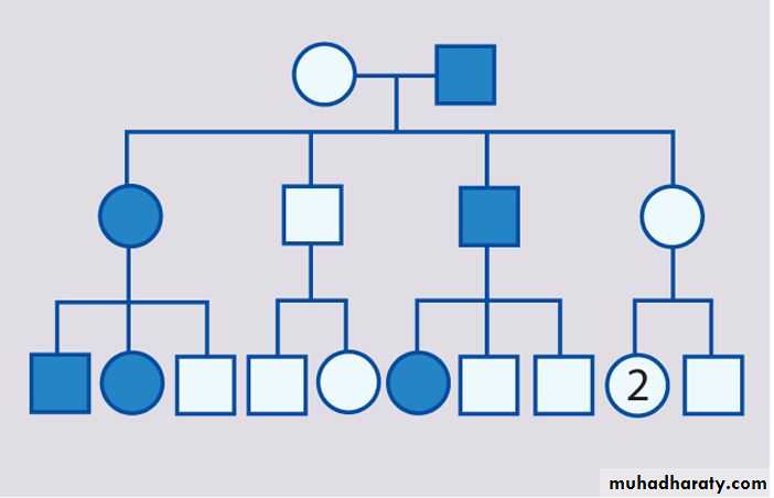
Trait appears in every generationEach child of an affected parent has a 1 in 2 chance of being affected
Males and females are equally affected
Male-to-male transmission occurs
Examples of Some Autosomal Dominant Disorders
Achondroplasia
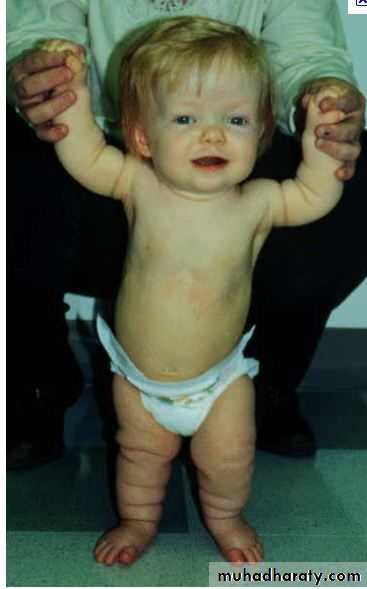
achondroplasia is the most common skeletal dysplasia in humans. The bony abnormalities in achondroplasia lead to short stature, macrocephaly, a flat midface with a prominent forehead, and rhizomelic shortening of the limbs.


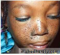
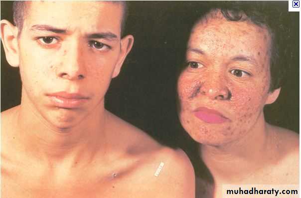
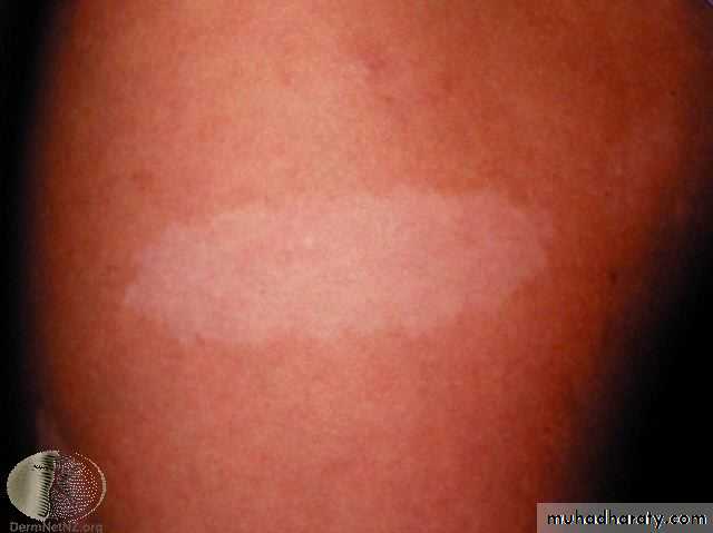
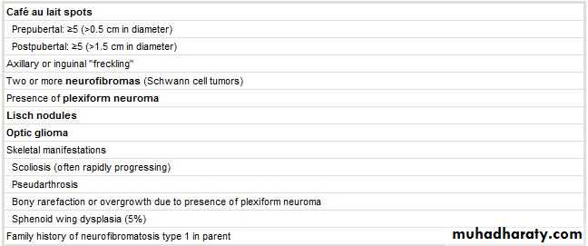
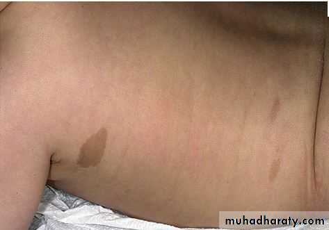
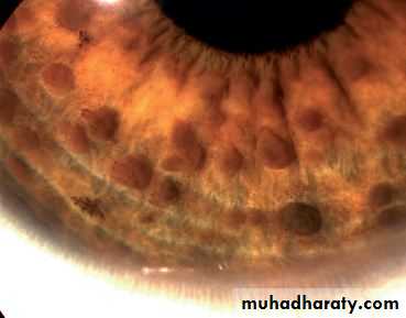
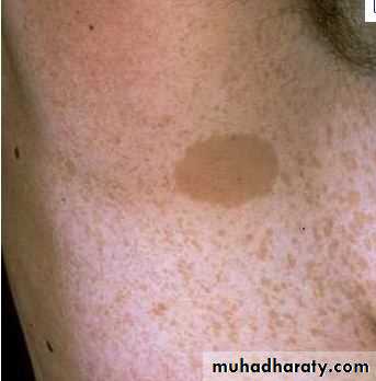
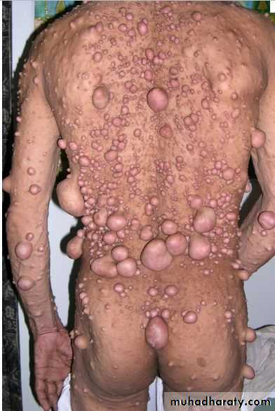
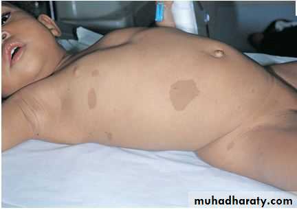
Neurofibromatosis Type 1
Marfan Syndrome
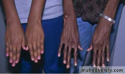
Clinical symptoms mostly involve three systems: cardiac, musculoskeletal, and ophthalmologic. Musculoskeletal findings include dolichostenomelia (a tall, thin body habitus), arachnodactyly (spider-like fingers and toes), abnormalities of the sternum (pectus excavatum or carinatum), kyphoscoliosis, pes planus, and joint laxityEye findings include high myopia, which eventually can lead to vitreoretinal degeneration; an abnormal suspensory ligament of the lens, which can lead to ectopia lentis (dislocation of the lens; in Marfan syndrome, the lens usually dislocates upward and outward); and cataracts. Cardiac findings include a weakened aortic wall, which leads to progressive dilation of the aortic root. Aortic insufficiency followed ultimately by aortic dissection is a common complication of this disorder