
1
Forth stage
Medicine
Lec-8
.د
رامي
1/1/2014
Pneumothorax
Pneumothorax is the presence of air in the pleural space,which can either occur
spontaneously, or result fromiatrogenic injury or trauma to the lung or chest wall.
Primary spontaneous pneumothorax occurs in patients with no history of lung
disease. Smoking, tall stature and the presence of apical subpleural blebs are risk factors.
Secondary pneumothorax affects patients with pre-existing lung disease and is
associated with higher mortality rates.
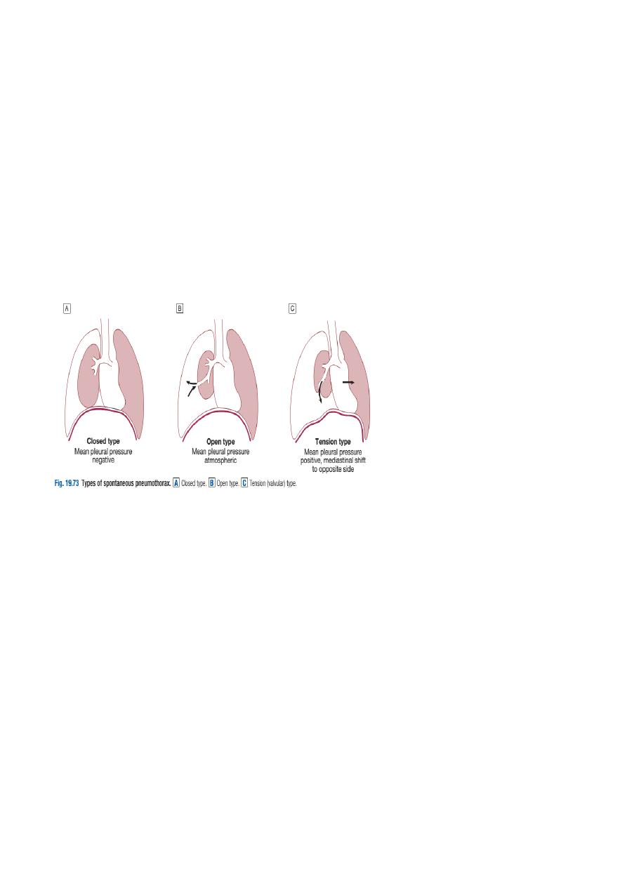
Where the communication between the airway and the pleural space seals off as the
lung deflates and does not re-open, the pneumothorax is referred to as ‘closed’.
The mean pleural pressure remains negative, spontaneous reabsorption of air and re-
expansion of the lung occur over a few days or weeks, and infection is uncommon. This
contrasts with an ‘open’ pneumothorax, where the communication fails to seal and
air continues to pass freely between the bronchial tree and pleural space.
Classification of pneumothorax:
1)Spontaneous:
A-Primary
•
No evidenceof overt lung disease.Air escapes from the lung into the pleural space
through rupture of a small pleural
bleb,or the pulmonary end of a pleural adhesion.
B-Secondary
•
Underlying lung disease,
most commonly COPD and tuberculosis; also
seen in
asthma,
lung abscess, pulmonary infarcts,
bronchgenic
carcinoma,and all forms of fibrotic and cystic lung disease.
2)Traumatic:
•
Iatrogenic (e.g. following thoracic surgery
or biopsy) or chest wall injury
2
An example of the latter is a bronchopleural fistula, which, if large, can facilitate the
transmission of infection from the airways into the pleural space, leading to empyema.
An open pneumothorax is commonly seen following rupture of an emphysematous
bulla, tuberculous cavity or lung abscess into the pleural space.
Occasionally, the communication between the airway and the pleural space acts as a one-
way valve, allowing air to enter the pleural space during inspiration but not to escape on
expiration. Large amounts of trapped air accumulate progressively in the pleural
space and the intrapleural pressure rises to well above atmospheric levels. This is a
tension pneumothorax. The pressure causes mediastinal displacement towards the
opposite side, with compression of the opposite normal lung and impairment of systemic
venous return, causing cardiovascular compromise.
Investigations:
The chest X-ray shows the sharply defined edge of the deflated lung with complete
translucency (no lung markings) between this and the chest wall.
Care must be taken to differentiate between a large preexisting emphysematous bulla
and a pneumothorax. CT is used in difficult cases to avoid misdirected attempts at
aspiration. X-rays may also show the extent of any mediastinal displacement and
reveal any pleural fluid or underlying pulmonary disease.
Management:
Primary pneumothorax, in which the lung edge is less than 2 cm from the chest wall and
the patient is not breathless, normally resolves without intervention. In young patients
presenting with a moderate or large spontaneous primary pneumothorax,
percutaneous needle aspiration of air is a simple and well-tolerated alternative to
intercostal tube drainage, with a 60–80% chance of avoiding the need for a chest drain.

3
In patients with significant underlying chronic lung disease ,however , secondary
pneumothorax may cause respiratory distress. In these patients, the success rate of
aspiration is much lower, and intercostal tube drainage and inpatient observation are
usually required, particularly in those individuals over 50 years old and those with
respiratory compromise
If there is a tension pneumothorax, immediate release of the positive pressure byinsertion
of a blunt cannula into the pleural space may be beneficial, allowing time to prepare for
chest drain insertion
When needed, intercostal drains are inserted in the 4th, 5th or 6th intercostal space in
the mid-axillary line, connected to an underwater seal or one-way Heimlich valve, and
secured firmly to the chest wall. Clamping of an intercostal drain is potentially dangerous
and rarely indicated. The drain should be removed the morning after the lung has
fully re-inflated and bubbling has stopped.
Continued bubbling after 5–7 days is an indication for surgery. If bubbling in the drainage
bottle stops before full re-inflation, the tube is either blocked, kinked or displaced.
Supplemental oxygen may speed resolution, as it accelerates the rate at which nitrogen is
reabsorbed by the pleura.
Patients with a closed pneumothorax should be advised not to fly, as the trapped
gas expands at altitude. After complete resolution, there is no clear evidence to
indicate how long patients should avoid flying, although guidelines suggest that waiting
1–2 weeks, with confirmation of full inflation prior to flight, is
prudent. Patients should also be advised to stop smoking and informed about the risks of a
recurrent pneumothorax. Diving is potentially dangerous after pneumothorax, unless a
surgical pleurodesis has sealed the lung to the chest wall.
Recurrent spontaneous pneumothorax:
After primary spontaneous pneumothorax, recurrence occurs within a year of either
aspiration or tube drainage in approximately 25% of patients, and should prompt
definitive treatment. Surgical pleurodesis is recommended in all patients following a
second pneumothorax and should be considered following the first episode of secondary
pneumothorax if low respiratory reserve makes recurrence hazardous.
Pleurodesis can be achieved by pleural abrasion or parietal pleurectomy at thoracotomy or
thoracoscopy