Pneumonia
Dr. Rami M Adil Al-HayaliAssistant professor in medicine
Definition
Pneumonia is an acute respiratory illness caused by an infection of the lung parenchyma, associated with recently developed radiological shadowing.This may take the form of:
Lobar pneumonia (or)
Bronchopneumonia
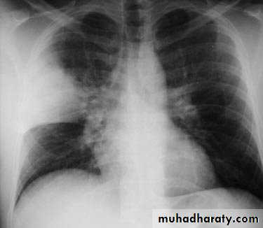
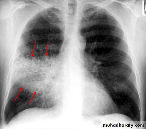
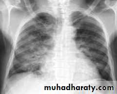
Lobar pneumonia: homogenous consolidation of one or more lung lobes, often with associated pleural effusion
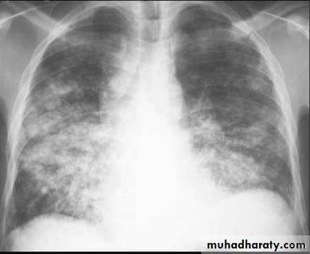
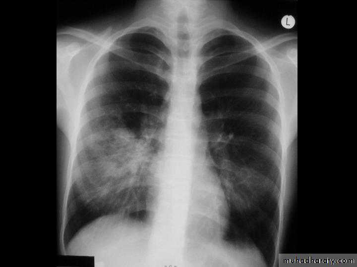
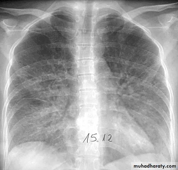
Bronchopneumonia: patchy alveolar consolidation associated with bronchial inflammation, often affecting lower lobes.
Classification
Pneumonia is best classified clinically according to the context in which it has developed:Community-acquired pneumonia(CAP)
Hospital-acquired pneumonia (HAP)
Pneumonia in immuno-compromised patient.
Community-acquired pneumoniaEpidemiology
Around 5-12/1000 adults suffer CAP each year in developed countries.Double incidence and higher mortality in the old.
Most cases are spread by droplet infection
Most cases occur in previously healthy individuals
Risk factors
Smoking
Upper respiratory tract infections
Alcohol drinking
Pre-existing lung disease
Corticosteroid therapy
Aetiology
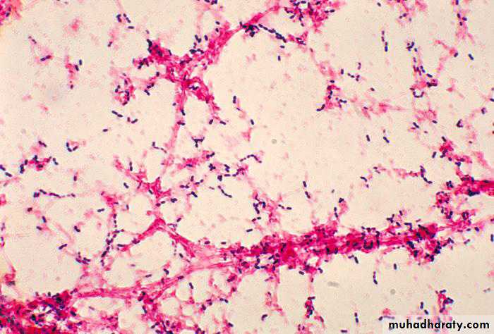
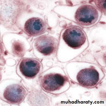
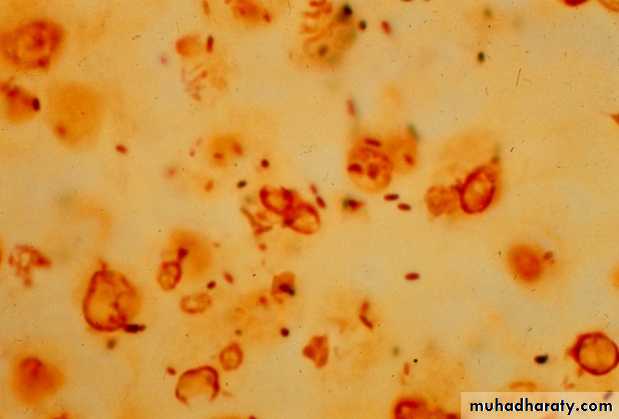
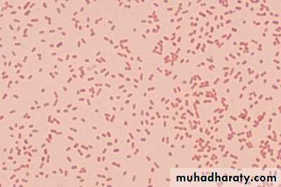
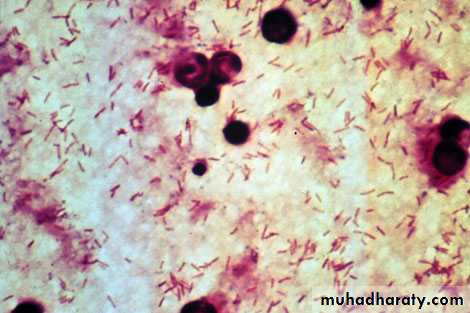
Extensive list of potential aetiological agents in CAP but most cases are caused by relatively few pathogensStreptococcus pneumoniae is the most common
Common pathogens also include Mycoplasma pneumoniae, Chlamydophila pneumoniae, Haemophilus influenzae and respiratory viruses.
Legionella pneumophila is also important (especially in inpatients).
Staphylococcus aureus (including MRSA) and Gram negative bacilli (Klebsiella pneumoniae and Pseudomonas aeroginosa) should be considered in selected cases.
Clinical features
Acute presentation with fever, shivering and/or rigor, vomiting, anorexia and headache.
Respiratory symptoms include cough, which is initially dry and painful, but later accompanied by mucoid, purulent or even bloody sputum. Pleuritic chest pain may be present.
The presence and degree of breathlessness depends on the severity of the disease.
Clinical features
On clinical examination, the patient is usually febrile with tachycardia, sweating or shivering.Tachypnoea and use of accessory muscle of respiration are markers of respiratory distress.
The patient may be cyanosed is severe cases.
Clinical features
Findings on chest examination depend on the degree of consolidation and the presence or absence of significant pleural effusion.Vocal fremitus may be increased or decreased
Percussion notes vary from impairment to stony dullness.
Crackles, bronchial breath sounds and friction rub may be heard on auscultation.
Clinical features
Elderly patients may present with confusion with few other respiratory manifestationsSeverely ill patients may have evidence of septic shock of organ failure.
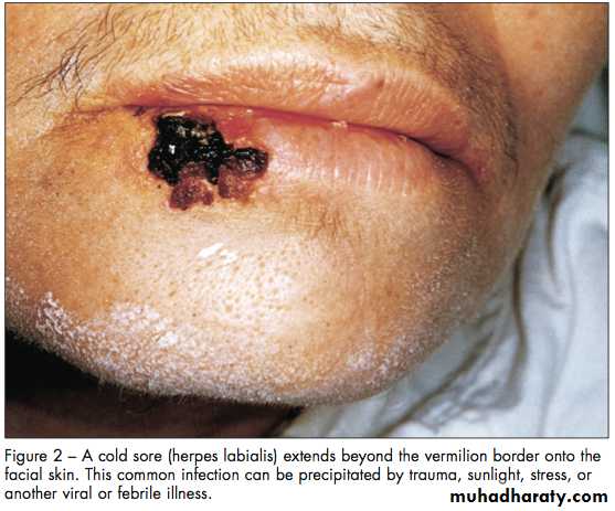
Streptococcus pneumoniae is the most common cause, characteristically has rapid onset, with high fever, pleuritic pain, rusty sputum, herpes labialis and lobar consolidation on chest X-ray.
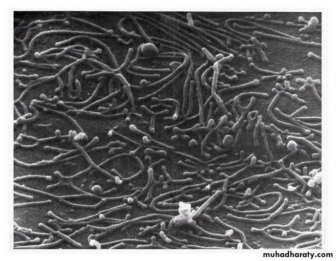
Mycoplasma pneumoniae is the second most common pathogen, especially affecting children and young adults.
It tends to occur in epidemics.
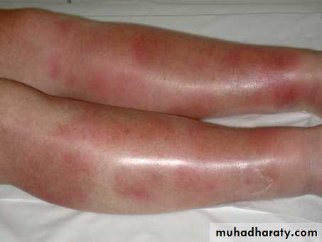
Unique complications include haemolytic anaemia, erythema multiforme, erythema nodosum, Guillain Barre syndrome as well as myocarditis, pericarditis and encephalitis.

Chlamydophila pneumoniae also affects young people with epidemic potential.
It is often mild and self limiting disease.Long duration of symptoms before presentation is characteristic.
H. influenzae is more common in old age and those with COPD or bronchiectasis.
It tends to cause bronchopneumonia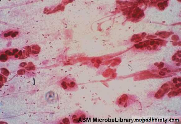
Viruses are responsible for about 18% of adult pneumonia.
It can be caused by influenza and parainfluenza (commonly complicated by secondary bacterial infections), respiratory syncytial virus and adenoviruses.Recently two coronaviruses were responsible for pandemic acute respiratory syndromes with viral pneumonia (SARS and MERS CoV).
Legionella pneumophila tends to affect middle and old aged
Usually as local epidemics around contaminated source (like cooling systems)Common associated symptoms include mental confusion and diarrhoea
Special lab abnormalities (hyponatraemia, elevated liver enzymes and hypoalbuminaemia)
Staphylococcus aureus occur in association with debilitating illness, or complicating influenza.
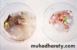
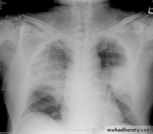
Multiple lobe involvement, cavitation, pneumatocoele and abscess formation are characteristic.
The infection may disseminate to other organs (as osteomyelitis)
Klebsiella pneumoniae is more common in alcoholic, diabetics and old men.
It usually affects upper lobesTendency to suppuration and abscess formation
Pseudomonas aeroginosa most commonly causes pneumonia in patients with bronchiectasis, cystic fibrosis and severe COPD
Differential diagnosis
Pneumonia should be differentiated from:Pulmonary infarction (due to pulmonary embolism)
Pulmonary and pleural tuberculosis
Radiation pneumonitis
Pulmonary oedema (can be unilateral)
Rare cases (as pulmonary oesinophilia and bronchoalveolar carcinoma)
Investigations
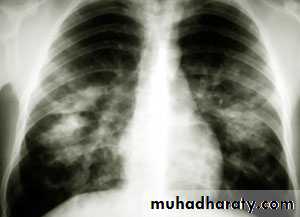
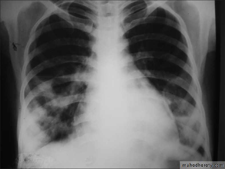
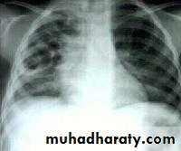
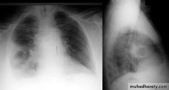
A chest X-ray is usually sufficient to confirm the clinical diagnosis of pneumonia.
In lobar pneumonia, a homogenous opacity localized to the affected lobe or segment usually appears within 12 – 18 hours of the onset of illness.
CT scan is rarely required, except for suspected underlying bronchial obstruction caused by tumour or foreign body.
Investigations
Chest X-ray helps in:• Differentiating CAP from other diagnosis
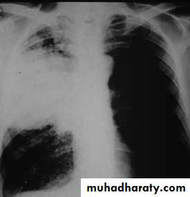
• Provide information about severity (cavitation and multilobar involvement)
• Detects complications (pleural effusion or abscess formation).
• It can occasionally suggest an aetiological agent (pneumatocoele in Staphylococcus aureus pneumonia).
Investigations
Pulse oximeter non-invasively assesses the arterial oxygen saturation (SpO2).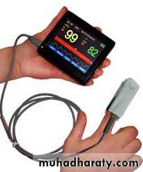
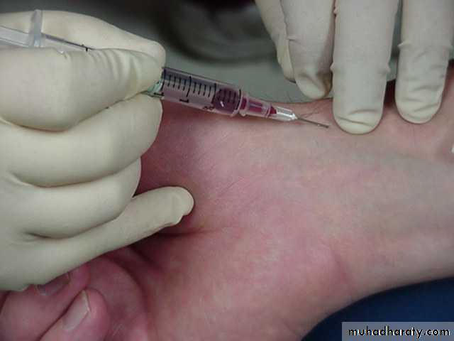
Arterial blood gas analysis is required when the SpO2<93% to assess the need for ventilator therapy.

Investigations
WBC is normal or only marginally raised in pneumonia caused by atypical pathogens (mycoplasma, chlamydia, legionella and viruses), whereas a neutrophilic leukocytosis of more than 15 109/L suggests a bacterial aetiology.Urea, electrolytes and liver function tests should be checked.
Investigations
Aetiologic diagnosis is not necessary in most cases.Microbiological tests are required in cases of severe pneumonia which include:
Sputum Gram and Zeihl Neelsen stains and sputum culture and sensitivity
Serology: antibody titres (mycoplasma, chlamydia and viruses), antigen detection in serum or urine (pneumococcus and legionella) and PCR (mycoplasma)
Assessment of disease severity
CURB-65 scoring system helps guiding antibiotic and admission policies and predicts prognosis. It encodes for:Confusion
Urea>7mmol/L
Respiratory rate>30/min
Blood pressure (systolic<90 mmHg or diastolic<60 mmHg)
Age>65.
Indications of ICU admission
ICU admission is also indicated in patients with:
• CURB 65 score of 4-5
• Persistent hypoxaemia
• Hypercapnoea and acidosis
• Septic shock
• Reduced conscious level
ManagementOxygen therapy
Oxygen therapy is indicated in patients with tachypnoea, hypoxaemia, hypotension or acidosis.PaO2 should be maintained around 60 mmHg (or SpO2 at 92%) giving high flow oxygen (>35%).
Mechanical ventilation should be considered in those who remain hypoxaemic despite oxygen therapy.
Non-invasive ventilation has limited role in pneumonia.
Management Fluid balance
Oral fluid intake should be encouraged.IV fluids are required in severely ill, old patients and those with vomiting.
Vasopressors may be required in patients with severe sepsis and septic shock.
Management Antibiotic therapy
Prompt institution of antibiotics improves outcome.The initial choice of antibiotics is primarily guided by the severity of illness.
Oral antibiotics are adequate in mild cases
The duration of treatment of uncomplicated pneumonia is 7-10 days
Management Antibiotic therapy
Outpatients:
Previously healthy patients, who have not received antibiotics during the preceding 3 months are treated with
Oral macrolide (clarithromycin 500mg twice daily, or azithromycin 500mg once daily)
Patients with co-morbidities (and those who had received antibiotics within 3 months) are better treated with:
Oral respiratory quinolone (moxifloxacin 400 mg once or levofloxacin 750mg once)
Management Antibiotic therapy
Inpatients:A respiratory quinolone (oral or IV) (OR)
A β-lactam (co-amoxiclav 1-2 gm three time daily IV or cefotaxime 1-2gm IV three time daily or ceftriaxone 1-2 gm IV daily) PLUS, a macrolide (oral or IV)
(If Staphylococcus aureus is suspected, add vancomycin or linezolid).
Management
Pleuritic pain is treated with paracetamol or NSAID. If not sufficient, opiates may be required (taking caution of possible respiratory depression)Physiotherapy may be required to assist expectoration.
Complications
Para-pneumonic effusion and empyemaSuppurative pneumonia and lung abscess
Lobar collapse due retained secretions
Pneumothorax (particularly in Staphylococcus aureus pnemonia)
Multi-organ failure (including ARDS and ATN)
DVT and pulmonary embolism
Prognosis
Most patients respond to antibiotic therapy.
Fever may persist for many days.Chest X-ray takes several weeks or months to resolve especially in elderly.
Delayed clinical recovery may suggest:
• Complications
• Incorrect diagnosis
• The pneumonia is secondary to proximal obstruction or recurrent aspiration
The mortality rate ranges from 1% in mild cases treated as outpatients to 50% in critically ill patients treated in the ICU.