1
Fifth stage
Surgery-Ortho
Lec-1
هشام القطان
18/10/2015
PRINCIPLE OF FRACTURES
References
Outline of orthopedics.
Mercers’ orthopaedic surgery.
Apley's system of orthopedics & fractures.
Fracture definition:-
It is a break in the structural continuity of bone .It may be no more than a crack, a
crumbling or splintering of the cortex ;more often the break is complete &the bone
fragment are displaced.
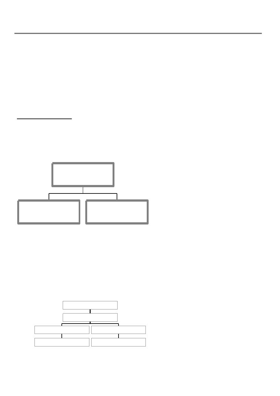
How fracture happen?
1.from single traumatic incident.
2.repetitive stress (fatigue).
3.abnormal weakening of the bone,(pathological fracture).
closed
compound
fracture
transverse fracture
tapping
comminuted fracture
crushing
direct force
traumatic incident
2
In cancellous bone trauma produce comminuted crush fracture.
Around joint pulling ligament and tendon produce avulsion fracture.
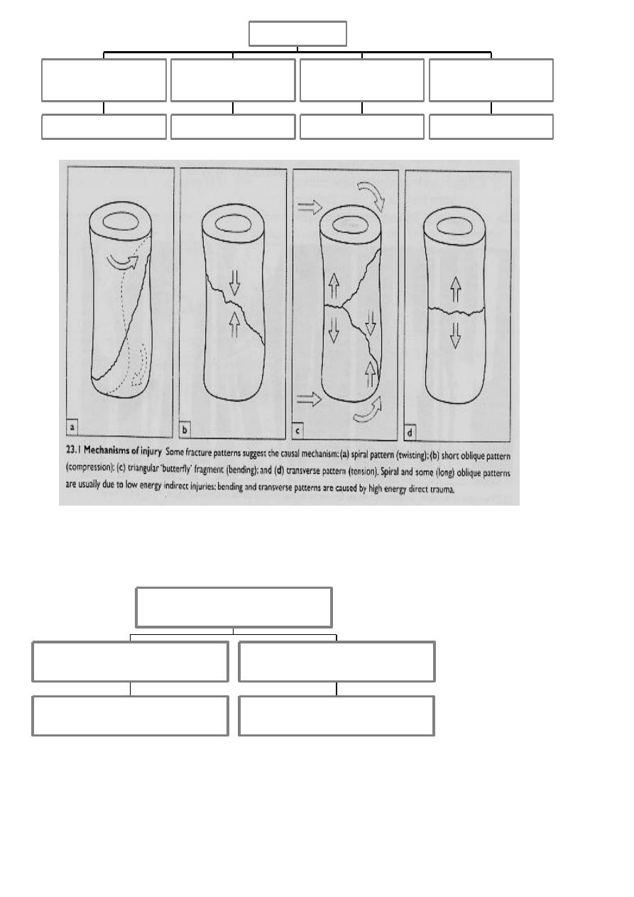
--How fracture are displaced?
Either by:-
spiral fracture
twisting
transverse
bending
transverse with butter fly
bending &compresion
short oblique
combination
bend.twist.comp.
indirect force
spiral
complete
green stick
incomplete
fracture

3
1-force of the injury.
2-Gravity.
3-Partly by pull of the muscle attached to them.
--How fracture heal?
Tissue destruction and haematoma formation.Inflammation and cellular proliferation
occurs within 8 hours. clotted haematoma is slowely absorbed and fine capillaries grow
into the area then (granulation tissues).
Callus formation is driven by inductive proteins.
Consolidation (woven bone is transformed into lamellar bone) takes several months.
Remodelling occurred over a period of months or even years.
Perkins’ time table
upper limb .
Spiral fracture
3 weeks united * 2 consolidation.
Lower limb * 2.
Transverse fracture * 2 again.
In children the time shorter, in elderly longer
OR THERE MUST BE CLINICAL AND RADIOLOGICAL evidence of consolidation before
full stress is permitted without splintage.
CLINICAL FEATURES:-
History.
General sign.
Local signs.
X-ray.
Special imaging.
History :-
There must be usually a history of injury ,followed by inability to use the injured limb.

4
BE WARE ……
The fracture is not always at the site of the injury,a blow to the knee may fracture the
patella ,femoral condyle ,even the acetabulum.
Pain are common symptom.
bruising ,swelling
Deformity.
Numbness,loss of movement ,
History of previous injury.
General medical history.
General signs:-
A broken bone part of a patient so look for .
Hemorrhage.
Associated damage to brain,spinal cord ,viscera.
Predisposing cause (pagets’ disease).
Local signs:-
Look :
swelling,
brushing,
deformity,
state of the skin.
Feel:-
Tenderness,
distal pulse,
sensation
Move:-
Crepitus
and abnormal movement,
movement of the joint distal to the injury.
5
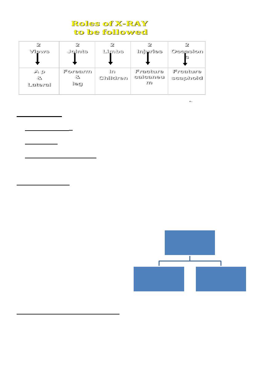
Special imaging:-
1.TOMOGRAPHY:-In spine or tibia condyle injure.
2.CT,M.R.I.:-In spine fracture which threatened the cord.
3.RADIO- ISOTOP scanning: -In stress fracture or undisplaced fracture.
Secondary injuries:-
Fracture spinecord injure.
Fracture pelvisabdominal viscera
Injuries(intestine, diaphragm).
Fracture ribslung ,heart
.Fracture and dislocation around
pectoral girdle--> brachial plexuses& vessels.
General treatment at the accident site
Air way .
Protection cervical spine.
Breathing.
Bleeding stoppage . by direct pressure OR by tourniquets. (time)
(Circulation) Fluid replacement
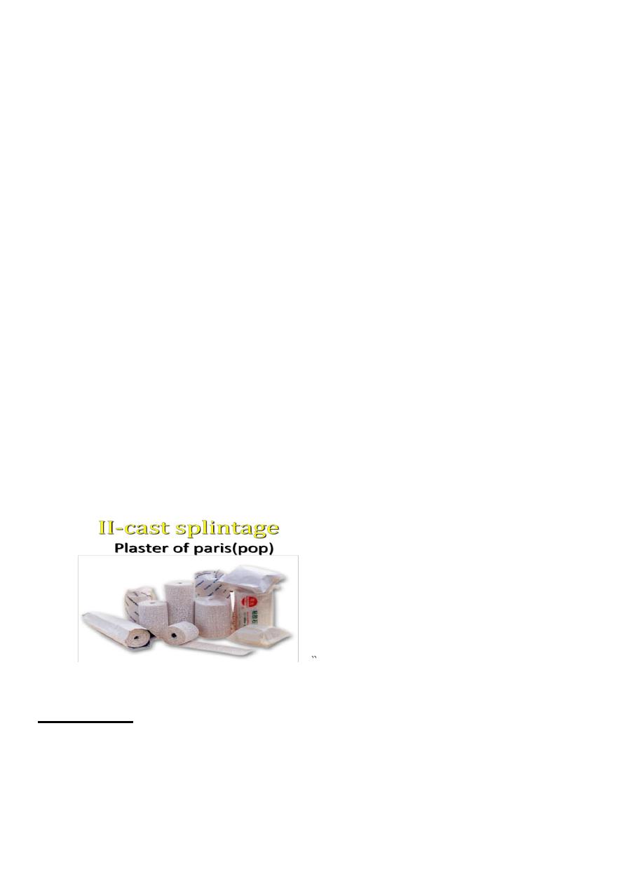
Treatment of
closed
fracture
General
treatment
Treatment of
fracture
6
Examination .
Analgesia.
Splintage to reduce pain, blood loss …….
Transport .by proper stretcher.
In the hospital:-
Rapid survey.
Constant re-evaluation.
Definitive care.
Reduction
Conservative (closed).
Operative (open).
Manipulation (reduction):-
A: closed reduction:
Under /anesthesia, with assistant.
Used in children fracture.
Minimally displaced fracture.
Fracture not unstable after reduction.
B:Open reduction.
If failure close.
Articular joint involvement.
Two bone fracture.
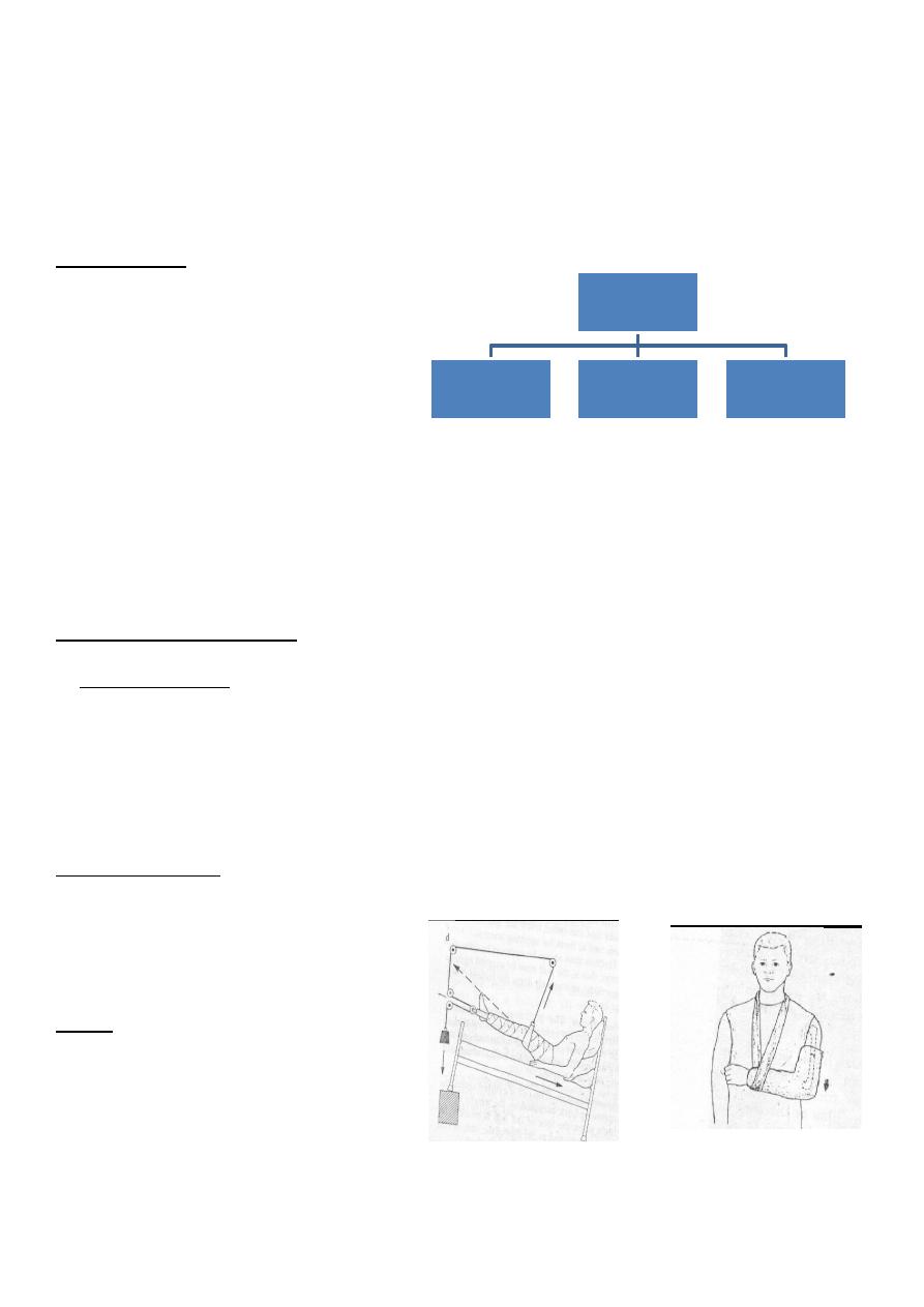
Hold
I. Continuous traction
1.Traction by the gravity:- **For upper limbs injuries.
treatment of
fracture
REDUCE
HOLD
PHYSIOTHERAPY
7
2.Skin traction:-
Not more (4-5KG) e.g.
fracture in children.
3.Skeletal traction:
By stein Mann pin or
Denham pin.
>5KG.
4.Fixed traction:-
5.Balanced traction:-
Over pulleys.
6.Combined traction:-
COMPLICATIONs of skin and skeletal:-
1.may constrict circulation (gallows traction).
2. Nerve injuries (peroneal).
3.compartment syndrome.
Complications:-
1.fracture disease:-stiffness, atrophy, osteoporosis,edema.
2.tight cast.
3.pressure sores.
8
4.Skin laceration
III-Functional bracing:-
Is one way of preventing joint stiffness while still
permitting fracture splintage and loading.
E.g.(fracture femur ,fracture tibia).
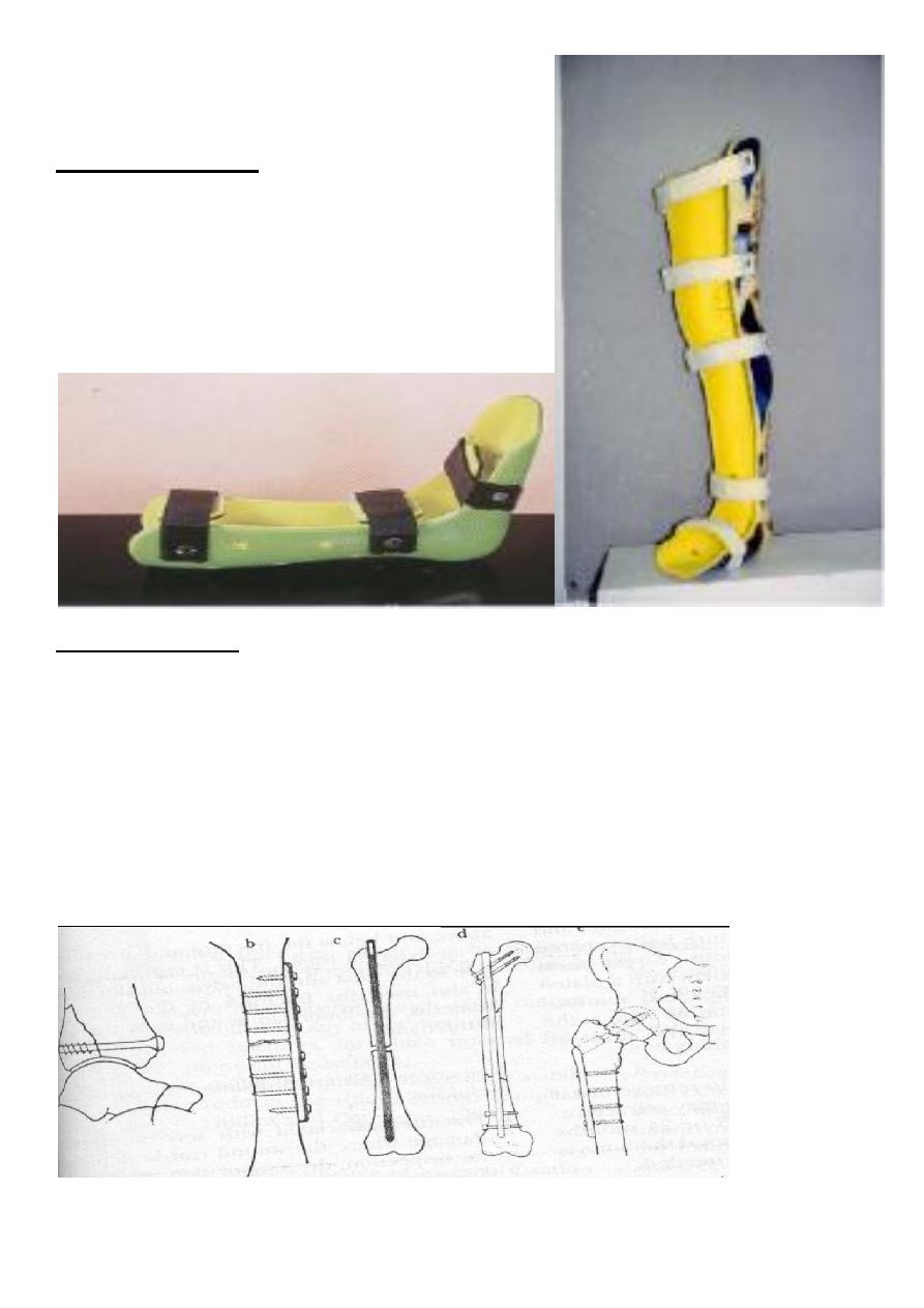
IV-internal fixation:-
Screw.
Plate .
Wire.
L-plate.
Compression screw.
K-nail.
9
Indications:-
1. Failure conservative.
2. Fracture two bones.
3. Intra articular fracture.
4. Pathological fracture.
5. In difficulty nursing patient.
Complications:-
1. Infection.
2. Non union.
3. Implant failure.
4. Re fracture.
**BE WHERE:-(fixation removal before one year at minimum and 18-24 months safer).
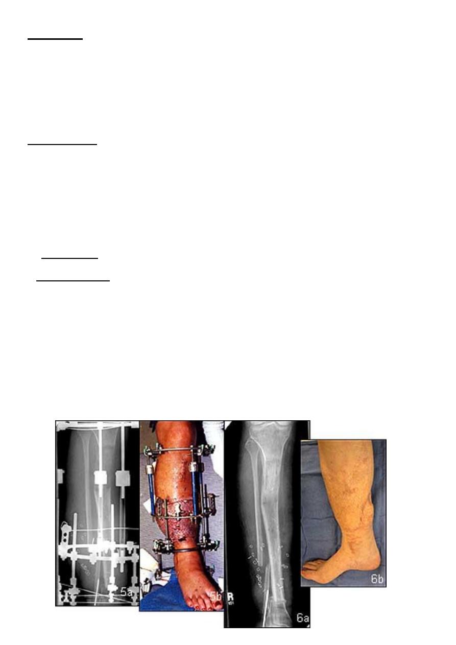
V.External fixation(indications):-
1. Fracture with severe soft tissue injuries.
2. Complicated fracture.
3. Infected non union.
4. Fracture pelvis.
5. Multiple injuries.
6. Bone lengthening.
7. Plastic surgery (flap).
8. Fracture neck femur.

11
Complications:-
Over distraction Reduce load transmission (so dynamization after 6-8 weeks or
remove).
Pin tract infection.
Exercise(physiotherapy):-
Prevention edema: by elevation.
Active exercise:So to pump edema fluid and stimulate circulation ,prevent soft tissue
adhesion &promotes fracture healing.
Assisted movement:By gentle movement only.
Functional activity:By improving patient mobility.
Photos www.muhadharaty.com/lecture/2828